salamander practical 2
1/22
There's no tags or description
Looks like no tags are added yet.
Name | Mastery | Learn | Test | Matching | Spaced | Call with Kai |
|---|
No analytics yet
Send a link to your students to track their progress
23 Terms

Salamander Buccal cavity: this is where the mouth would be opening from. Most anterior part of the body and sort of large. Shown on the ventral side.


Gular Fold: A flap of skin on the ventral side of the salamander

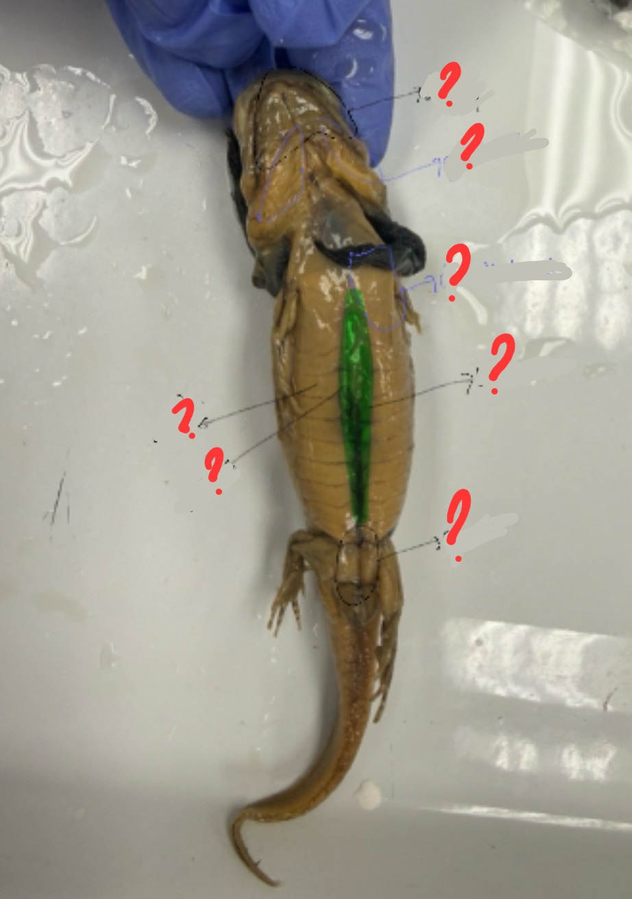
Dorsalis Trunci: This is on the dorsal side of the salamander. It is on the anterior portion of the body on each side of the middorsal septum.


Linea Alba: This is a very noticeable dark line on the ventral side of the salamander


Cloaca: This is found in between the legs of the pelvic girdle. It is on the ventral side.


External Oblique: This is on the ventral side of the Salamander, proximal to the rectus abdominis
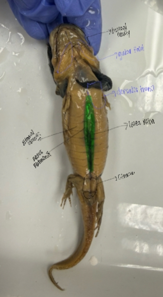

Rectus Abdominis: This is on the ventral side of the Salamander and the rectus abdominis is proximal to the linea alba.


External Gills: This is mostly on the lateral and ventral sides. It has attached parts that are very long.
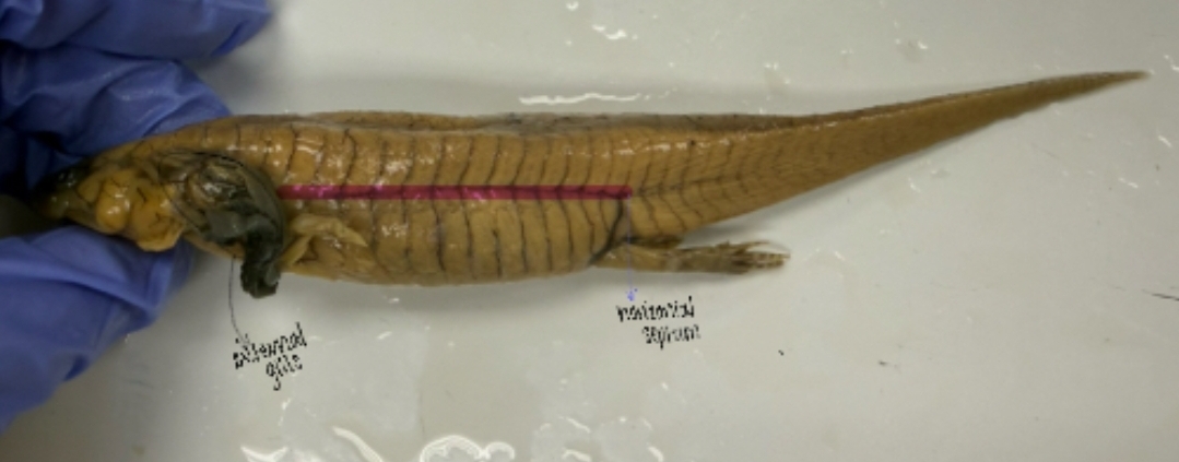
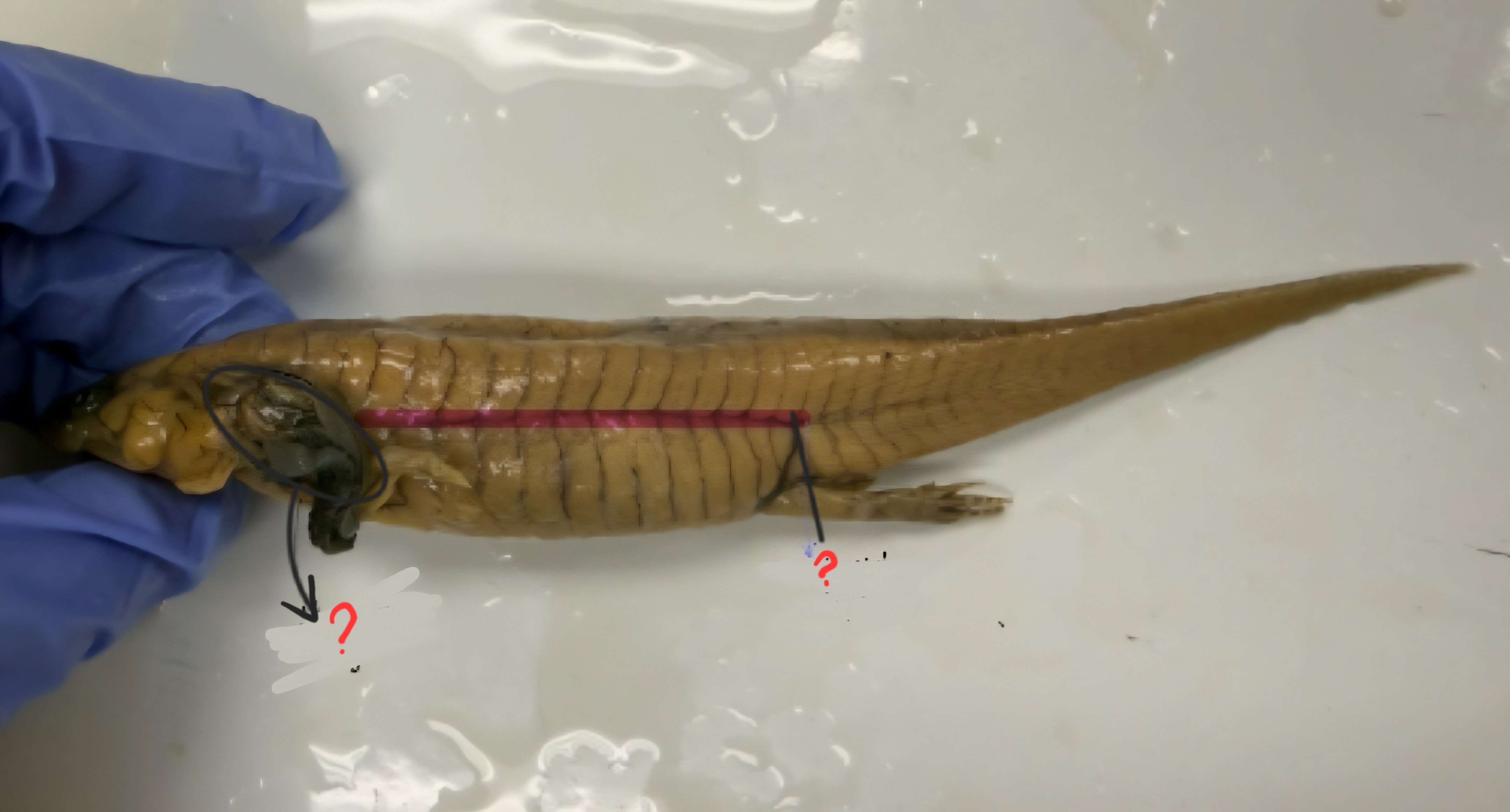
Horizontal Septum: The line on the lateral side of the salamander

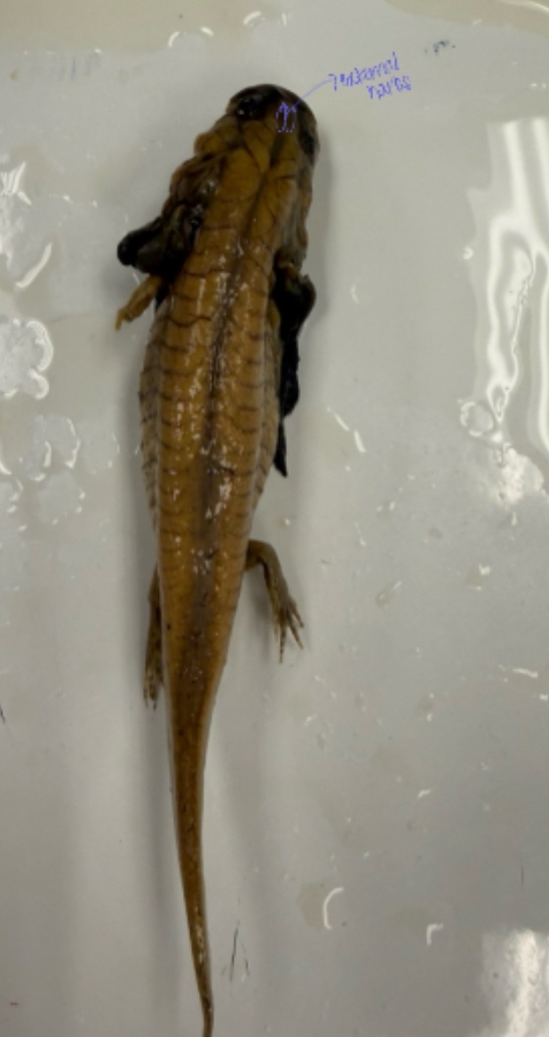
External Nares: This is found on the anterior part of the body with two tiny but long holes.
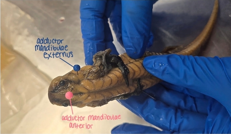
adductor mandibulae anterior: small, anterior jaw closing muscle, near front of skull
adductor mandibulae externus: larger, more lateral/dorsal jaw closing muscle, bulk of cheek region
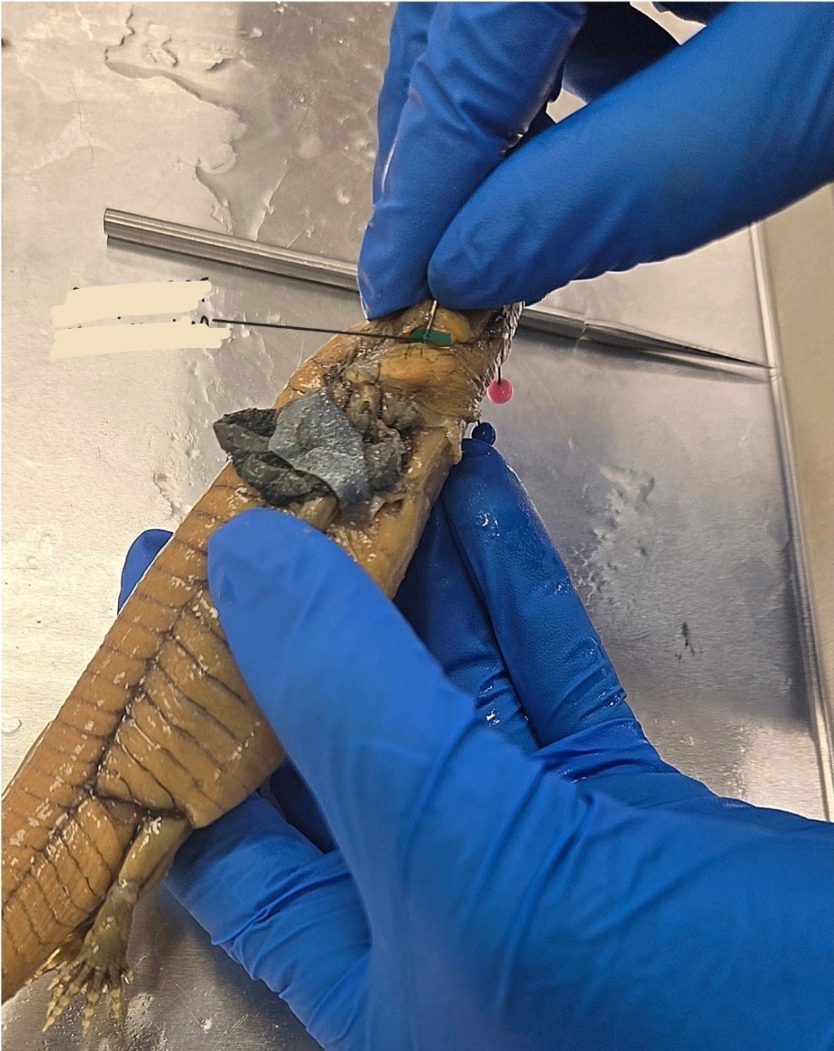
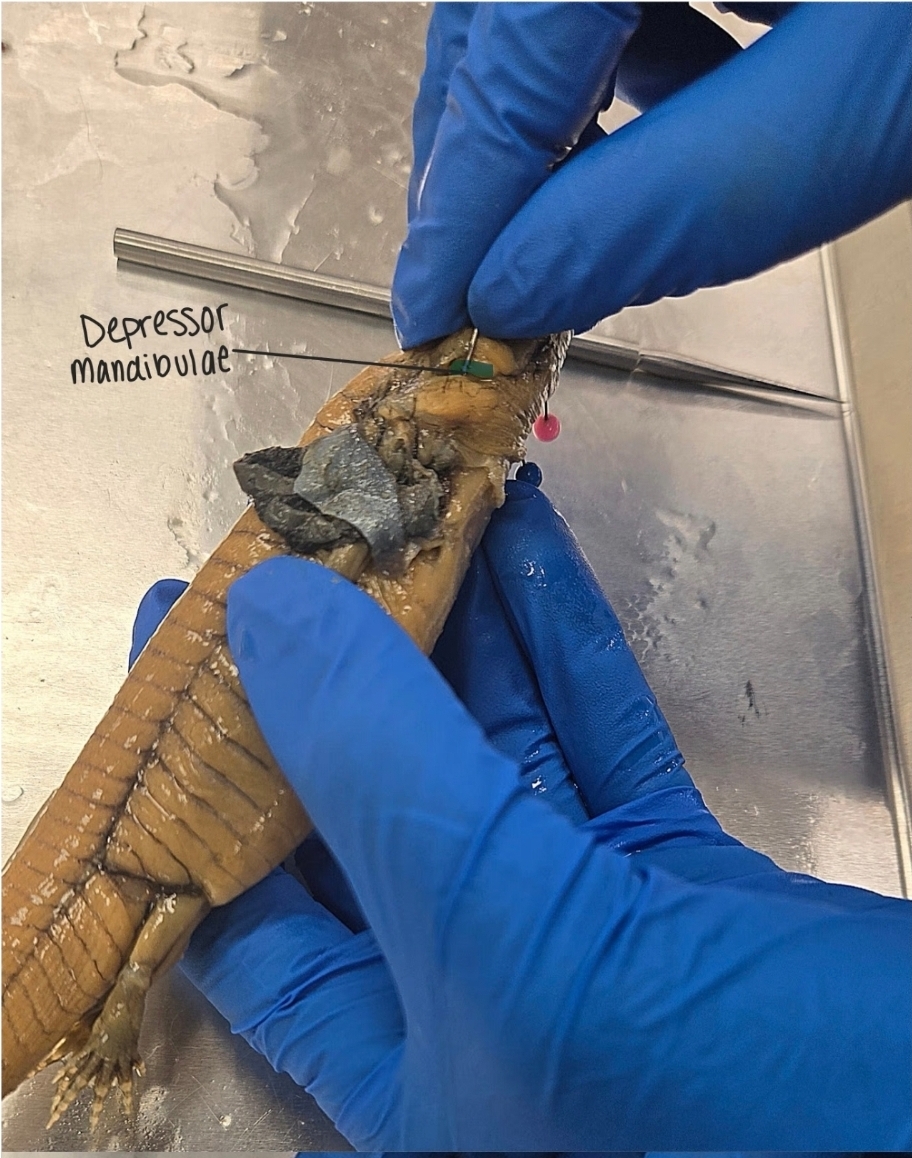
depressor mandibulae: posterior muscle; opens jaw
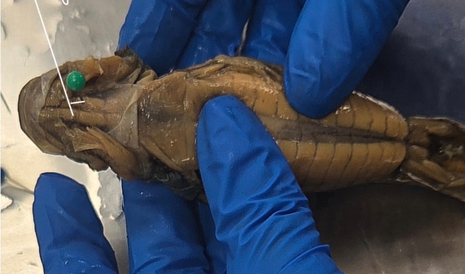
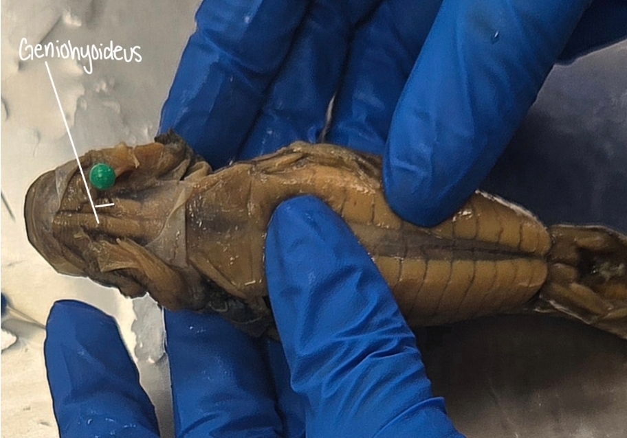
geniohyoideus: thin, runs from chin to hyoid; midline, anterior throat

interhyoideus: broader, more posterior throat sheet


intermandibularis: very superficial sheet between lower jaws (ventral floor of mouth)
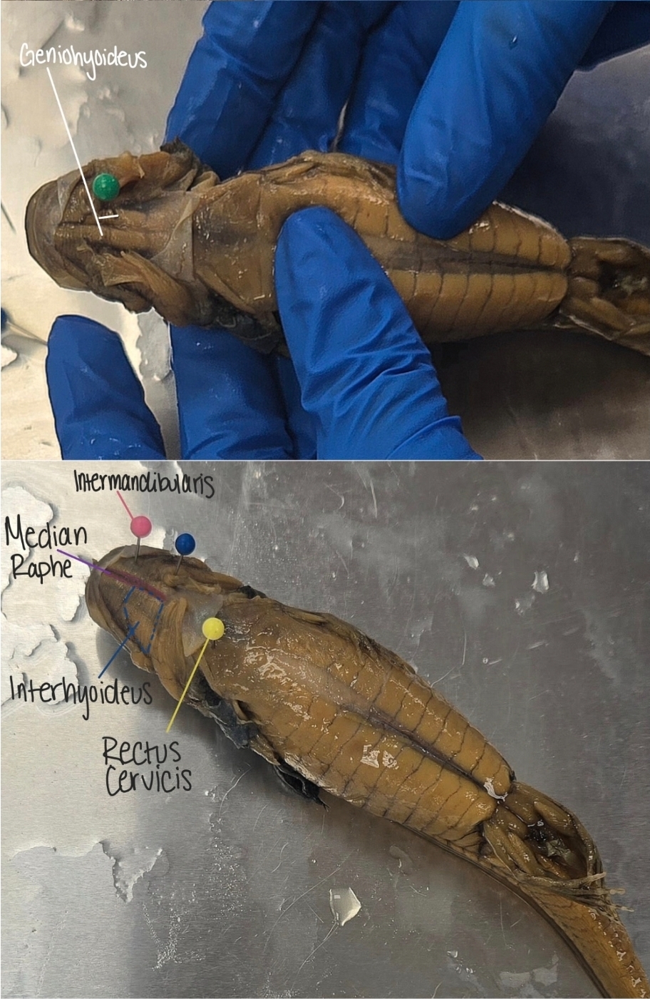
rectus cervicis: long strap-like muscle running posteriorly along midline of neck
rectus cervicis vs geniohyoideus:
rectus cervicis is more posterior (farther back), longer, extends into neck/trunk region, runs from hyoid to pectoral girdle, and looks like a deep, posterior strap
geniohyoideus is more anterior (under the jaw), shorter, runs from mandible to hyoid, and sits closwer to midline and under the mouth


median raphe: not a muscle; thin/faint midline seem where left/right muscles meet (especially visible in intermandibularis); this is connective tissue

A and B?
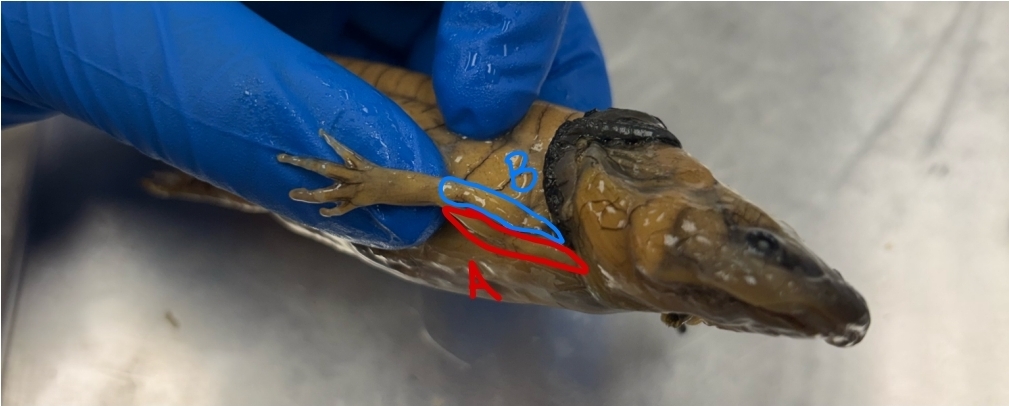
A: humeroantebrachialis
B: triceps brachii
A, B, C?
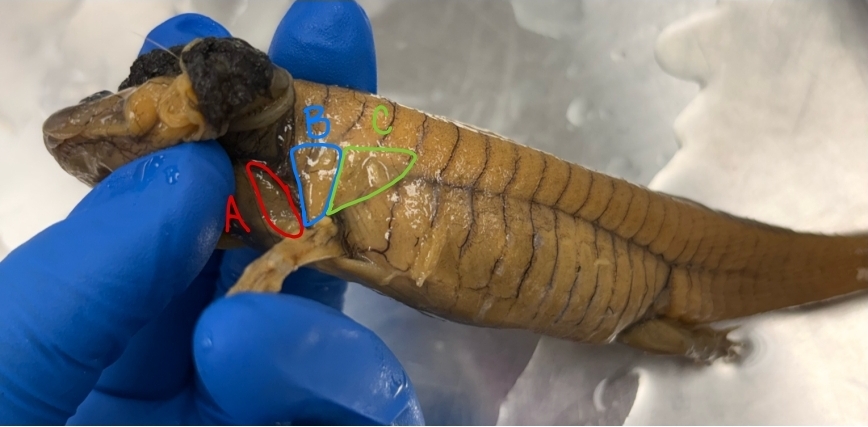
A: cucullaris
B: dorsalis scapulae
C: latissimus dorsi
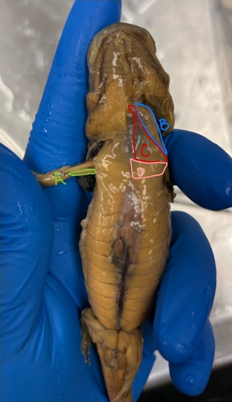
A, B, C, D?
A: coracobrachialis
B: procoracohumeralis
C: supracoracoideus
D: pectoralis

Levatores Arcuum- Fan shaped paired muscles connecting to gills
Origin: Fascia of the dorsalis trunci
Insertion: Epibranchial cartilages
Action: Gill elevators


Dilatator Laryngis Arcuum- An shaped muscle anterior to Levatores Arcuum
Origin: Fascia of the dorsalis trunci
Insertion: Cartilages of the larynx
Action: Expands the larynx

Branchiohyoideus- Cheek muscle directly ventral to Levatores Arcuum
Origin: Ceratobranchial of visceral arch III
Insertion: Ceratohyal (visceral arch II)
Action: Retracts the hyoid apparatus
