Looks like no one added any tags here yet for you.
mandibular foramen (picture)
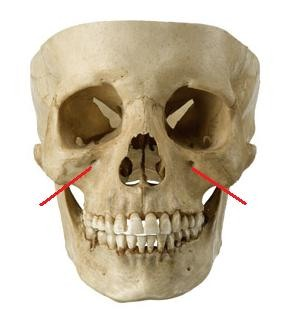
Zygomatic bone
Forms the cheek bones
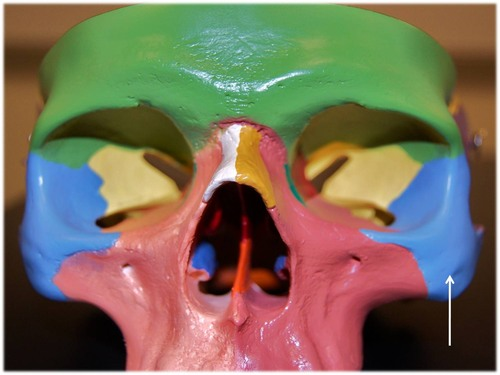
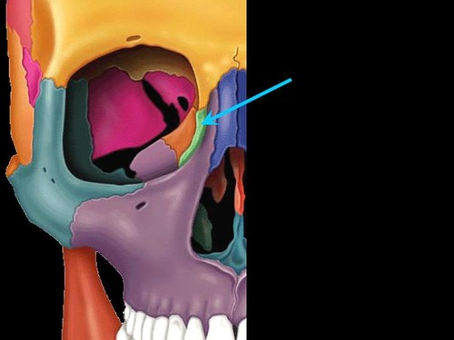
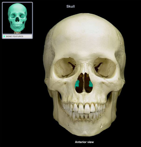
lacrimal bone
medial side of the orbit has a groove where the lacrimal sac is located
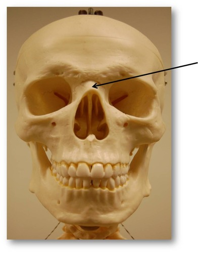
nasal bone
forms the bridge of your nose
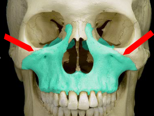
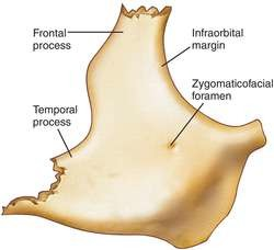
maxilla bone
contains (sockets) for the upper teeth
has infraorbital foramen
helps form the anterior part of the hard palate (roof of the mouth)
vomer
it forms the bony aspect of the nasal septum
interior nasal conchae
small hook like bones (most inferior) that project into the nasal cavity
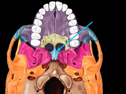
palatine
forms the posterior part of the hard palate
helps form part of the orbit
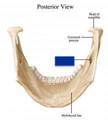
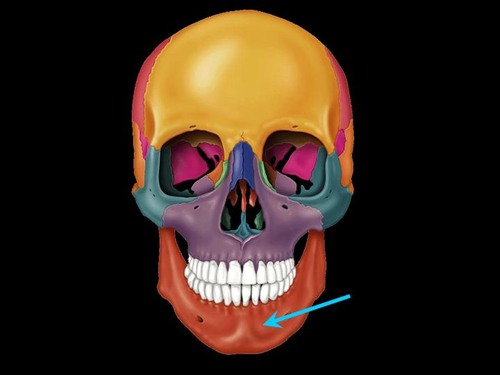
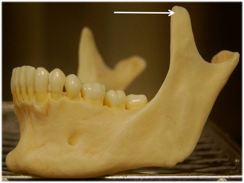
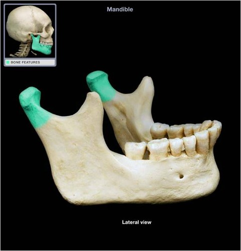
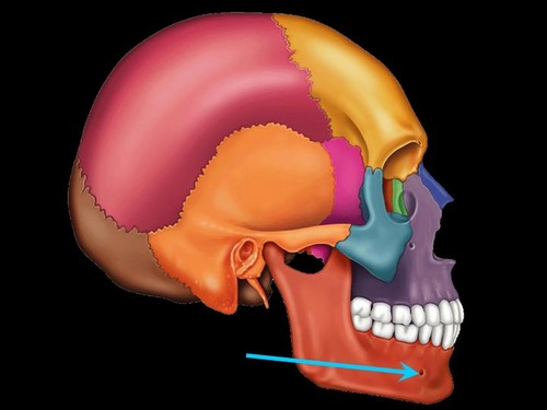
mandible
aka jaw
has sockets for the lower teeth
has a condylar process, coronoid process, mandibular foramen, mental foramen
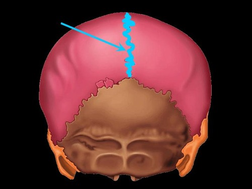
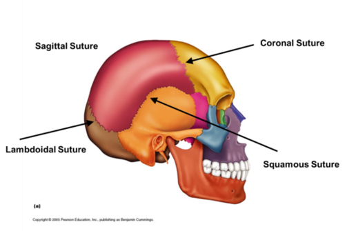
sagittal suture
joins the parietal bones
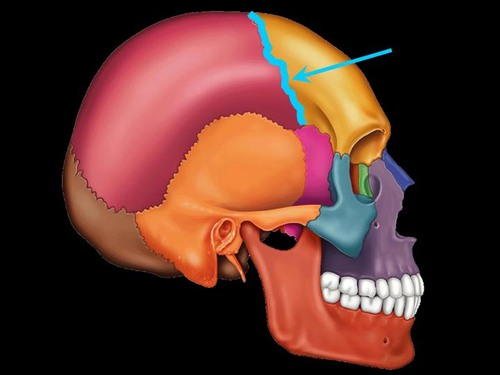
coronal suture
joins frontal and parietal bones
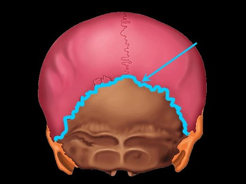
lambdoidal suture
joins parietal and occipital bones
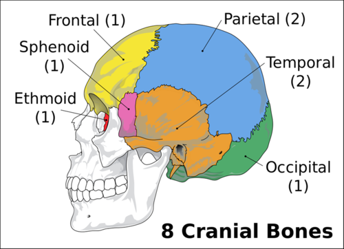
frontal bones
contains supraorbital foramen-hole where nerves and blood vessels pass through
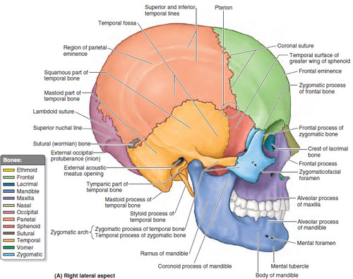
landmarks of temporal bone
masteroid process, external auditory meatus, styloid process
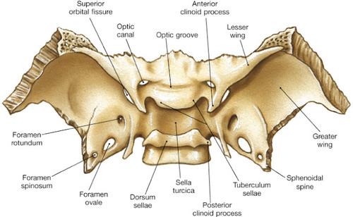
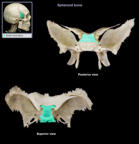
significance of sphenoid bone
aka keystone bone
articulates with all other cranial bones
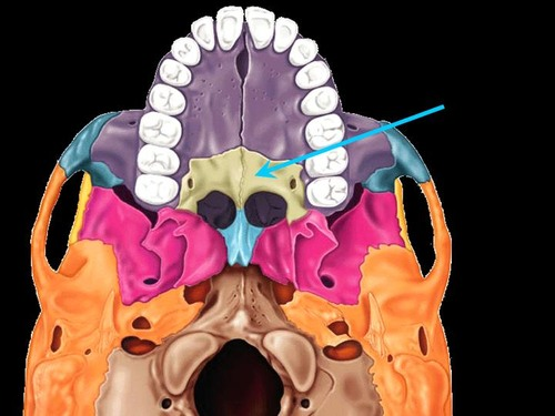
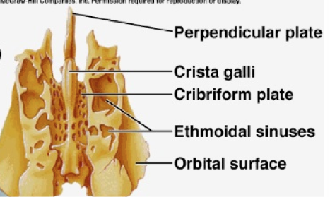
landmakrs of ethmoid bone
contains cribform plate and crista galli
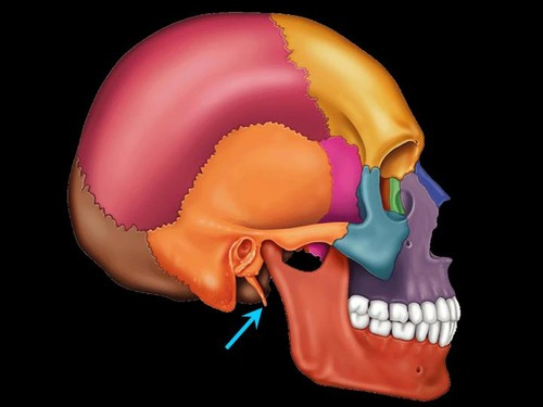
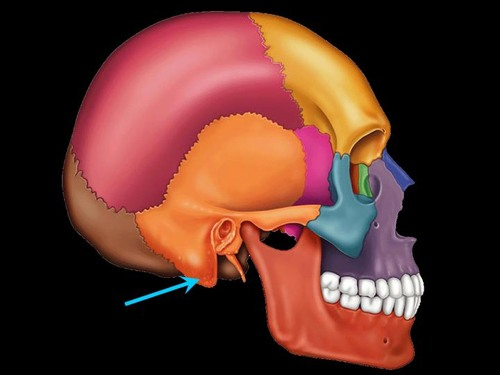
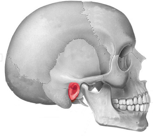
styloid process
skinny process that extends from the undersurface of the temporal bone
mastoid process
round projection on the temporal bone behind the ear
sella turcica
depression in the sphenoid bone where the pituitary gland is located
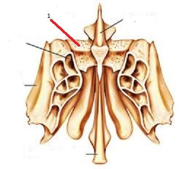
cribform plate of ethmoid
forms the roof of the nasal cavity and has many foramen (openings) through olfactory nerves pass
pituitary gland
brain part that sits in the sella turcica of the sphenoid bone
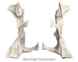
palatine bone
either of two irregulary shaped bones (similar to goal posts) that form the back of the hard palate and help us to form the floor of the orbits
vomer
nostril separator
coronoid process of mandible (jaw bone)
bone part
condylar process (mandibular condyle)
bone part
mental foramen
zygomatic process of maxilla
articulates with zygomatic bone
auditory meatus
infraorbital foramen
zygomatic bone
cheek bone
cranial bones
frontal, parietal (2), temporal (2), occipital, sphenoid, ethmoid
inferior nasal concha
facial bone
palatine
cranial sutures all in one pictures
coronal, sagittal, lambdoid, squamous
articulation
bone term for place where bones come together
process
bone term for a projecting part of a bone
condyle
bone term for a large rounded protuberance at the end of the bone
suture
bone term for a fibrous joint (fixed) between bones of the skull
fossa
bone term for a depression in bone
foramen
bone term for an opening through a bone
meatus
bone term for a tube like opening
sinus
a chamber, hollow space or cavity in a bone
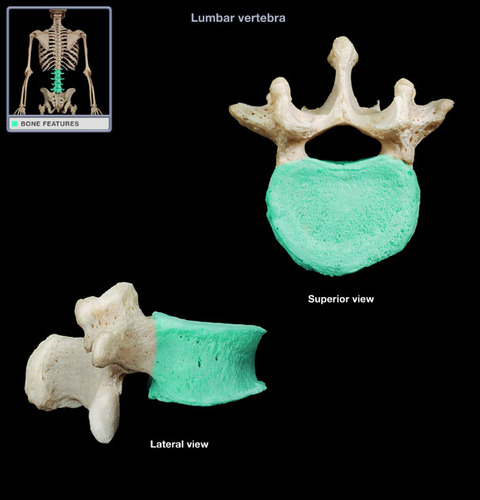
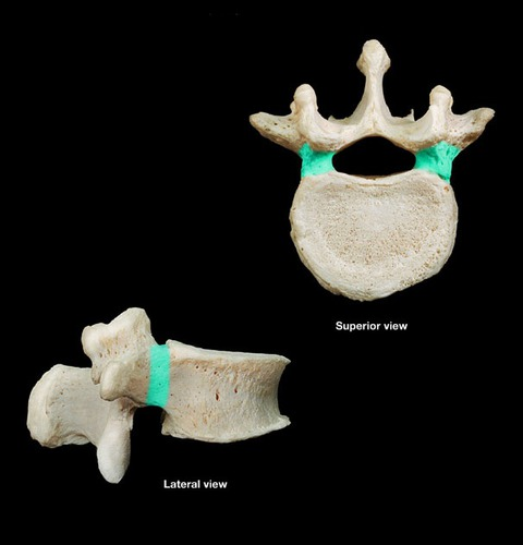
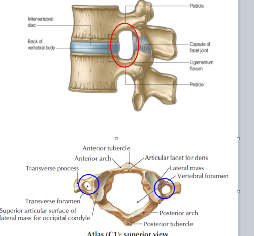
body
pedicles
lamina
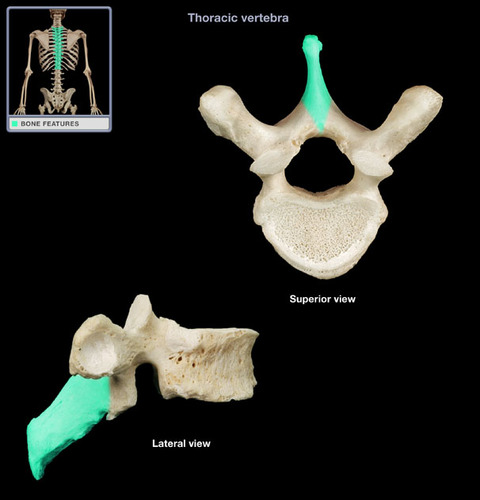
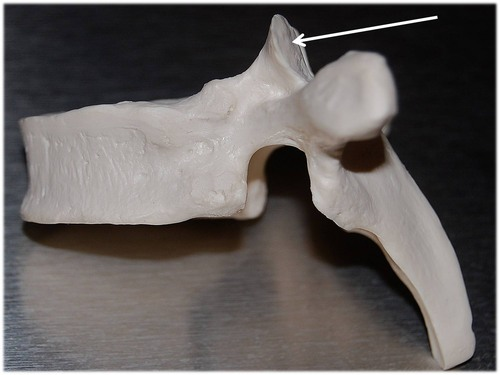
spineous process
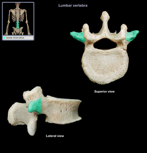
transverse process
superior articular process - facet
inferior articular process facet
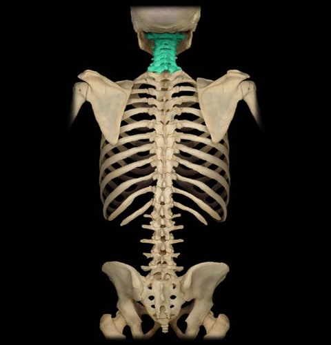
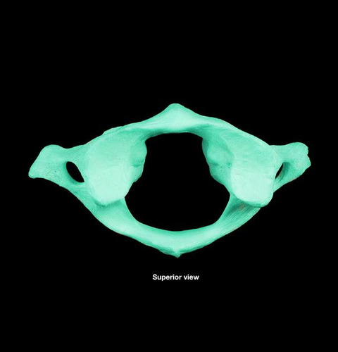
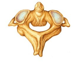
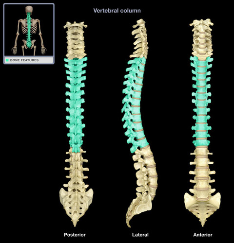
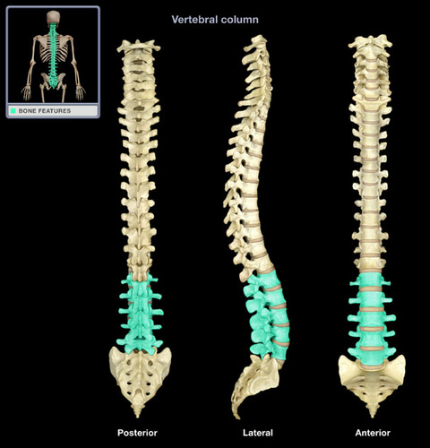
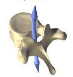
7 cervicals
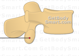
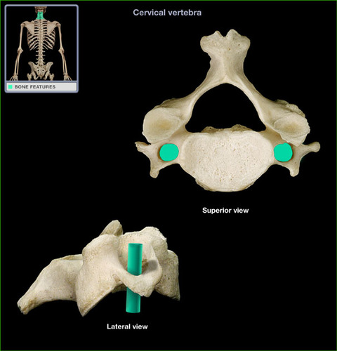
transverse foramen
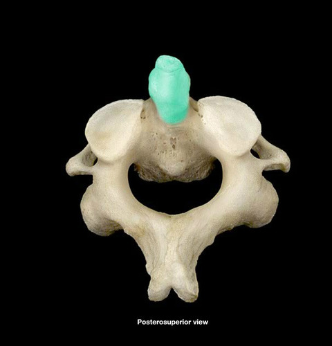
atlas
axis
dens
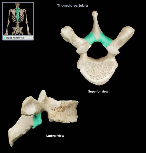
12 thoracic
5 lumbar
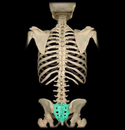
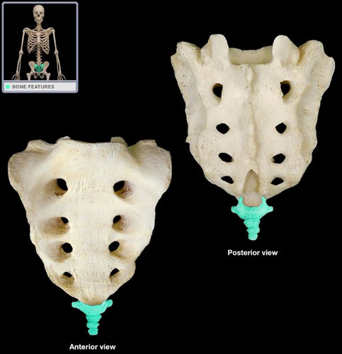
sacrum
coccyx
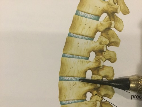
intervetebral disc
intervetebral foramen
vertebral canal
4 categories of tissues
epithelial, connective, muscular, nervous
epithelias characteristics
cellularity
special cell contacts
polarity
supppered by connective tissue
avascular but innervated
regeneration
epithelial - cellularity
almost entirely cell with very little extraceulluar matrix between cells
epithelial - special cell contacts
desmosomes and tight junctions bind adjacent epithelial cells together
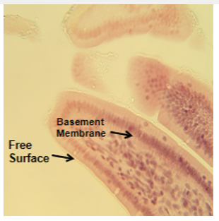
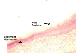
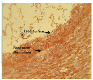
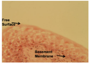
epithelial - polarity
epithelial cells have apical or free surfaces that is not associated with other cells and most also have a basement membrane that helps attach them to underlying tissues
epithelial - suppoerted by connective tissue
reticular lamina, basal laminal forms the basement membrane of epithelial
epithelial - avascular but innervated
blood vessels do not penetrate the basement membrane to reach the epithelium, but contains nerve supply
epithelial - regeneration
epithelial cells can undergo mitosis and replace themselves
epithelial - where its found
covers surfaces such as digestive tract, outside of body, can also form glands, membrane
lamina lucida
hellps anchor cells
lamina densa
provides strength
reticular lamina
attached to underlying connective tissue
functions of epithelium - simple
diffusion, filtration, secretion, absorption
functions of epithelium - stratified
protection
functions of epithelium - squamous
thing and flat and therefore allow substances to diffuse through them or act as filters
functions of epithelium - cubidal or columnar
generally cells that secrete or absorb substances because they have a greater cytoplasmic volume than squamous cells
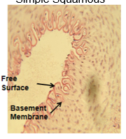
simple squamous
structure: flat or oval with central nucleus
location: places with little wear and tear, places where substances are diffused and filtered (capillaries, enothelium, alveoli, glomeruli, lens of eye)
function: diffusion, filtration, secretion
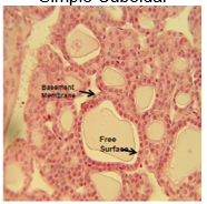
simple cuboidal
structure: cube shaped with a central nucleus
location: in tubules where they can reabsorb substances, collecting ducts of kidneys, ducts, surface of ovaries
function: mainly absoprtion, active transport, secretion
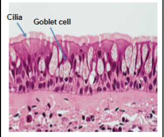
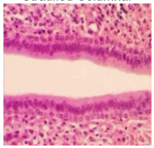
pseudostratified
structure: all cells touch basement membrane but not all reach the free surface
location: respiratory tract, male urethra
function: specialized goblet cells release mucous that coat passageways (is able to expand)
simple columnar
structure: nucleus near bottom of column shaped cells
location: inside lining of digestive tract, also contain goblet cells
function: secretion of mucous, absoprtion
stratified squamous
structure: many layers with outermost layer being flat and scale-like, underlying layers more cuboidal or columnar
location: anyway the outer layers can be abraded and generally wherever protection is needed
epidermis of skin, inside oral cavity, esophagus, vaginal canal
function: protection against abrasion, keratin also acts as waterproofing agent
stratified cubodial
structure: cuboidal cells but in layers
location: wherever protection is needed
sweat glands, ovarian follicles
function: protection and secretion
lots of layers of cuboidal cells, lots of layers of cells
stratified columnar
kidney ducts, apical lauer is composed of columnar shaped cells
basal layer is usually cuboidal cells
large ducts of cells
transitional
structure: looks like stacks of cuboidal cells when in relaxed state, layers closest to apical surface can distend as bladder fills
location: bladder, urethra, ureters
function: allows stretching of urinary bladder as it fills
connective tissue - general characteristics
all connective tissues develop from an embryonic tissue called mesenchyme
abundant extracellular matrix separates cells
protein fibers
ground substances of nonfibrous proteins and molecules
fluid
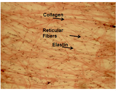
connective tissue - protein fibers of the extracellular matrix
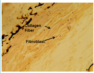
collagen: rope-like and strong but inflexible
reticular fibers - tiny collagen fibers that act like a web between tissues
elastin - elastic proteins that can be stretched and return to their original shape
substances in connective tissue
proteoglycans: aid in water retention by connective tissue, make it able to withstand pressure
glycosaminoglygans: help to hold water
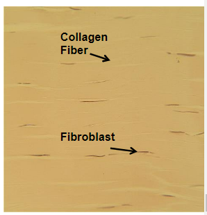
loose connective tissue
usually vascular (vessel and blood adjacent) with nervous innervation
main cells: fibroblasts, fibrocytes, a few fibroclasts
areolar connective tissue
structure: collagen, elastin, retuclar fibers loosel arranged in the EM
location: widespread throughout body, organs, surrounding capillaries, between tissues
function: acts to anchor skin to underlying tissues and also to anchor tissues together
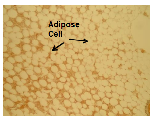
adipose tissue
connective tissue
structure: spherical or round, 95% fats, nucleus pushed to one side, highly vascular
location: under skin, around kidneys, eyeballs, other organs
function: cushioning, insulation, energy storage
dense ct
avascular and has no nerve input
often referred to as fibrous connective tissue
main cells are fibroblasts
regular and irregular
dense regular white fibrous ct
densley packed fibers, primarily collagen, arranged parallel and unidirectional, nuceli are also parallel
location: tendons, ligaments, aponeuroses
function: attach muscles to bones (tendons), attach bones to bones
dense irregular ct
irregular/random arrangement of fibers densley packed in multiple directions
location: mostly dermis of skin, capsules around organs and joints
function: strong in all directions
cartilage
avascular and lacks nerve fibers
has tensile and compressive strength
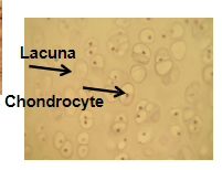
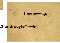
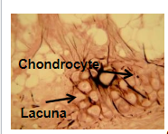
chondroblasts, chondrocytes, chondroclasts are the main cell types
chondro: relates to the cartilage
hyaline cartilage
structure: collagenous fibers not really visible, but chondrocytes which are cartilage producing cells are visible in the hollow spaces
function: provides support and reinforcements, also covers joint surfaces and secretes fluid (lubricates joint surface and protects the ends of the long bones)
location: covering articular surfaces, at the ends of ribs, epiphyseal plates
fibrocartilage
structure: scattering of chondrocytes among a dense network of visible collagenous fibers
function: can absorb large amounts of compressive force, found in areas under great stress
location: pubic symphysis, intervertebral disks, discs of the knee and jaw joint
elastic cartilage
structure: chondrocytes in a matrix of mainly elastic tissue
function: gives support while remaining flexible
location: larynx, nose, external ear
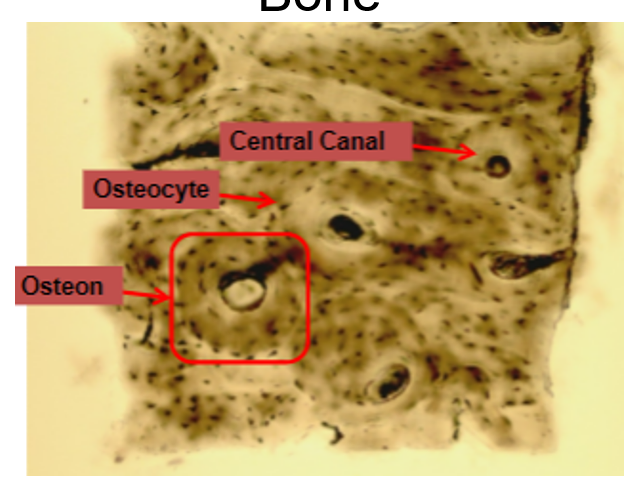
bone
consist of mineralized EM which is hydroxyapatite and living cells (osteocytes and collagen)
holes in matrix are called lucane this is where osteocyte production occuring
very vascular and heals readily
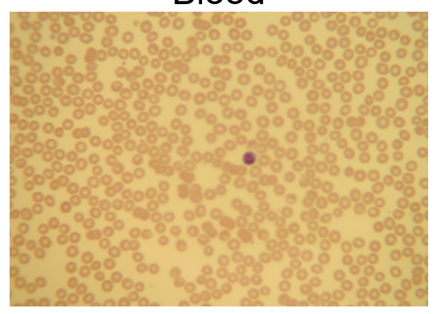
blood cells
red and white blood cells are unique because they are suspended in a fluid matrix
classified as CT because it derives from mesenchyme