Week 22a: Muscles
0.0(0)
Studied by 7 peopleCard Sorting
1/111
There's no tags or description
Looks like no tags are added yet.
Last updated 2:23 PM on 4/7/23
Name | Mastery | Learn | Test | Matching | Spaced | Call with Kai |
|---|
No analytics yet
Send a link to your students to track their progress
112 Terms
1
New cards
^^Sarcomeres^^
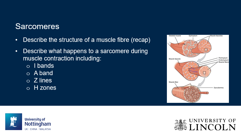
Can you label, describe and explain what this diagram is/shows?
Can you label, describe and explain what this diagram is/shows?
2
New cards
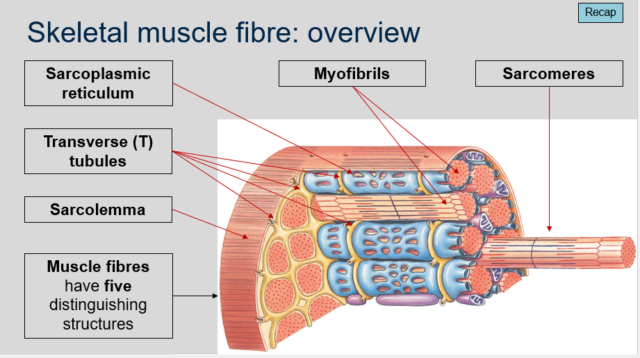
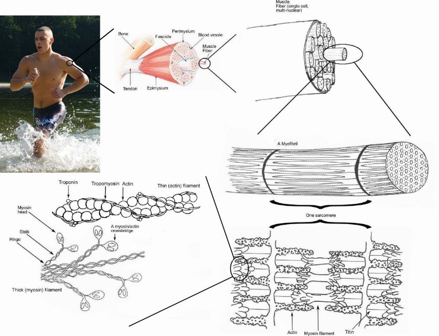
^^Skeletal muscle fibre: Overview^^
1. What is the synonym for muscle fibre?
2. What are the two key differences between skeletal muscle fibers and other cells?
3. How much larger are muscle fibers compared to other cells?
4. What is meant by the term "multinucleate" in relation to muscle fibers?
1. What is the synonym for muscle fibre?
2. What are the two key differences between skeletal muscle fibers and other cells?
3. How much larger are muscle fibers compared to other cells?
4. What is meant by the term "multinucleate" in relation to muscle fibers?
1. Muscle cell.
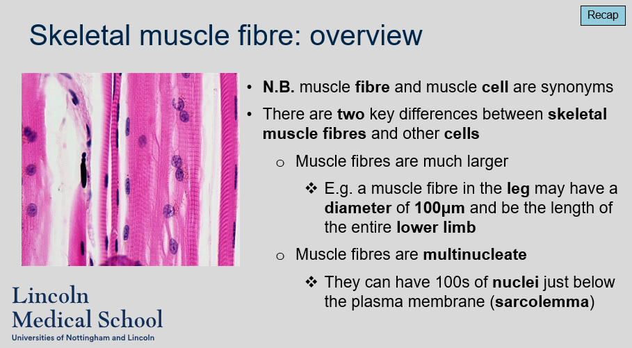
2. The two key differences between skeletal muscle fibers and other cells are that muscle fibers are much larger and multinucleate.
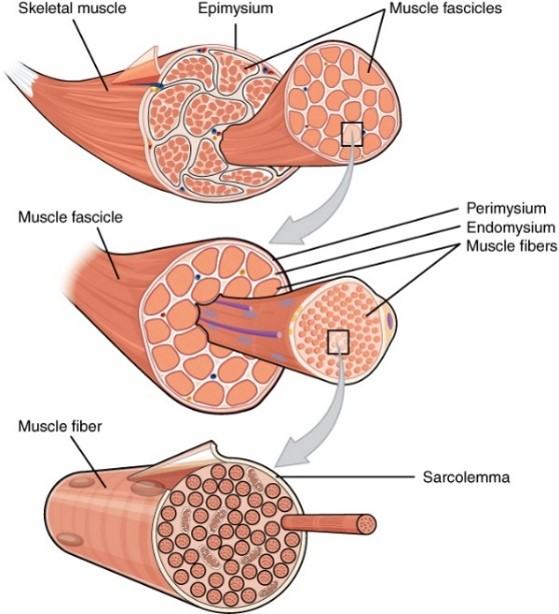
3. Muscle fibers can be much larger than other cells. For example, a muscle fiber in the leg may have a diameter of 100μm and be the length of the entire lower limb.
4. Muscle fibers are multinucleate, which means they can have hundreds of nuclei just below the plasma membrane (sarcolemma).
3
New cards
^^Skeletal muscle fibre: Overview^^
Can you describe/explain what this image is/shows?
Can you describe/explain what this image is/shows?

4
New cards
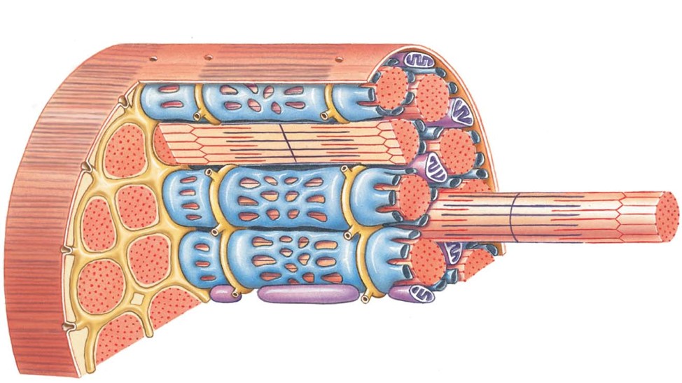
^^Skeletal muscle fibre: Overview^^
Can you label, describe and explain what this image is/shows?
Can you label, describe and explain what this image is/shows?
5
New cards
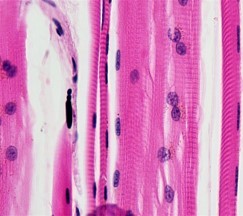
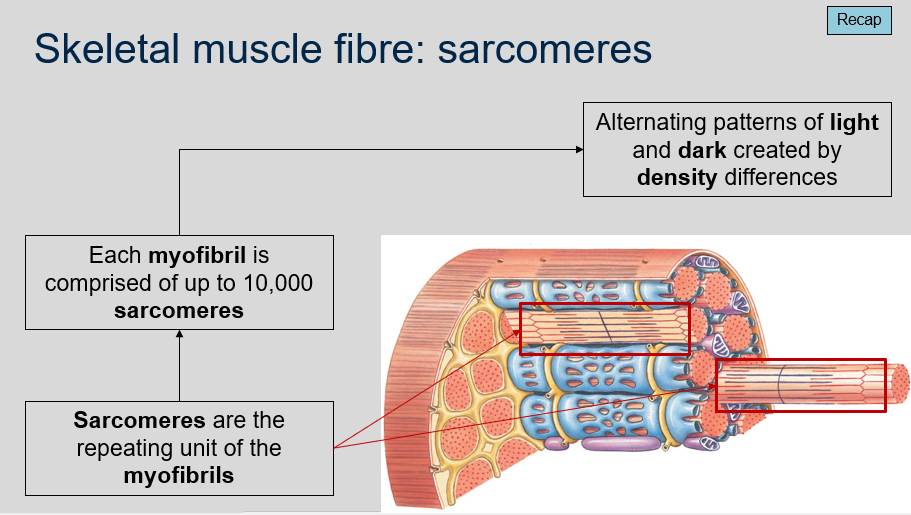
^^Skeletal muscle fibre: Sarcomeres^^
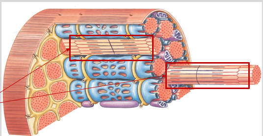
1. What are sarcomeres in relation to myofibrils?
2. How many sarcomeres are in a myofibril?
3. What creates the alternating patterns of light and dark in sarcomeres?
1. What are sarcomeres in relation to myofibrils?
2. How many sarcomeres are in a myofibril?
3. What creates the alternating patterns of light and dark in sarcomeres?
1. Sarcomeres are the repeating units of the myofibrils. They are the fundamental unit of muscle contraction.
2. Each myofibril can be comprised of up to 10,000 sarcomeres.
3. The alternating patterns of light and dark in sarcomeres are created by density differences. The lighter bands are called I-bands and are composed of thin actin filaments, while the darker bands are called A-bands and are composed of thick myosin filaments. The area where the two overlap is called the H-zone.
6
New cards
^^Skeletal muscle fibre: Sarcomeres^^
Can you label, describe and explain what this image is/shows?
Can you label, describe and explain what this image is/shows?

7
New cards
^^Sarcomeres: Overview^^
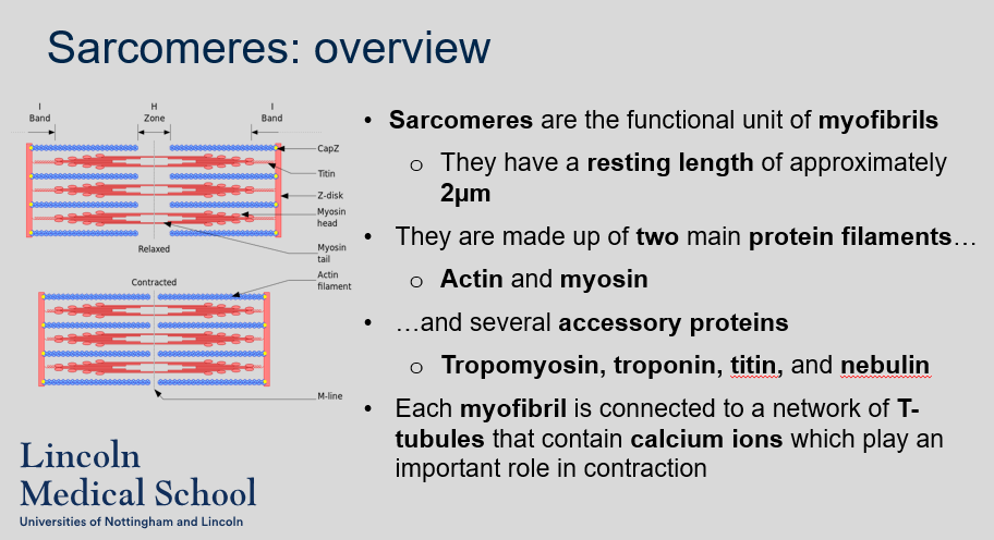
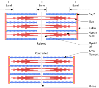
1. What is the functional unit of myofibrils?
2. What is the resting length of a sarcomere?
3. What are the two main protein filaments that make up sarcomeres?
4. What are some of the accessory proteins that are found in sarcomeres?
5. What is the role of T-tubules in muscle contraction?
1. What is the functional unit of myofibrils?
2. What is the resting length of a sarcomere?
3. What are the two main protein filaments that make up sarcomeres?
4. What are some of the accessory proteins that are found in sarcomeres?
5. What is the role of T-tubules in muscle contraction?
1. The functional unit of myofibrils is the sarcomere.
2. The resting length of a sarcomere is approximately 2µm.
3. The two main protein filaments that make up sarcomeres are actin and myosin.
4. Some of the accessory proteins found in sarcomeres include tropomyosin, troponin, titin, and nebulin.
5. T-tubules are connected to each myofibril and contain calcium ions, which play an important role in muscle contraction. The release of calcium ions from the T-tubules triggers a series of events that result in the sliding of actin and myosin filaments and muscle contraction.
8
New cards
^^Sarcomeres: Overview^^
Can you label, describe and explain what this image is/shows?
Can you label, describe and explain what this image is/shows?

9
New cards
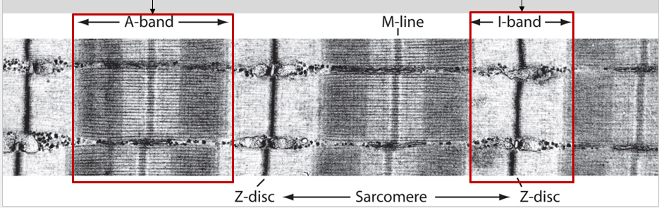
^^Sarcomeres: Structure (banding)^^
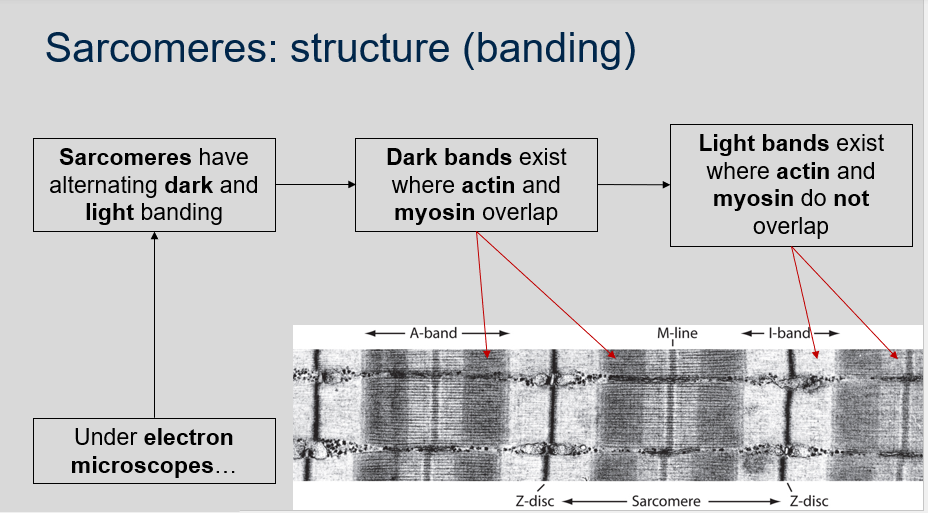
1. What is the appearance of sarcomeres under electron microscopes?
2. What causes the dark banding in sarcomeres?
3. What causes the light banding in sarcomeres?
1. What is the appearance of sarcomeres under electron microscopes?
2. What causes the dark banding in sarcomeres?
3. What causes the light banding in sarcomeres?
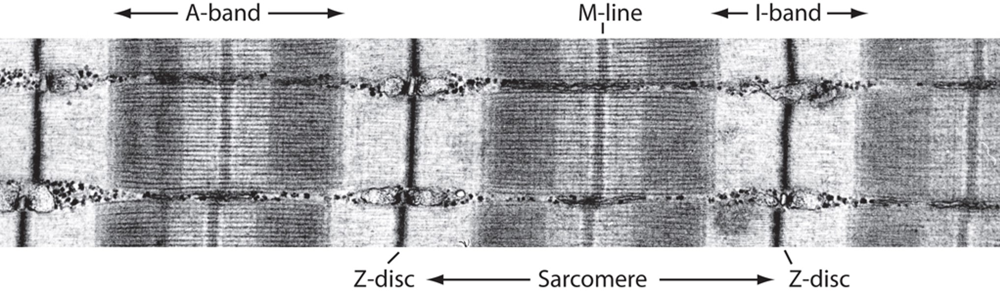
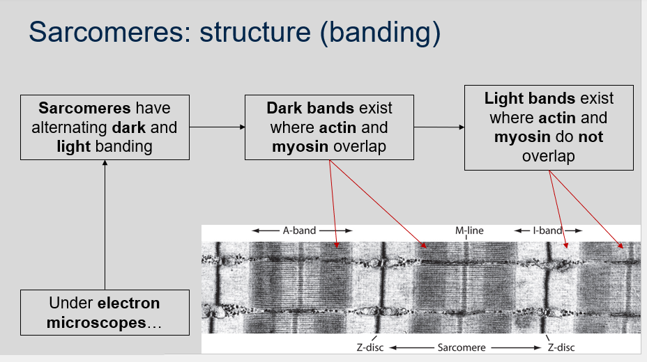
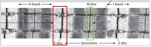
1. Under electron microscopes, sarcomeres have alternating dark and light banding. The dark bands, called A-bands, exist where actin and myosin filaments overlap. The light bands, called I-bands, exist where actin and myosin filaments do not overlap.
2. The dark banding in sarcomeres is caused by the overlap of actin and myosin filaments.
3. The light banding in sarcomeres is caused by the absence of myosin filaments in the region where only actin filaments exist.
10
New cards
^^Sarcomeres: Structure (banding)^^
Can you label, describe and explain what this image is/shows?
Can you label, describe and explain what this image is/shows?
11
New cards
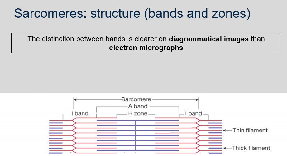
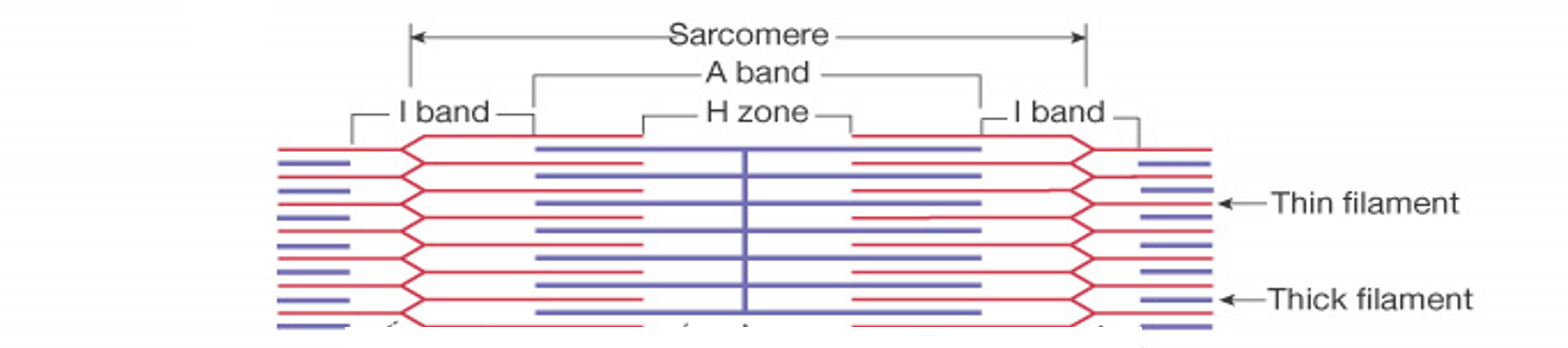
^^Sarcomeres: Structure (bands and zones)^^
1. Where does the H-zone exist in sarcomeres?
2. What is the composition of the H-zone?
3. What is the A-band in sarcomeres?
4. Why is the I-band lighter than the H-zone in sarcomeres?
5. What is the composition of the I-band in sarcomeres?
1. Where does the H-zone exist in sarcomeres?
2. What is the composition of the H-zone?
3. What is the A-band in sarcomeres?
4. Why is the I-band lighter than the H-zone in sarcomeres?
5. What is the composition of the I-band in sarcomeres?
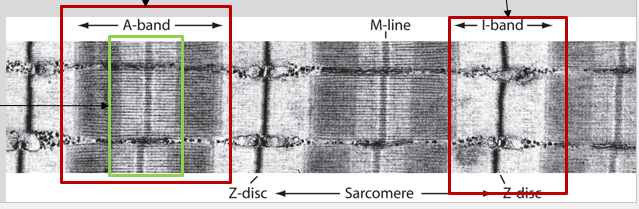
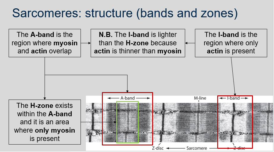
1. The H-zone exists within the A-band in sarcomeres.
2. The H-zone is an area where only myosin filaments are present.
3. The A-band is the region where myosin and actin filaments overlap in sarcomeres.
4. The I-band is lighter than the H-zone in sarcomeres because actin filaments are thinner than myosin filaments, resulting in less density.
5. The I-band is the region where only actin filaments are present in sarcomeres.

12
New cards
^^Sarcomeres: Structure (bands and zones)^^
Can you label, describe and explain what this diagram is/shows?
Can you label, describe and explain what this diagram is/shows?
13
New cards
^^Sarcomeres: Structure (bands and zones)^^
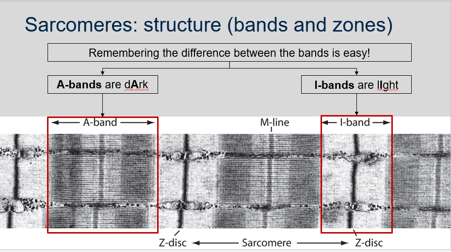
What is an easy way to remember the difference between the A-band and I-band in sarcomeres?
What is an easy way to remember the difference between the A-band and I-band in sarcomeres?
An easy way to remember the difference between the A-band and I-band in sarcomeres is to think of A-bands as "dArk" because they contain both actin and myosin filaments, and to think of I-bands as "lIght" because they only contain actin filaments.

14
New cards
^^Sarcomeres: Structure (bands and zones)^^
Can you label, describe and explain what this diagram is/shows?
Can you label, describe and explain what this diagram is/shows?
15
New cards
^^Sarcomeres: Structure (bands and zones)^^
Is the distinction between the different bands in sarcomeres clearer on diagrammatical images or on electron micrographs?
Is the distinction between the different bands in sarcomeres clearer on diagrammatical images or on electron micrographs?
The distinction between the different bands in sarcomeres is usually clearer on diagrammatical images than on electron micrographs. Diagrammatical images often use contrasting colors to highlight the different regions of the sarcomere, whereas electron micrographs show the actual structures of the sarcomeres without any coloration.
16
New cards
^^Sarcomeres: Structure (bands and zones)^^
Can you label, describe and explain what this diagram is/shows?
Can you label, describe and explain what this diagram is/shows?

17
New cards
^^Sarcomeres: Structure (during contraction)^^
1. What happens to the sarcomere during contraction?
2. Why does the I-band become shorter during contraction?
3. Why does the A-band stay the same length during contraction?
4. Why do the Z-lines move closer together during contraction?
5. Why does the H-zone become narrower during contraction?
1. What happens to the sarcomere during contraction?
2. Why does the I-band become shorter during contraction?
3. Why does the A-band stay the same length during contraction?
4. Why do the Z-lines move closer together during contraction?
5. Why does the H-zone become narrower during contraction?
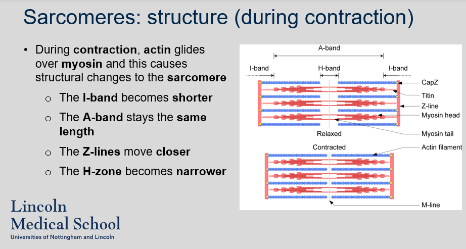
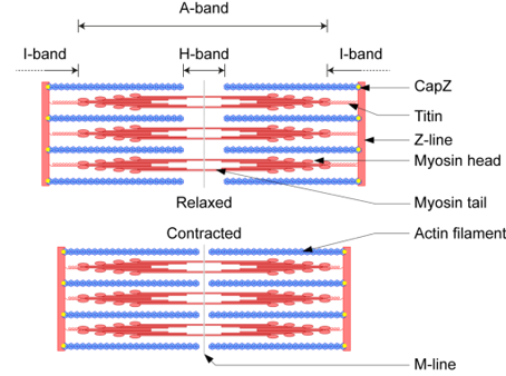
1. During contraction, actin filaments slide over myosin filaments causing structural changes to the sarcomere. Specifically, the I-band becomes shorter, the A-band stays the same length, the Z-lines move closer together, and the H-zone becomes narrower.
2. The I-band becomes shorter during contraction because the actin filaments slide towards the center of the sarcomere and overlap more with the myosin filaments, reducing the length of the region where only actin is present.
3. The A-band stays the same length during contraction because the length of the myosin filaments does not change.
4. The Z-lines move closer together during contraction because the actin filaments slide towards the center of the sarcomere, pulling the Z-lines towards each other.
5. The H-zone becomes narrower during contraction because the actin filaments slide towards the center of the sarcomere and overlap more with the myosin filaments, reducing the length of the region where only myosin is present.
18
New cards
^^Sarcomeres: Structure (during contraction)^^
Can you label, describe and explain what this diagram is/shows?
Can you label, describe and explain what this diagram is/shows?

19
New cards
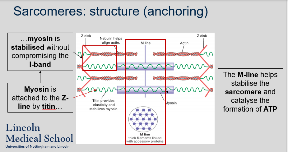
^^Sarcomeres: Structure (anchoring)^^
1. What are Z-lines and what is their role in the sarcomere?
2. Where is the M-line located in the sarcomere and what is its function?
3. What is the H-zone in the sarcomere?
1. What are Z-lines and what is their role in the sarcomere?
2. Where is the M-line located in the sarcomere and what is its function?
3. What is the H-zone in the sarcomere?
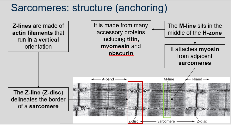
1. Z-lines, also known as Z-discs, are thin, vertical structures made of actin filaments that mark the border of a sarcomere. They play an important role in anchoring the actin filaments and maintaining the structural integrity of the sarcomere during muscle contraction.
2. The M-line sits in the middle of the H-zone. It attaches myosin from adjacent sarcomeres. It is made from many accessory proteins including titin, myomesin and obscurin.
3. The H-zone is the central region of the A-band in the sarcomere where only myosin filaments are present. During muscle contraction, the H-zone becomes narrower as actin filaments slide towards the center of the sarcomere and overlap more with the myosin filaments.

20
New cards
^^Sarcomeres: Structure (anchoring)^^
Can you label, describe and explain what this diagram is/shows?
Can you label, describe and explain what this diagram is/shows?
21
New cards
^^Sarcomeres: Structure (anchoring)^^
1. How is myosin attached to the Z-line in a sarcomere?
2. What is the function of the M-line in a sarcomere?
1. How is myosin attached to the Z-line in a sarcomere?
2. What is the function of the M-line in a sarcomere?
1. Myosin is attached to the Z-line by titin. Myosin is stabilised without compromising the I-band.
2. The M-line helps stabilise the sarcomere and catalyse the formation of ATP.

22
New cards

^^Sarcomeres: Structure (anchoring)^^
Can you label, describe and explain what this diagram is/shows?
Can you label, describe and explain what this diagram is/shows?
23
New cards
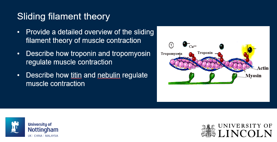
^^Sliding filament theory^^
Can you label, describe and explain what this diagram is/shows?
Can you label, describe and explain what this diagram is/shows?
24
New cards
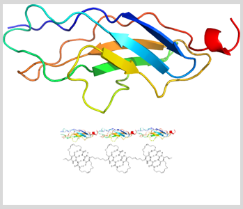
^^Sarcomeres: Titin^^
1. What is titin?
2. What is the function of titin in muscle cells?
3. How many domains does titin have and how are they held together?
1. What is titin?
2. What is the function of titin in muscle cells?
3. How many domains does titin have and how are they held together?
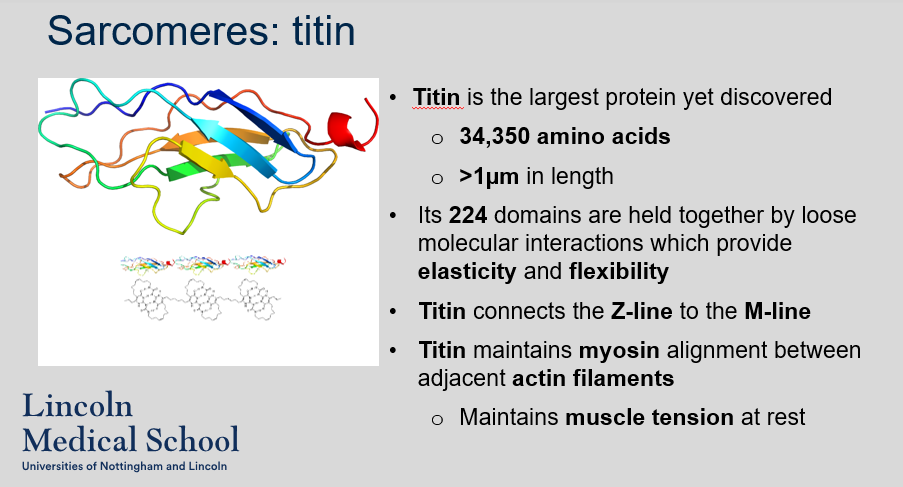
1. Titin is the largest protein known, consisting of 34,350 amino acids and measuring over 1 µm in length.
2. Titin has several important functions in muscle cells. It connects the Z-line to the M-line, maintaining myosin alignment between adjacent actin filaments. It also provides elasticity and flexibility to the sarcomere through its loosely interacting domains. Additionally, titin helps maintain muscle tension at rest.
3. Titin has 224 domains, which are held together by loose molecular interactions that allow for its elasticity and flexibility.

25
New cards
^^Sarcomeres: Titin^^
Can you describe/explain what this image is/shows?
Can you describe/explain what this image is/shows?
26
New cards
^^Sarcomeres: Nebulin^^
What is nebulin and what is its role in the sarcomere?
What is nebulin and what is its role in the sarcomere?
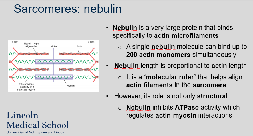
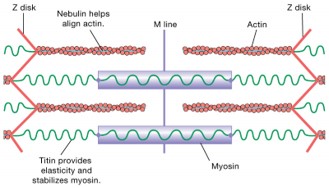
Nebulin is a very large protein that binds specifically to actin microfilaments. A single nebulin molecule can bind up to 200 actin monomers simultaneously Nebulin length is proportional to actin length. It is a ‘molecular ruler’ that helps align actin filaments in the sarcomere. However, its role is not only structural Nebulin inhibits ATPase activity which regulates actin-myosin interactions.
27
New cards
^^Sarcomeres: Nebulin^^
Can you label, describe and explain what this diagram is/shows?
Can you label, describe and explain what this diagram is/shows?

28
New cards
^^Sarcomeres: Myosin and actin interaction^^
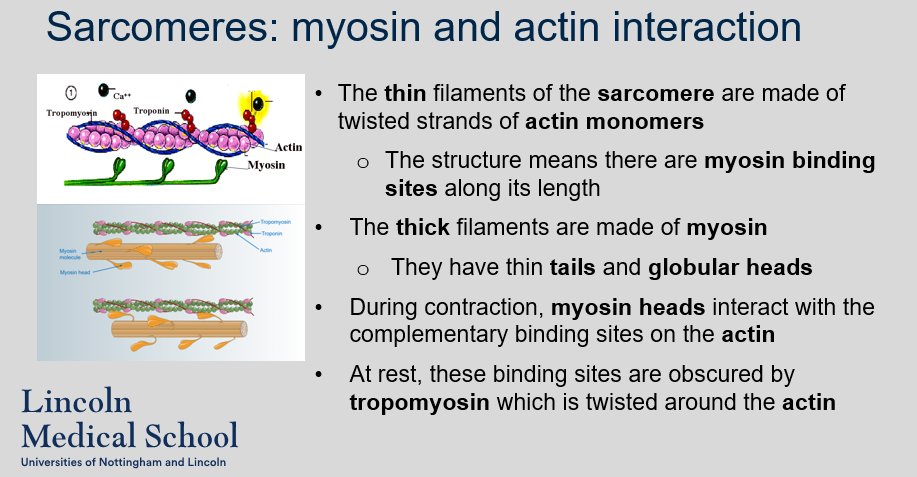
1. What are the thin filaments of the sarcomere made of?
2. What is the structure of the thin filaments of the sarcomere?
3. What are the thick filaments of the sarcomere made of?
4. What is the structure of the thick filaments of the sarcomere?
5. What happens during contraction in the sarcomere?
6. What happens to the binding sites on actin during rest?
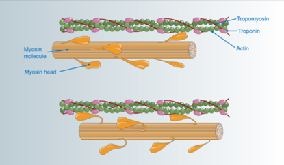
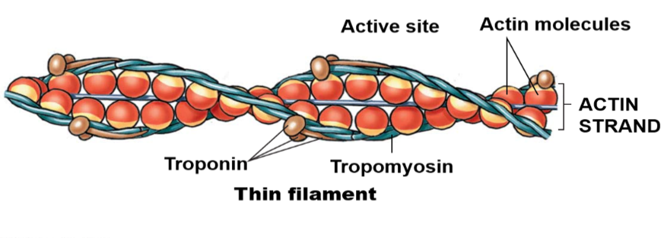
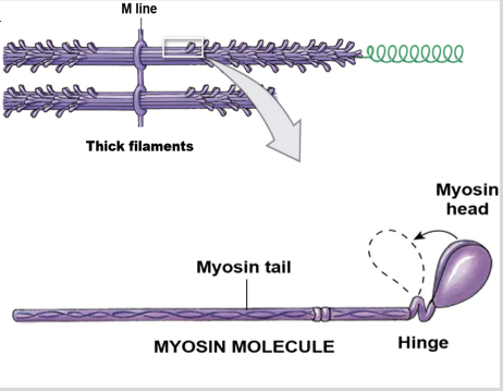
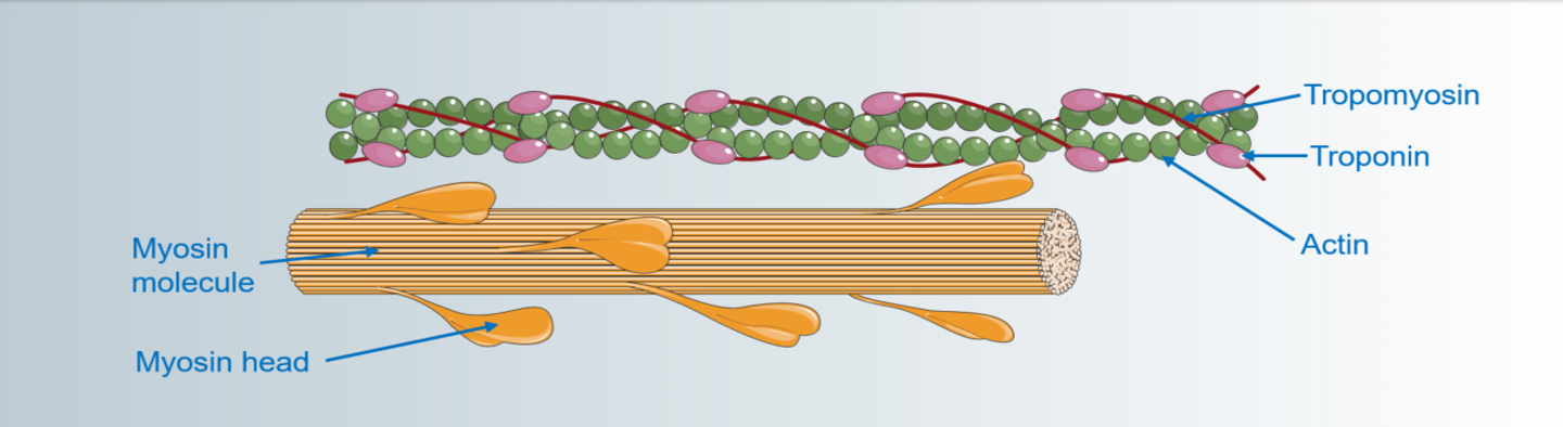
1. The thin filaments of the sarcomere are made of twisted strands of actin monomers.
2. The structure of the thin filaments of the sarcomere means there are myosin binding sites along its length.
3. The thick filaments of the sarcomere are made of myosin.
4. The thick filaments of the sarcomere have thin tails and globular heads.
5. During contraction, myosin heads interact with the complementary binding sites on the actin.
6. At rest, the binding sites on actin are obscured by tropomyosin which is twisted around the actin.

29
New cards
^^Sarcomeres: Myosin and actin interaction^^
Can you label, describe and explain what this diagram is/shows?
Can you label, describe and explain what this diagram is/shows?
30
New cards
^^Sarcomeres: Myosin and actin interaction^^
Can you label, describe and explain what this diagram is/shows?
Can you label, describe and explain what this diagram is/shows?

31
New cards
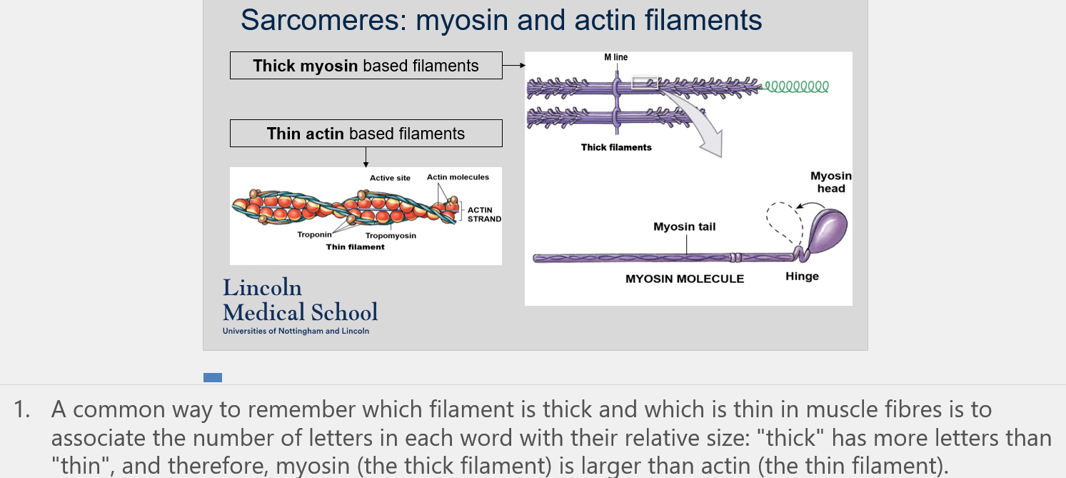
^^Sarcomeres: Myosin and actin filaments^^
What is a common way to remember which filament is thick and which is thin in muscle fibers?
What is a common way to remember which filament is thick and which is thin in muscle fibers?
One common way is to associate the number of letters in each word with their relative size: "thick" has more letters than "thin", and therefore, myosin (the thick filament) is larger than actin (the thin filament).

32
New cards
^^Sarcomeres: Myosin and actin filaments^^
Can you label, describe and explain what this diagram is/shows?
Can you label, describe and explain what this diagram is/shows?
33
New cards
^^Sarcomeres: Myosin and actin filaments^^
Can you label, describe and explain what this diagram is/shows?
Can you label, describe and explain what this diagram is/shows?

34
New cards
^^Sarcomeres: Tropomyosin and troponin^^
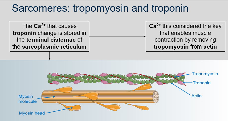
What is the function of tropomyosin in muscle contraction?
What is the function of tropomyosin in muscle contraction?
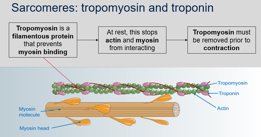
Tropomyosin is a filamentous protein that prevents myosin binding, which stops actin and myosin from interacting at rest. Therefore, the function of tropomyosin is to regulate muscle contraction by inhibiting the interaction between actin and myosin until it is removed prior to contraction.

35
New cards
^^Sarcomeres: Tropomyosin and troponin^^
Can you label, describe and explain what this diagram is/shows?
Can you label, describe and explain what this diagram is/shows?
36
New cards
^^Sarcomeres: Tropomyosin and troponin^^
1. What is troponin?
2. What is the role of troponin at rest?
3. What happens to troponin when it binds to Ca2+?
1. What is troponin?
2. What is the role of troponin at rest?
3. What happens to troponin when it binds to Ca2+?
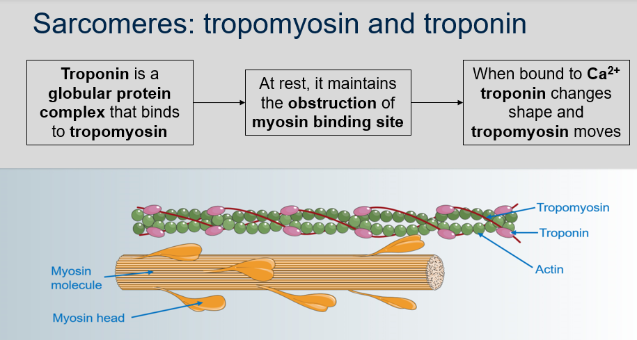
1. Troponin is a globular protein complex that binds to tropomyosin.
2. At rest, troponin maintains the obstruction of the myosin binding site by interacting with tropomyosin.
3. When troponin binds to Ca2+, it undergoes a conformational change that causes tropomyosin to move and expose the myosin binding sites on the actin filament, allowing for muscle contraction.
37
New cards

^^Sarcomeres: Tropomyosin and troponin^^
Can you label, describe and explain what this diagram is/shows?
Can you label, describe and explain what this diagram is/shows?

38
New cards
^^Sarcomeres: Tropomyosin and troponin^^
1. Where is the Ca2+ stored that is necessary for muscle contraction?
2. What is the role of Ca2+ in muscle contraction?
1. Where is the Ca2+ stored that is necessary for muscle contraction?
2. What is the role of Ca2+ in muscle contraction?
1. The Ca2+ that causes troponin change is stored in the terminal cisternae of the sarcoplasmic reticulum.
2. Ca2+ is considered the key that enables muscle contraction by removing tropomyosin from actin.
39
New cards

^^Sarcomeres: Tropomyosin and troponin^^
Can you label, describe and explain what this diagram is/shows?
Can you label, describe and explain what this diagram is/shows?

40
New cards
^^Sliding filament theory^^
1. What is the sliding filament theory?
2. What are the steps involved in the sliding filament theory?
1. What is the sliding filament theory?
2. What are the steps involved in the sliding filament theory?
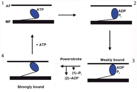
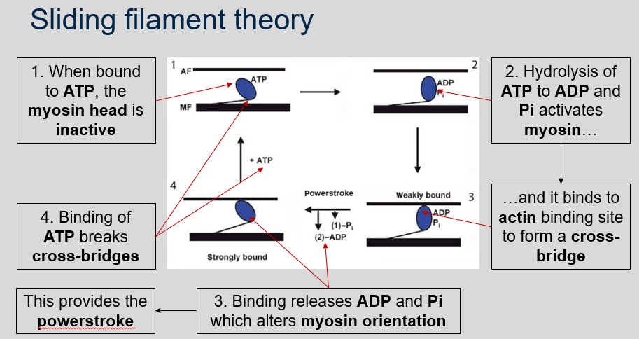
1. The sliding filament theory is a model of muscle contraction that explains how actin and myosin filaments slide past each other to generate muscle movement.
2. The steps involved in the sliding filament theory are as follows:
* When bound to ATP, the myosin head is inactive.
* Hydrolysis of ATP to ADP and Pi activates myosin and it binds to actin binding site to form a cross-bridge.
* Binding releases ADP and Pi which alters myosin orientation. This provides the powerstroke.
* Binding of ATP breaks cross-bridges.

41
New cards
^^Sliding filament theory^^
Can you label, describe and explain what this diagram is/shows?
Can you label, describe and explain what this diagram is/shows?
42
New cards
^^Sliding filament theory^^
What is the sliding filament theory in muscle contraction?
What is the sliding filament theory in muscle contraction?
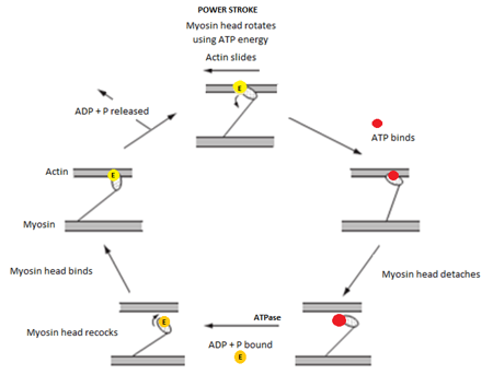
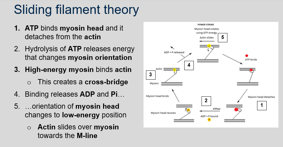
The sliding filament theory is the explanation for how muscle contraction occurs at the molecular level. According to this theory:
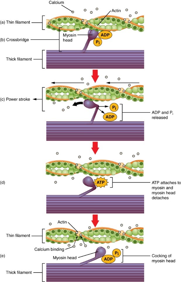
1. When ATP binds to myosin head, it detaches from the actin.
2. Hydrolysis of ATP releases energy that changes myosin orientation to high-energy position.
3. High-energy myosin binds to actin and creates a cross-bridge.
4. Binding releases ADP and Pi and orientation of myosin head changes to low-energy position.
5. Actin slides over myosin towards the M-line.
6. Binding of ATP breaks cross-bridges and the cycle can repeat if Ca2+ is still present.

43
New cards
^^Sliding filament theory^^
Can you label, describe and explain what this image is/shows?
Can you label, describe and explain what this image is/shows?
44
New cards
^^Sliding filament theory^^
1. What is the ratchet movement in the sliding filament theory?
2. How is energy provided for the sliding filament theory?
3. How many heads do myosin filaments have, and how many cross-bridges do they form per second?
4. What limits the rate of muscle contraction in the sliding filament theory?
1. What is the ratchet movement in the sliding filament theory?
2. How is energy provided for the sliding filament theory?
3. How many heads do myosin filaments have, and how many cross-bridges do they form per second?
4. What limits the rate of muscle contraction in the sliding filament theory?
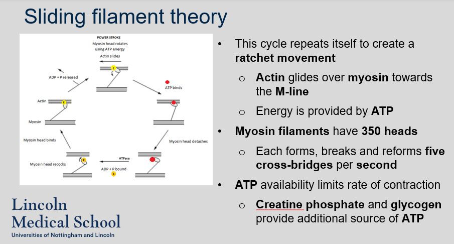
1. The ratchet movement refers to the repeated cycling of myosin heads binding to actin and then releasing, which causes actin to slide over myosin towards the M-line in a stepwise manner.
2. Energy is provided by ATP. When ATP binds to the myosin head, it becomes energized and able to bind to actin, initiating the powerstroke.
3. Myosin filaments have 350 heads, and each head forms, breaks, and reforms five cross-bridges per second.
4. The rate of muscle contraction is limited by the availability of ATP. However, additional sources of ATP such as creatine phosphate and glycogen can provide extra energy for muscle contraction.
45
New cards

^^Sliding filament theory^^
Can you label, describe and explain what this diagram is/shows?
Can you label, describe and explain what this diagram is/shows?

46
New cards
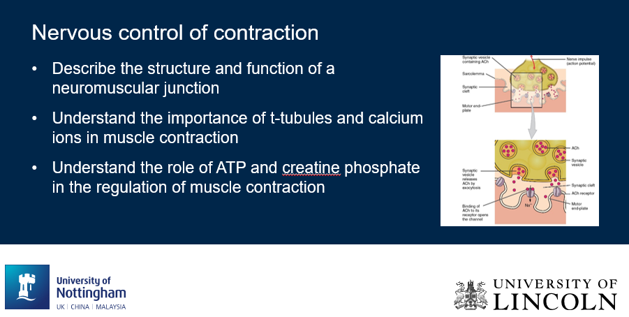
^^Nervous control of contraction^^
Can you label, describe and explain what this image is/shows?
Can you label, describe and explain what this image is/shows?
47
New cards

^^Overview of muscle contraction^^
Can you label, describe and explain what this diagram is/shows?
Can you label, describe and explain what this diagram is/shows?
48
New cards

^^Overview of muscle contraction^^
Can you label, describe and explain what this diagram is/shows?
Can you label, describe and explain what this diagram is/shows?
49
New cards
^^Neuromuscular junctions: axon terminal^^
1. What is the innervation pattern of muscle fibres?
2. What is the structure of the axon terminal at the neuromuscular junction?
1. What is the innervation pattern of muscle fibres?
2. What is the structure of the axon terminal at the neuromuscular junction?
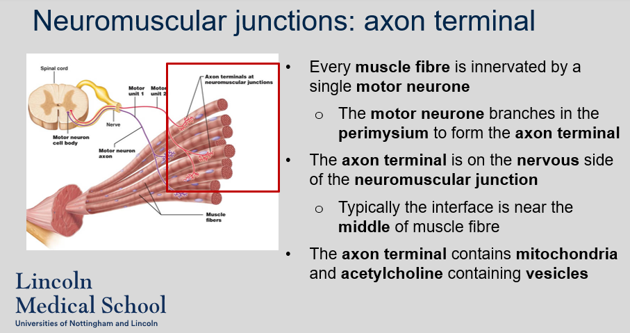
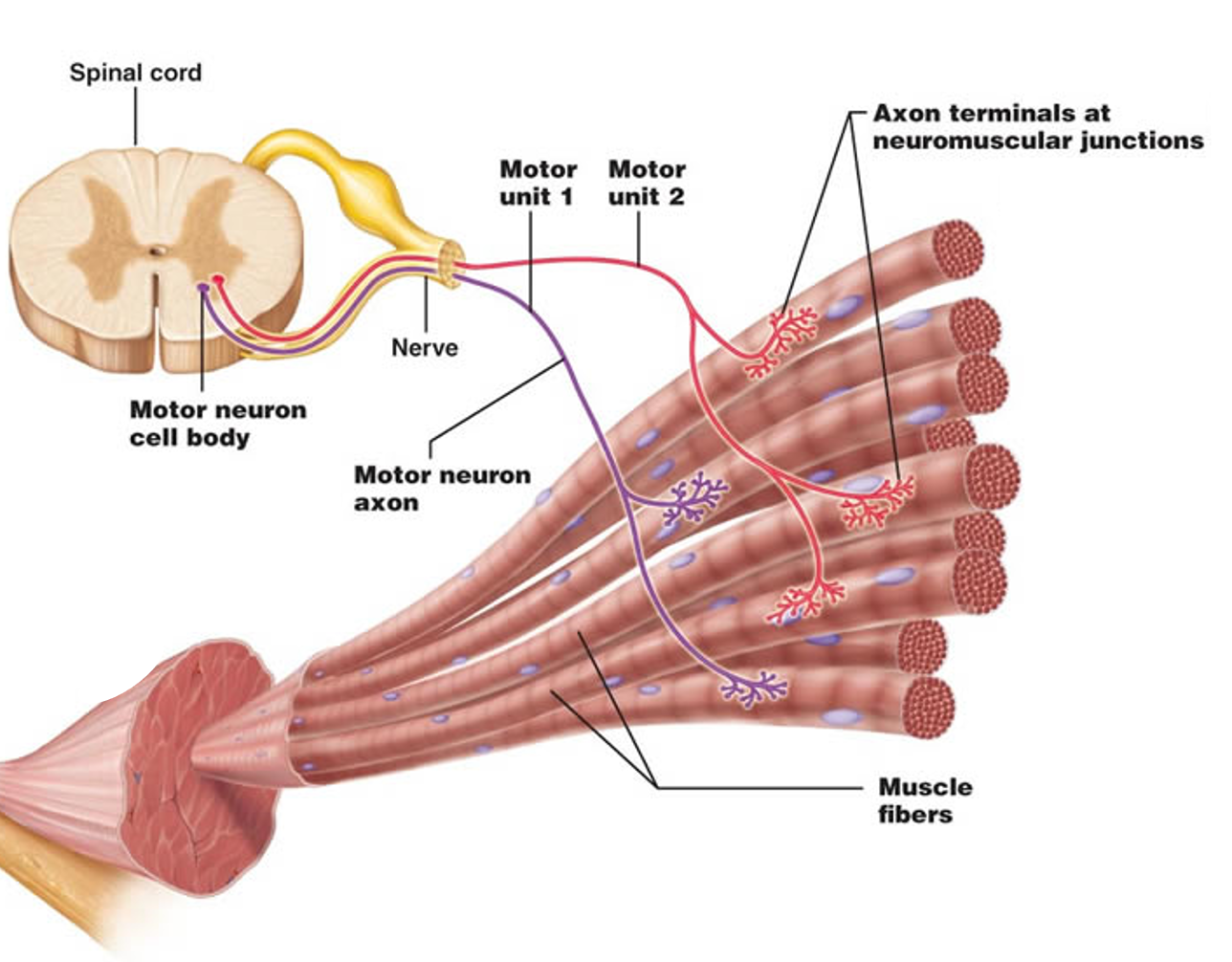
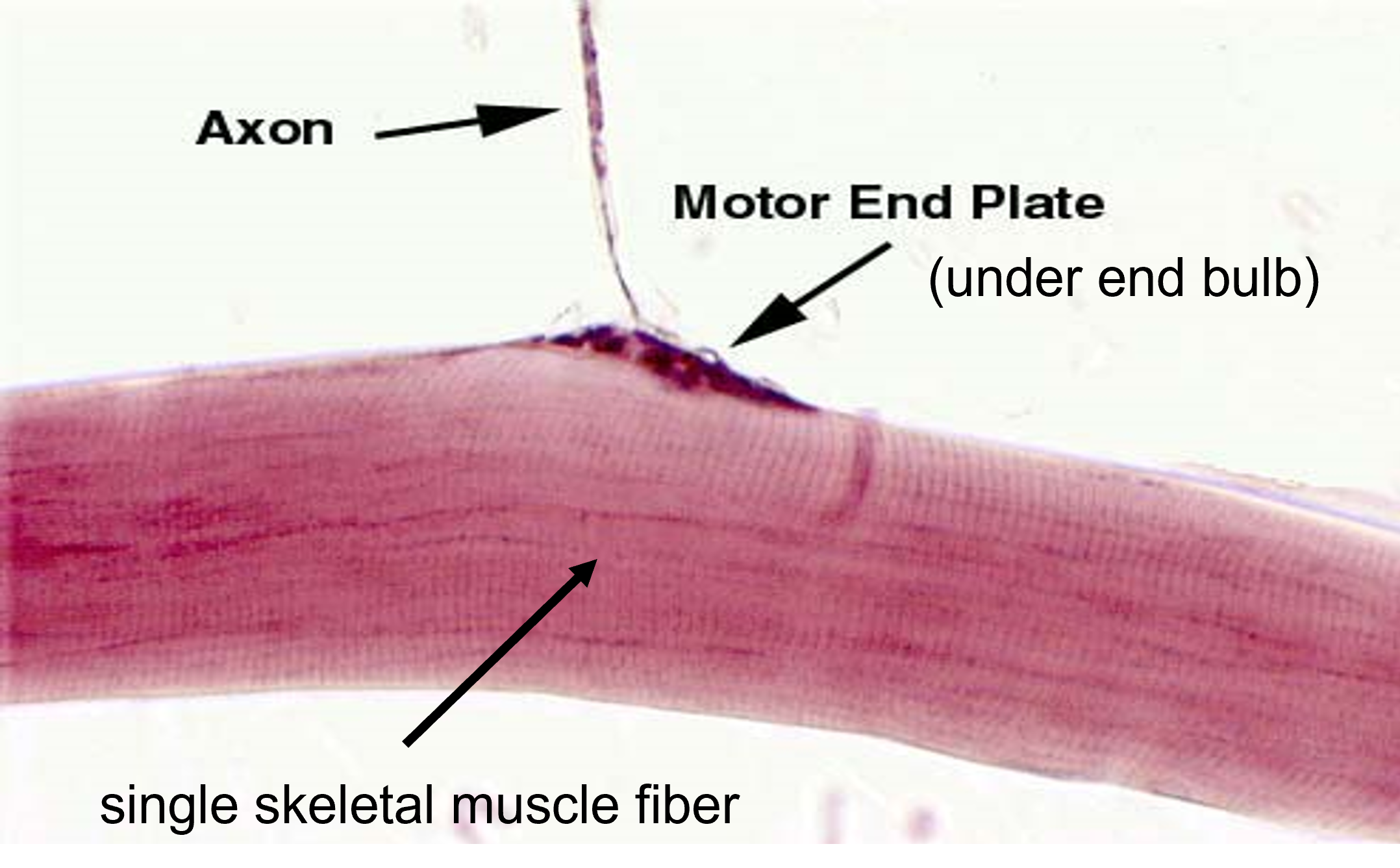
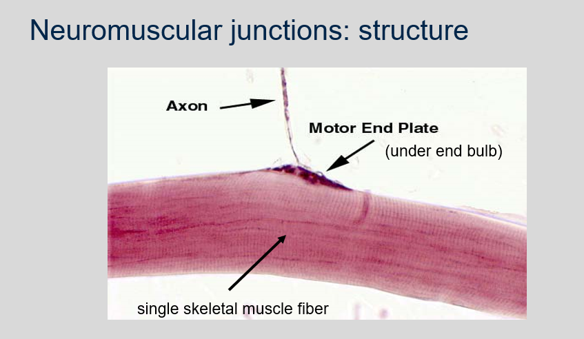
1. Every muscle fibre is innervated by a single motor neurone.
2. The motor neurone branches in the perimysium to form the axon terminal. The axon terminal is on the nervous side of the neuromuscular junction. Typically the interface is near the middle of muscle fibre. The axon terminal contains mitochondria and acetylcholine containing vesicles.
50
New cards
^^Neuromuscular junctions: axon terminal^^
Can you label, describe and explain what this diagram is/shows?
Can you label, describe and explain what this diagram is/shows?

51
New cards
^^Neuromuscular junctions: motor end plate^^
1. What is the motor end plate in the neuromuscular junction?
2. What are the characteristics of the motor end plate?
1. What is the motor end plate in the neuromuscular junction?
2. What are the characteristics of the motor end plate?
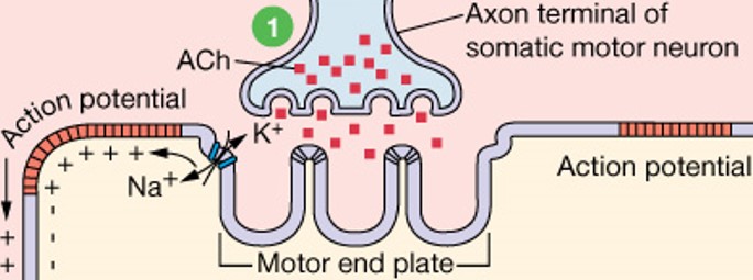
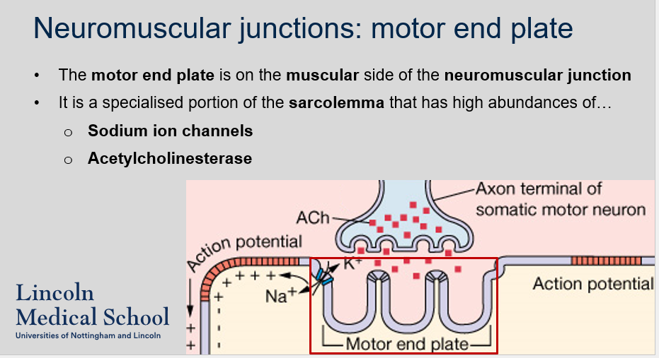
1. The motor end plate is a specialized region of the sarcolemma on the muscular side of the neuromuscular junction.
2. The motor end plate has a high abundance of sodium ion channels. The sodium ion channels allow the influx of sodium ions, which depolarizes the muscle fibre.

52
New cards
^^Neuromuscular junctions: motor end plate^^
Can you label, describe and explain what this diagram is/shows?
Can you label, describe and explain what this diagram is/shows?
53
New cards
^^Neuromuscular junctions: structure^^
Can you label, describe and explain what this diagram is/shows?
Can you label, describe and explain what this diagram is/shows?
54
New cards
^^Neuromuscular junctions: activity^^
What happens during activity at the neuromuscular junction according to the sliding filament theory?
What happens during activity at the neuromuscular junction according to the sliding filament theory?
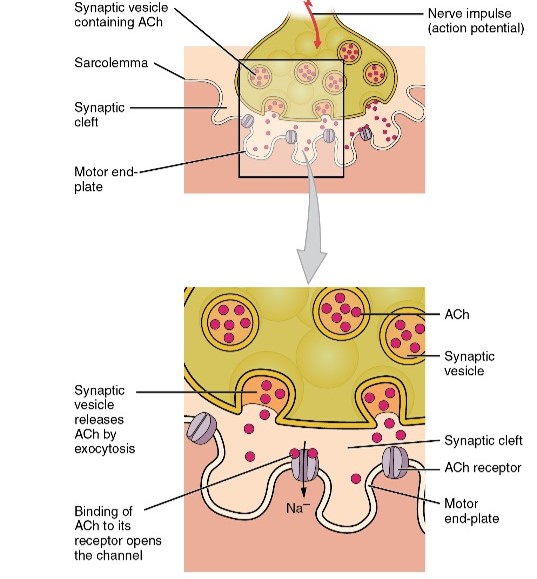
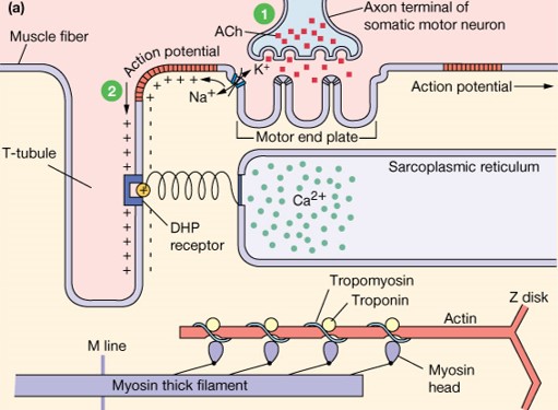
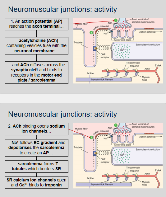
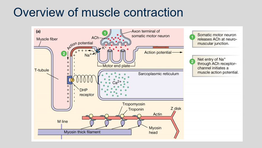
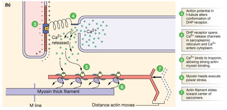
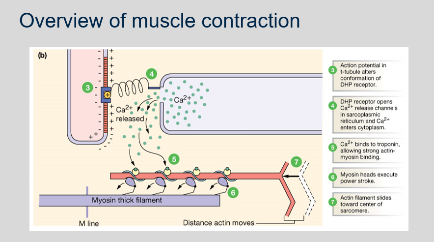
During activity at the neuromuscular junction, the following events occur according to the sliding filament theory:
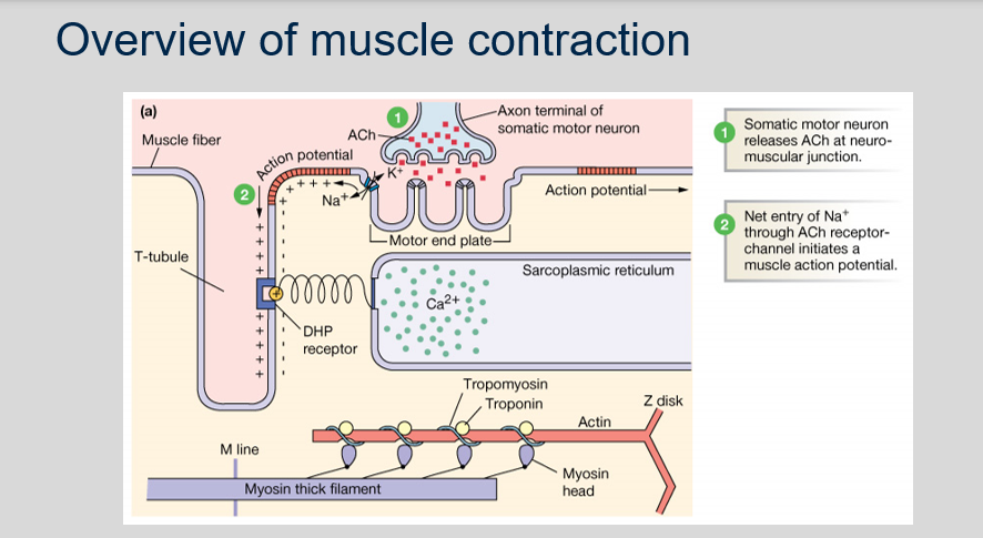
1. An action potential (AP) reaches the axon terminal.
2. Acetylcholine (ACh)-containing vesicles fuse with the neuronal membrane and ACh diffuses across the synaptic cleft and binds to receptors in the motor end plate/sarcolemma.
3. ACh binding opens sodium ion channels.
4. Na+ follows its electrochemical gradient and depolarizes the sarcolemma, creating an AP.
5. The sarcolemma forms T-tubules which border the sarcoplasmic reticulum (SR).
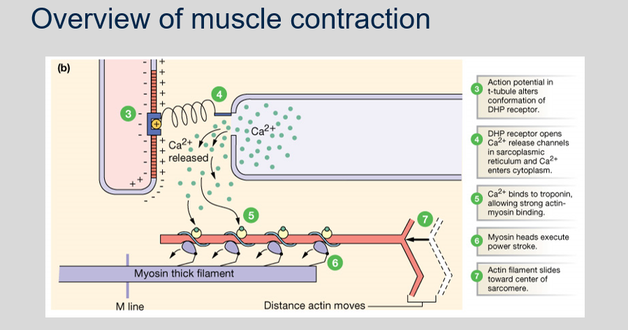
6. SR calcium ion channels open and Ca2+ binds to troponin, causing a conformational change in the troponin-tropomyosin complex, which exposes the myosin-binding sites on actin.
1. An action potential (AP) reaches the axon terminal.
2. Acetylcholine (ACh)-containing vesicles fuse with the neuronal membrane and ACh diffuses across the synaptic cleft and binds to receptors in the motor end plate/sarcolemma.
3. ACh binding opens sodium ion channels.
4. Na+ follows its electrochemical gradient and depolarizes the sarcolemma, creating an AP.
5. The sarcolemma forms T-tubules which border the sarcoplasmic reticulum (SR).
6. SR calcium ion channels open and Ca2+ binds to troponin, causing a conformational change in the troponin-tropomyosin complex, which exposes the myosin-binding sites on actin.

55
New cards
^^Neuromuscular junctions: activity^^
Can you label, describe and explain what this diagram is/shows?
Can you label, describe and explain what this diagram is/shows?
56
New cards
^^Transverse tubules (t-tubules)^^
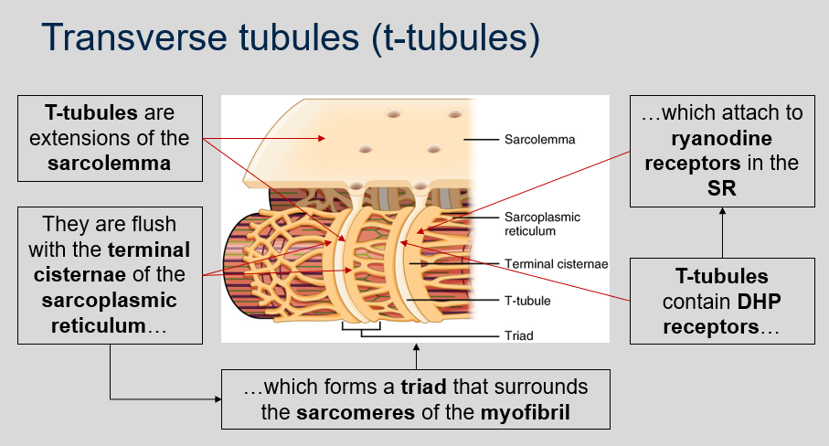
What are T-tubules in muscle fibers and what is their relationship with the sarcoplasmic reticulum?
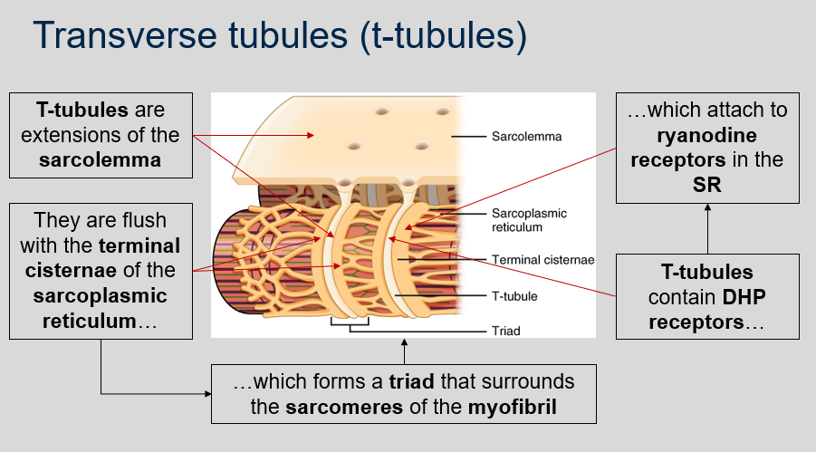
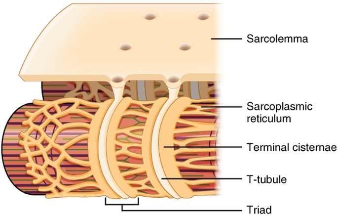
What are T-tubules in muscle fibers and what is their relationship with the sarcoplasmic reticulum?
T-tubules are extensions of the sarcolemma that are flush with the terminal cisternae of the sarcoplasmic reticulum, which forms a triad that surrounds the sarcomeres of the myofibril, and they contain DHP receptors that attach to ryanodine receptors in the SR. This coupling allows for the release of calcium ions from the sarcoplasmic reticulum into the cytosol of the muscle fiber, which triggers muscle contraction.
57
New cards
^^Transverse tubules (t-tubules)^^
Can you label, describe and explain what this diagram is/shows?
Can you label, describe and explain what this diagram is/shows?
58
New cards
^^Sarcoplasmic reticulum (SR)^^
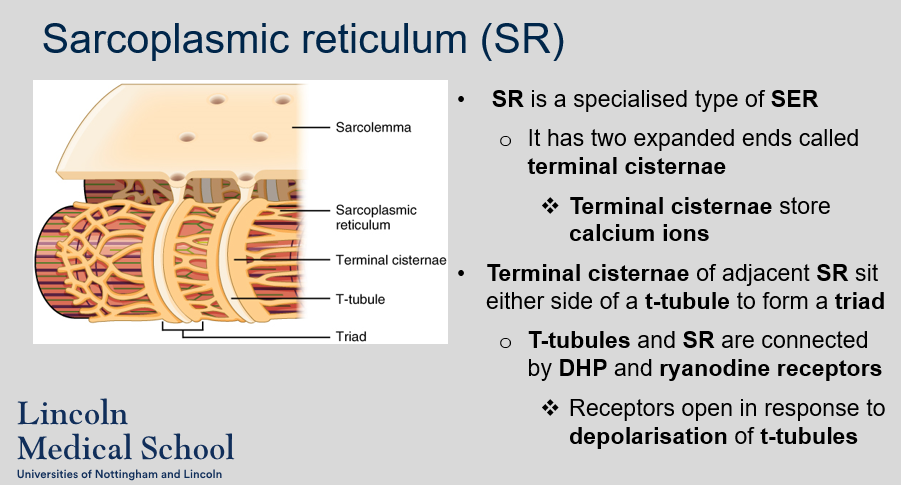
What is the sarcoplasmic reticulum (SR) and how is it connected to the T-tubules?
What is the sarcoplasmic reticulum (SR) and how is it connected to the T-tubules?
The SR is a specialized type of smooth endoplasmic reticulum (ER) in muscle cells that stores and releases calcium ions. It has two expanded ends called terminal cisternae, which store calcium ions. The terminal cisternae of adjacent SR sit on either side of a T-tubule, forming a triad. The T-tubules and SR are connected by dihydropyridine (DHP) receptors on the T-tubule membrane and ryanodine receptors on the SR membrane. These receptors open in response to depolarization of the T-tubules, allowing calcium ions to be released from the SR into the cytoplasm of the muscle cell.
59
New cards
^^DHP receptors and ryanodine receptors^^
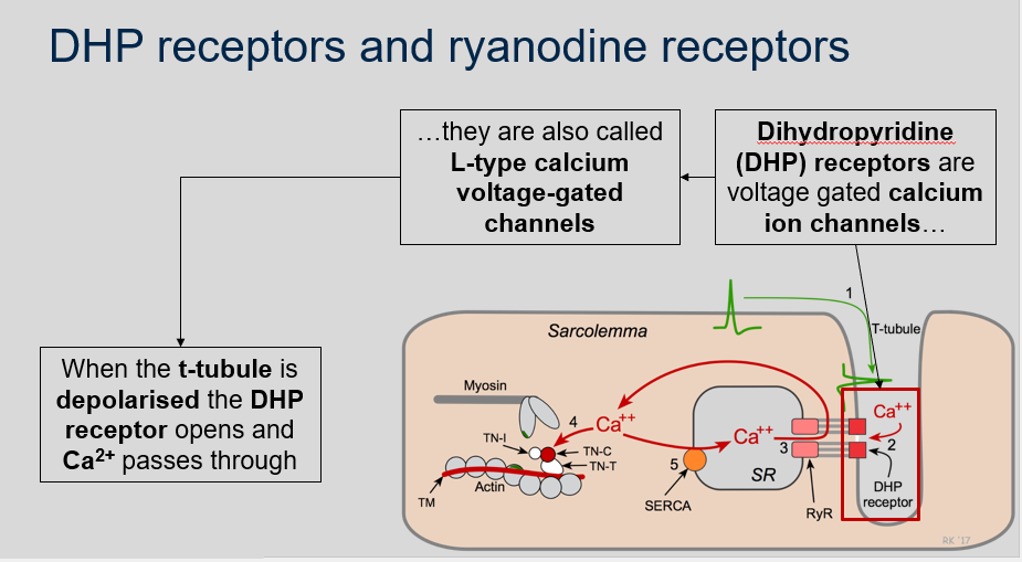
1. What are dihydropyridine (DHP) receptors?
2. What happens when the t-tubule is depolarised in relation to DHP receptors?
1. What are dihydropyridine (DHP) receptors?
2. What happens when the t-tubule is depolarised in relation to DHP receptors?
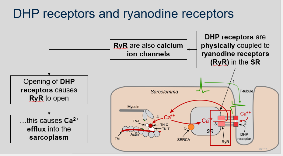
1. Dihydropyridine (DHP) receptors are voltage-gated calcium ion channels. They are also called L-type calcium voltage-gated channels.
2. When the t-tubule is depolarised, the DHP receptor opens and Ca2+ passes through.

60
New cards
^^DHP receptors and ryanodine receptors^^
Can you label, describe and explain what this diagram is/shows?
Can you label, describe and explain what this diagram is/shows?
61
New cards
^^DHP receptors and ryanodine receptors^^
How are DHP receptors and ryanodine receptors (RyR) in the SR connected?
What are RyR and what is their function in muscle contraction?
How are DHP receptors and ryanodine receptors (RyR) in the SR connected?
What are RyR and what is their function in muscle contraction?
DHP receptors are physically coupled to ryanodine receptors (RyR) in the SR.
RyR are calcium ion channels in the sarcoplasmic reticulum (SR) and they play a crucial role in muscle contraction. When DHP receptors open in response to depolarization of t-tubules, this triggers the opening of RyR, which causes Ca2+ efflux from the SR into the sarcoplasm.
RyR are calcium ion channels in the sarcoplasmic reticulum (SR) and they play a crucial role in muscle contraction. When DHP receptors open in response to depolarization of t-tubules, this triggers the opening of RyR, which causes Ca2+ efflux from the SR into the sarcoplasm.
62
New cards
^^Calcium ions and SERCAs^^
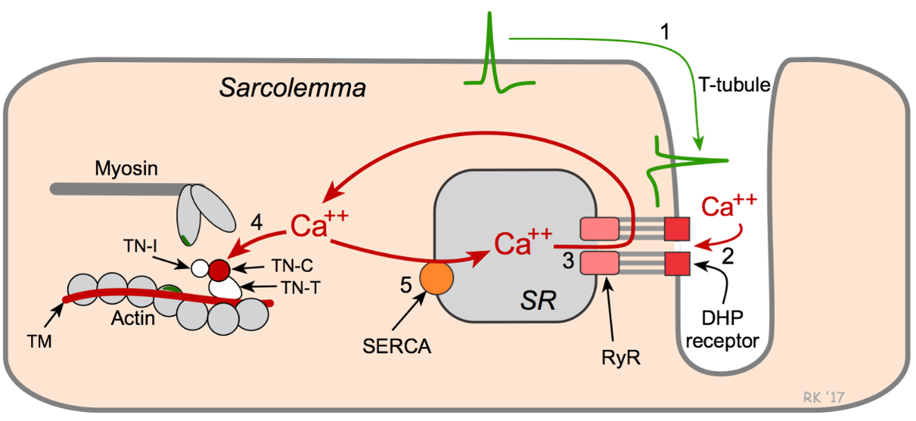
What is the role of Ca2+ in muscle contraction and what is the role of SERCAs?
What is the role of Ca2+ in muscle contraction and what is the role of SERCAs?
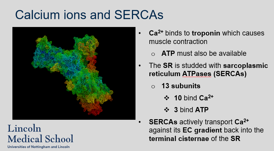
Ca2+ plays a crucial role in muscle contraction by binding to troponin and exposing the myosin-binding sites on actin. This allows the myosin heads to bind to actin and form cross-bridges, which generate force and cause the muscle to contract. ATP is also required for the cross-bridge cycle to occur.
The SR is studded with sarcoplasmic reticulum ATPases (SERCAs). SERCAs are responsible for actively transporting Ca2+ against its electrochemical gradient back into the terminal cisternae of the sarcoplasmic reticulum (SR) after muscle contraction. This process requires ATP hydrolysis and is crucial for muscle relaxation and preventing sustained contraction. SERCAs consist of 13 subunits, where 10 subunits bind to Ca2+ and 3 subunits bind to ATP.
The SR is studded with sarcoplasmic reticulum ATPases (SERCAs). SERCAs are responsible for actively transporting Ca2+ against its electrochemical gradient back into the terminal cisternae of the sarcoplasmic reticulum (SR) after muscle contraction. This process requires ATP hydrolysis and is crucial for muscle relaxation and preventing sustained contraction. SERCAs consist of 13 subunits, where 10 subunits bind to Ca2+ and 3 subunits bind to ATP.
63
New cards

^^Overview of muscle contraction^^
Can you label, describe and explain what this diagram is/shows?
Can you label, describe and explain what this diagram is/shows?
64
New cards
^^Overview of muscle contraction^^
Can you label, describe and explain what this diagram is/shows?
Can you label, describe and explain what this diagram is/shows?
\
65
New cards
^^Role of ATP^^
What is the role of ATP in muscle contraction?
What is the role of ATP in muscle contraction?
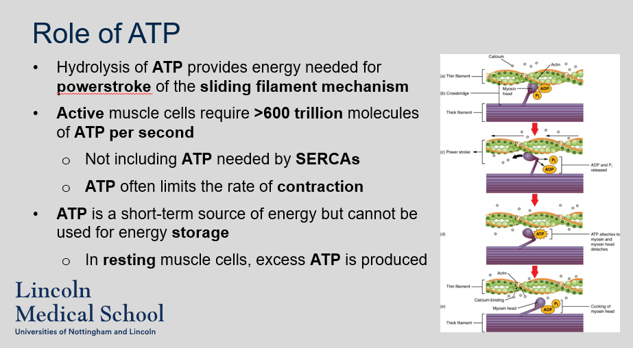
Hydrolysis of ATP provides energy needed for powerstroke of the sliding filament mechanism. Active muscle cells require >600 trillion molecules of ATP per second. Not including ATP needed by SERCAs. ATP often limits the rate of contraction. ATP is a short-term source of energy but cannot be used for energy storage. In resting muscle cells, excess ATP is produced
66
New cards
^^Role of ATP^^
Can you label, describe and explain what this diagram is/shows?
Can you label, describe and explain what this diagram is/shows?

67
New cards
^^Role of ATP^^
1. What are the two roles of ATP in muscle contraction?
2. What are the three sources of ATP in muscle cells?
1. What are the two roles of ATP in muscle contraction?
2. What are the three sources of ATP in muscle cells?
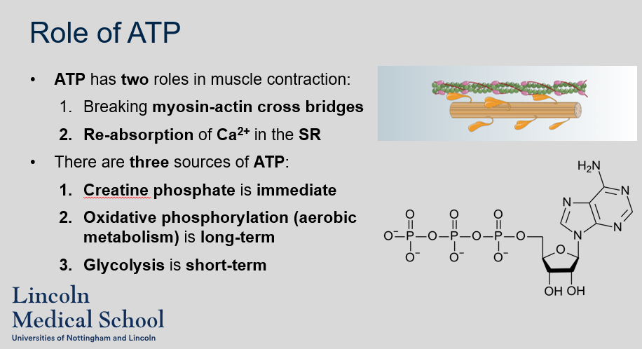
1. The two roles of ATP in muscle contraction are breaking myosin-actin cross bridges and re-absorption of Ca2+ in the SR.
2. The three sources of ATP in muscle cells are creatine phosphate (immediate), oxidative phosphorylation (aerobic metabolism, long-term), and glycolysis (short-term).
68
New cards

^^Role of ATP^^
Can you describe/explain what this image is/shows?
Can you describe/explain what this image is/shows?

69
New cards
^^Sources of ATP: creatine phosphate^^
Why is creatine phosphate important in muscle contraction?
Why is creatine phosphate important in muscle contraction?
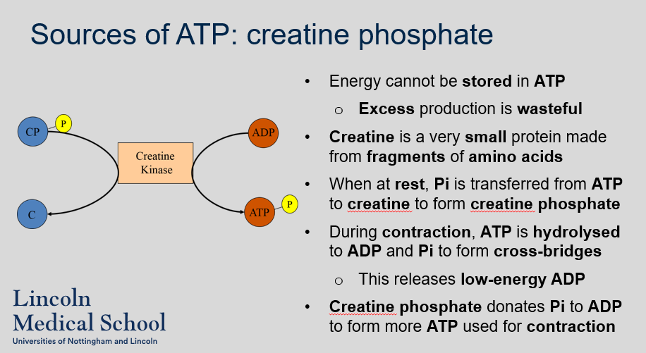
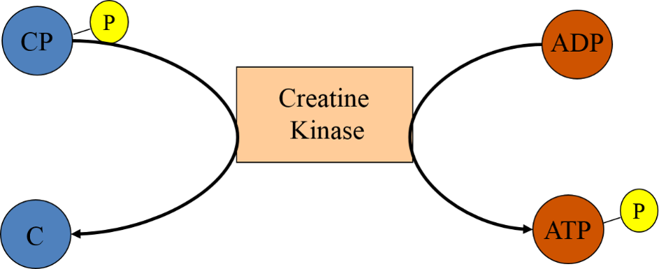
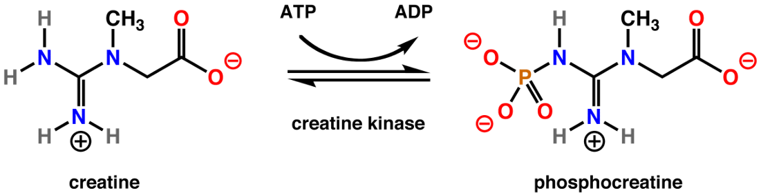
Energy cannot be stored in ATP. Excess production is wasteful. Creatine is a very small protein made from fragments of amino acids. When at rest, Pi is transferred from ATP to creatine to form creatine phosphate. During contraction, ATP is hydrolysed to ADP and Pi to form cross-bridges. This releases low-energy ADP. Creatine phosphate donates Pi to ADP to form more ATP used for contraction.
70
New cards
^^Sources of ATP: creatine phosphate^^
Can you label, describe and explain what this diagram is/shows?
Can you label, describe and explain what this diagram is/shows?

71
New cards
^^Sources of ATP: creatine phosphate^^
What is the role of creatine phosphate in muscle contraction, and how is it regulated?
What is the role of creatine phosphate in muscle contraction, and how is it regulated?
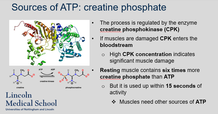
Creatine phosphate plays a critical role in muscle contraction by donating a phosphate group to ADP to form ATP, which is used to power the sliding filament mechanism. The process is regulated by the enzyme creatine phosphokinase (CPK), which facilitates the transfer of phosphate from creatine phosphate to ADP. If muscles are damaged, CPK enters the bloodstream, and high CPK concentration indicates significant muscle damage. Resting muscle contains six times more creatine phosphate than ATP, but it is used up within 15 seconds of activity, and muscles need other sources of ATP, including oxidative phosphorylation (aerobic metabolism) and glycolysis.

72
New cards
^^Sources of ATP: creatine phosphate^^
Can you label, describe and explain what this diagram is/shows?
Can you label, describe and explain what this diagram is/shows?
73
New cards
^^Rhabdomyolysis^^
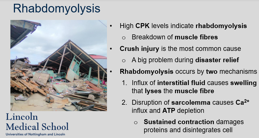
What does high CPK levels indicate and what is rhabdomyolysis?
What does high CPK levels indicate and what is rhabdomyolysis?
High levels of CPK indicate significant muscle damage (Rhabdomyolysis). Rhabdomyolysis is the breakdown of muscle fibers, and it is the most common cause of high CPK levels. Crush injuries are a frequent cause of rhabdomyolysis. Rhabdomyolysis can occur by two mechanisms: (1) the influx of interstitial fluid causes swelling that lyses the muscle fiber, and (2) disruption of the sarcolemma causes Ca2+ influx and ATP depletion, leading to sustained contraction, damage to proteins, and disintegration of the cell.
74
New cards
^^Sources of ATP: aerobic metabolism^^
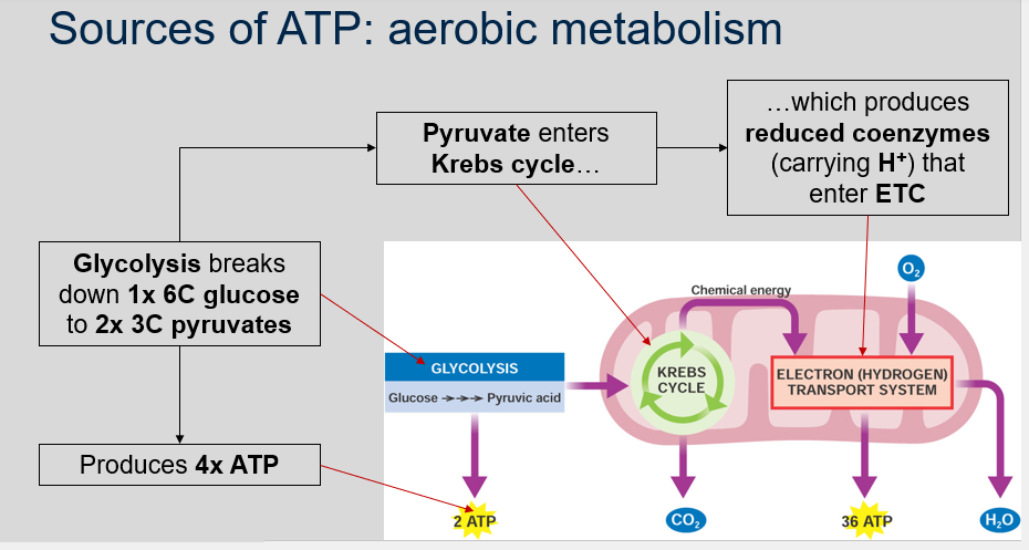
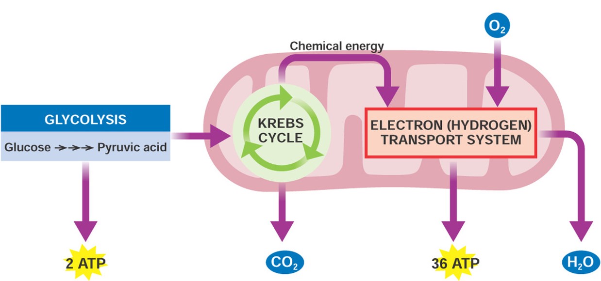
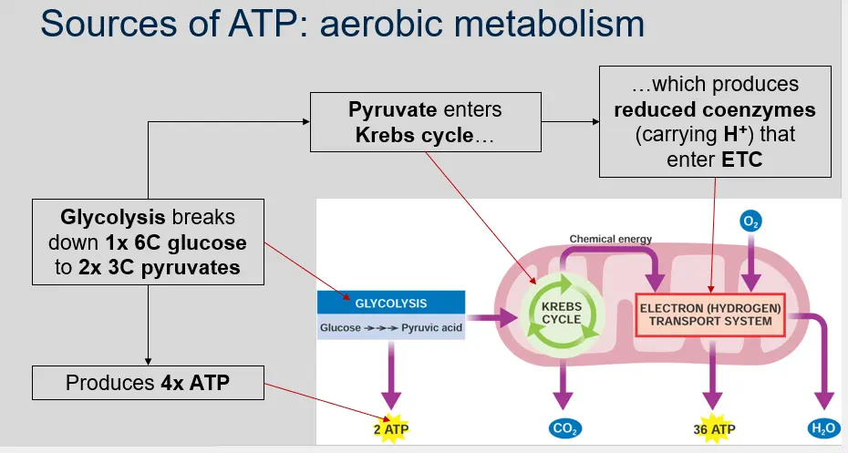
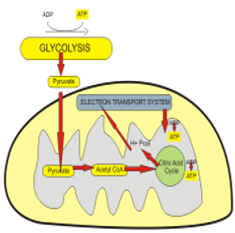
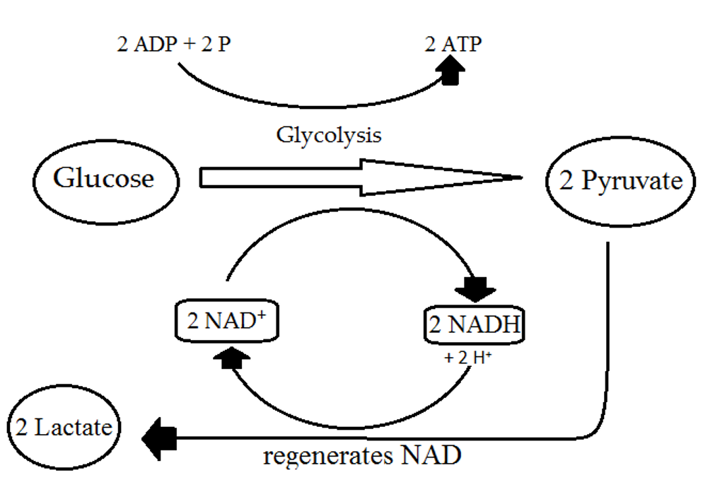
What is the process by which glucose is broken down into pyruvate, and what happens to pyruvate after glycolysis?
What is the process by which glucose is broken down into pyruvate, and what happens to pyruvate after glycolysis?
Glycolysis breaks down 1x 6C glucose to 2x 3C pyruvates and produces 4x ATP. Pyruvate enters Krebs cycle which produces reduced coenzymes (carrying H+) that enter ETC.
75
New cards
^^Sources of ATP: aerobic metabolism^^
Can you label, describe and explain what this diagram is/shows?
Can you label, describe and explain what this diagram is/shows?
76
New cards
^^Sources of ATP: aerobic metabolism^^
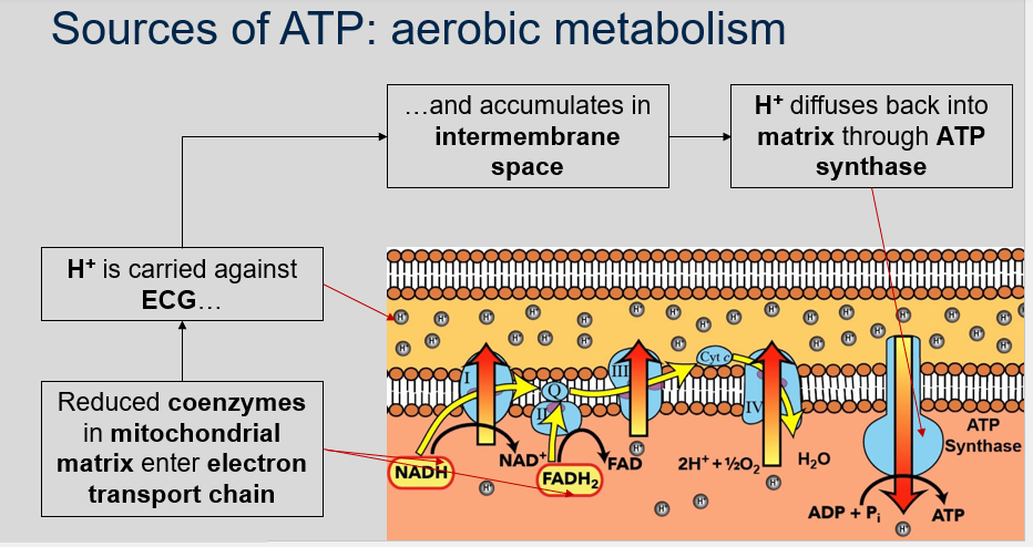
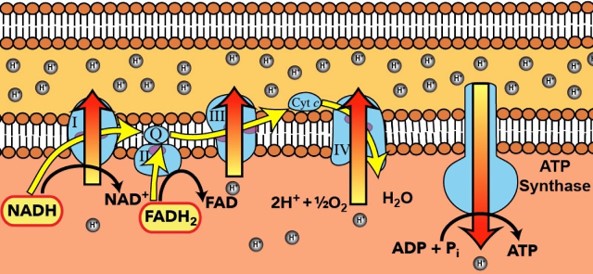
What happens to reduced coenzymes in the mitochondrial matrix?
What happens to reduced coenzymes in the mitochondrial matrix?
Reduced coenzymes in mitochondrial matrix enter the electron transport chain. H+ is carried against ECG and accumulates in the intermembrane space where H+ diffuses back into the matrix through ATP synthase.
77
New cards
^^Sources of ATP: aerobic metabolism^^
Can you label, describe and explain what this diagram is/shows?
Can you label, describe and explain what this diagram is/shows?

78
New cards
^^Sources of ATP: aerobic metabolism^^
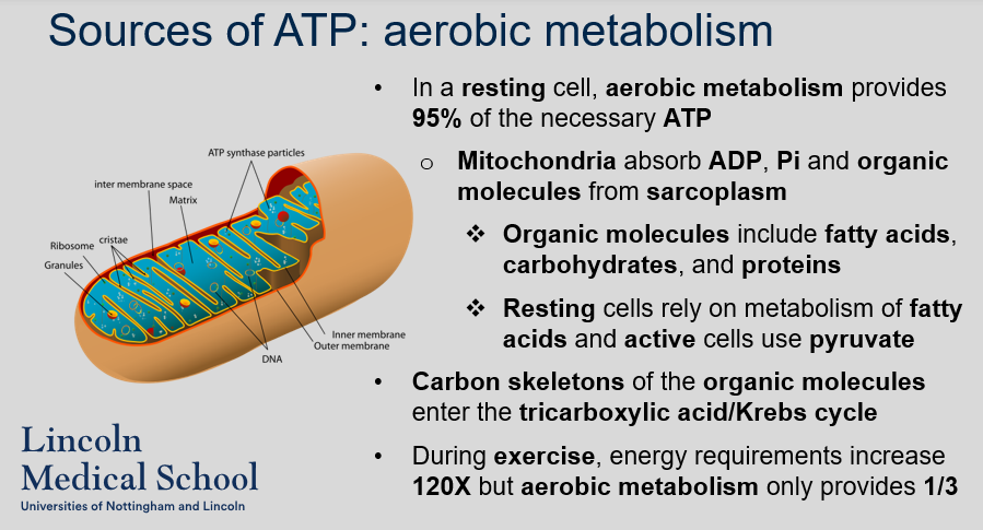
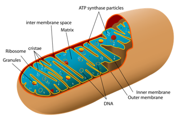
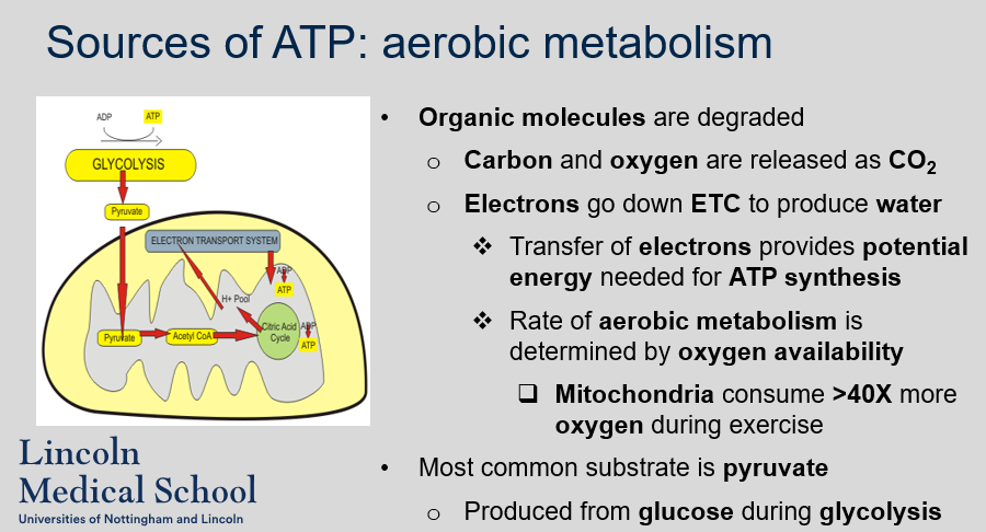
What is the role of aerobic metabolism in providing ATP in muscle cells? What organic molecules are used as a source of energy in resting and active muscle cells?
What is the role of aerobic metabolism in providing ATP in muscle cells? What organic molecules are used as a source of energy in resting and active muscle cells?
Aerobic metabolism provides 95% of the necessary ATP in resting muscle cells. Mitochondria absorb ADP, Pi, and organic molecules such as fatty acids, carbohydrates, and proteins from the sarcoplasm. Resting muscle cells rely on the metabolism of fatty acids as a source of energy, while active muscle cells use pyruvate. Carbon skeletons of the organic molecules enter the tricarboxylic acid/Krebs cycle to produce ATP. However, during exercise, energy requirements increase 120 times but aerobic metabolism only provides one-third of the necessary ATP.
79
New cards
^^Sources of ATP: aerobic metabolism^^
Can you label, describe and explain what this diagram is/shows?
Can you label, describe and explain what this diagram is/shows?

80
New cards
^^Sources of ATP: aerobic metabolism^^
How is ATP produced through aerobic metabolism in muscle cells?
How is ATP produced through aerobic metabolism in muscle cells?
ATP is primarily produced through the degradation of organic molecules, such as fatty acids, carbohydrates, and proteins, in the mitochondria of muscle cells. During aerobic metabolism, carbon and oxygen are released as CO2, and electrons go down the electron transport chain (ETC) to produce water. The transfer of electrons provides the potential energy needed for ATP synthesis. The rate of aerobic metabolism is determined by the availability of oxygen, and mitochondria consume over 40 times more oxygen during exercise. The most common substrate for ATP production during aerobic metabolism in muscle cells is pyruvate, which is produced from glucose during glycolysis.
81
New cards
^^Sources of ATP: aerobic metabolism^^
Can you label, describe and explain what this diagram is/shows?
Can you label, describe and explain what this diagram is/shows?

82
New cards
^^Sources of ATP: glycolysis^^
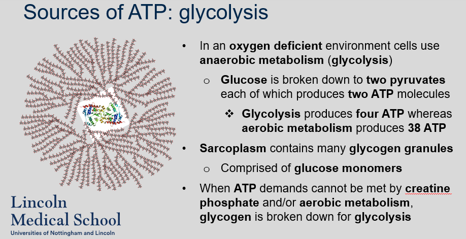
What happens to cells when there is an oxygen deficient environment?
What happens to cells when there is an oxygen deficient environment?
In an oxygen deficient environment, cells use anaerobic metabolism (glycolysis). Glucose is broken down to two pyruvates, each of which produces two ATP molecules. Glycolysis produces four ATP, whereas aerobic metabolism produces 38 ATP. Sarcoplasm contains many glycogen granules, which are comprised of glucose monomers. When ATP demands cannot be met by creatine phosphate and/or aerobic metabolism, glycogen is broken down for glycolysis.
83
New cards
^^Sources of ATP: glycolysis (lactic acid)^^
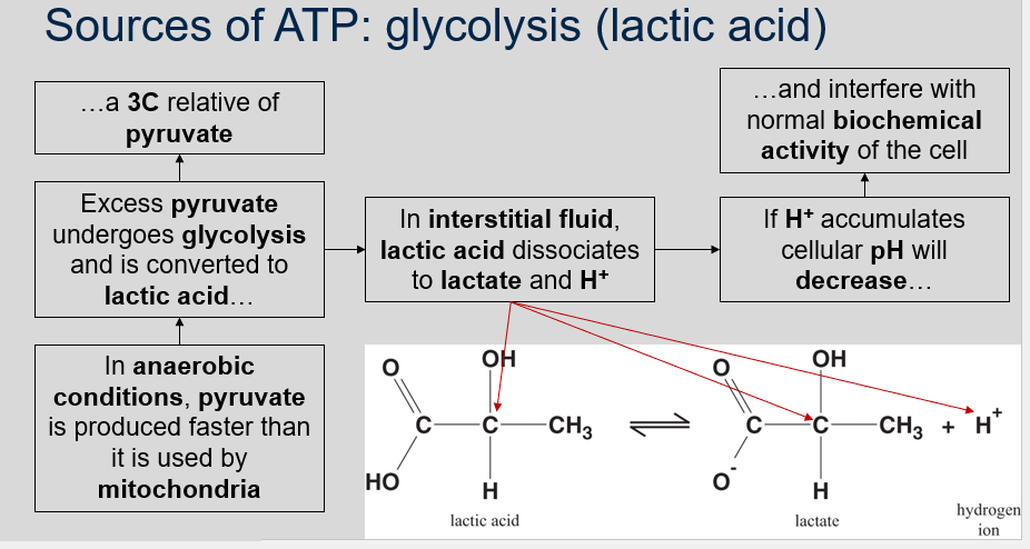
1. What happens to excess pyruvate in anaerobic conditions?
2. What happens to lactic acid in the interstitial fluid?
3. What happens if H+ accumulates in cells during anaerobic conditions?
1. What happens to excess pyruvate in anaerobic conditions?
2. What happens to lactic acid in the interstitial fluid?
3. What happens if H+ accumulates in cells during anaerobic conditions?
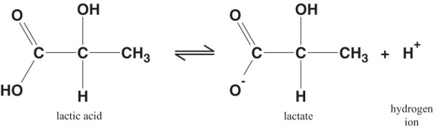
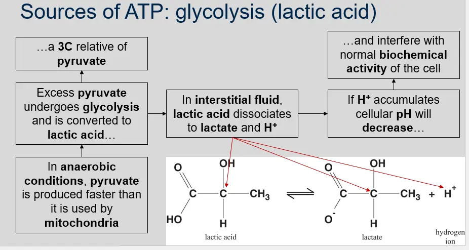
1. In anaerobic conditions, pyruvate is produced faster than it is used by mitochondria Excess pyruvate undergoes glycolysis and is converted to lactic acid a 3C relative of pyruvate.
2. In the interstitial fluid, lactic acid dissociates to lactate and H+.
3. If H+ accumulates in cells during anaerobic conditions, cellular pH will decrease and interfere with normal biochemical activity of the cell.
84
New cards
^^Sources of ATP: glycolysis (lactic acid)^^
Can you label, describe and explain what this equation is/shows?
Can you label, describe and explain what this equation is/shows?
85
New cards
^^Sources of ATP: muscle fatigue^^
What are the sources of muscle fatigue?
What are the sources of muscle fatigue?
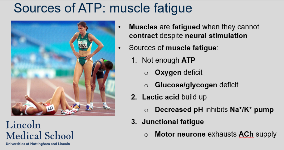
The sources of muscle fatigue are:
1. Not enough ATP
* Oxygen deficit
* Glucose/glycogen deficit
2. Lactic acid build up
* Decreased pH inhibits Na+/K+ pump
3. Junctional fatigue
* Motor neurone exhausts ACh supply
1. Not enough ATP
* Oxygen deficit
* Glucose/glycogen deficit
2. Lactic acid build up
* Decreased pH inhibits Na+/K+ pump
3. Junctional fatigue
* Motor neurone exhausts ACh supply
86
New cards
^^Sources of ATP: muscle fatigue (‘carb loading’)^^
What is the relationship between aerobic metabolism, glucose, and carb loading?
What is the relationship between aerobic metabolism, glucose, and carb loading?
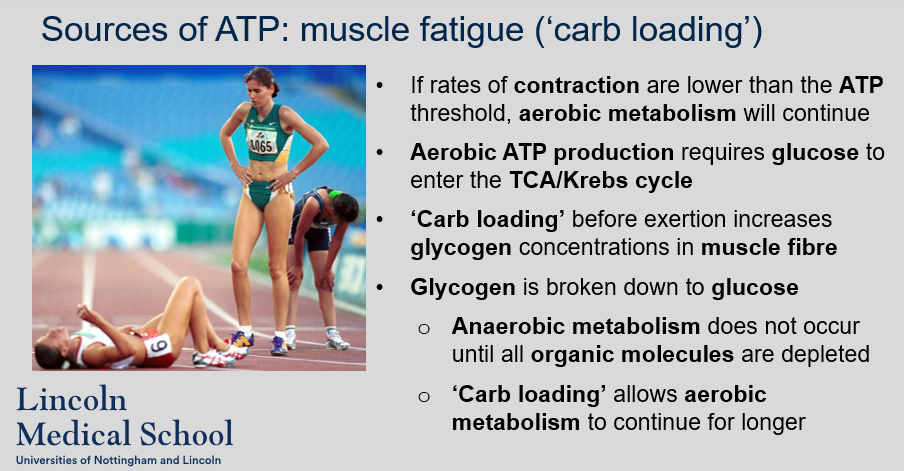
If rates of contraction are lower than the ATP threshold, aerobic metabolism will continue. Aerobic ATP production requires glucose to enter the TCA/Krebs cycle. ‘Carb loading’ before exertion increases glycogen concentrations in muscle fibre. Glycogen is broken down to glucose. Anaerobic metabolism does not occur until all organic molecules are depleted. ‘Carb loading’ allows aerobic metabolism to continue for longer
87
New cards
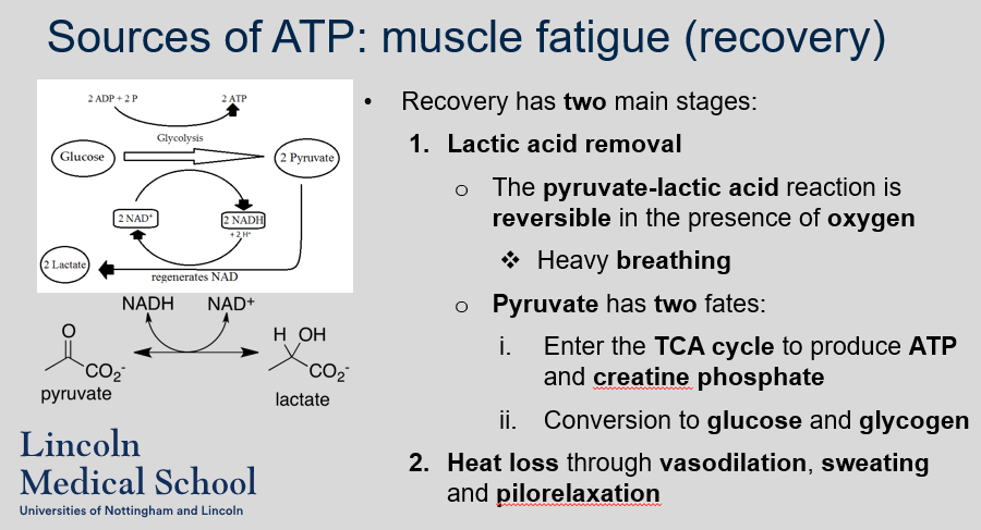
^^Sources of ATP: muscle fatigue (recovery)^^
1. What are the two main stages of recovery after exercise?
2. How is lactic acid removed during recovery?
3. What are the two fates of pyruvate during recovery?
4. How is heat loss promoted during recovery?
1. What are the two main stages of recovery after exercise?
2. How is lactic acid removed during recovery?
3. What are the two fates of pyruvate during recovery?
4. How is heat loss promoted during recovery?
1. The two main stages of recovery after exercise are lactic acid removal and restoration of energy reserves.
2. Lactic acid is removed during recovery through the pyruvate-lactic acid reaction, which is reversible in the presence of oxygen. Heavy breathing helps to increase oxygen levels and promote this reaction.
3. Pyruvate can either enter the TCA cycle to produce ATP and creatine phosphate or be converted to glucose and glycogen.
4. Heat loss is promoted during recovery through vasodilation, sweating, and pilorelaxation.
88
New cards
^^Sources of ATP: muscle fatigue (recovery)^^
Can you label, describe and explain what this diagram is/shows?
Can you label, describe and explain what this diagram is/shows?

89
New cards

^^Sources of ATP: muscle fatigue (recovery)^^
Can you label, describe and explain what this diagram is/shows?
Can you label, describe and explain what this diagram is/shows?

90
New cards
^^Sliding filament theory: cramp^^
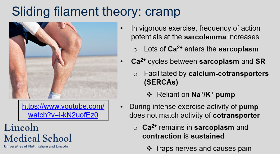
What happens to Ca2+ levels in the sarcoplasm during vigorous exercise?
What happens to Ca2+ levels in the sarcoplasm during vigorous exercise?
In vigorous exercise, frequency of action potentials at the sarcolemma increases thus lots of Ca2+ enters the sarcoplasm. Ca2+ cycles between the sarcoplasm and SR which is facilitated by calcium-cotransporters (SERCAs). However, during intense exercise, the activity of the Na+/K+ pump, which is reliant on the activity of cotransporter, does not match the activity of the cotransporter, causing Ca2+ to remain in the sarcoplasm and sustain contraction. This can trap nerves and cause pain.
91
New cards
^^Sliding filament theory: rigor mortis^^
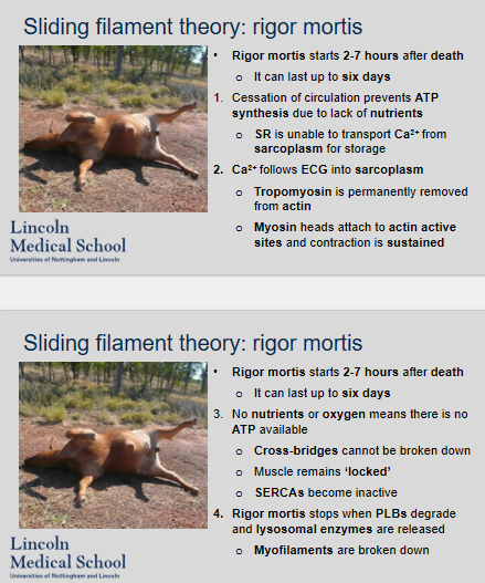
1. When does rigor mortis occur and how long can it last?
2. What causes rigor mortis?
3. How does rigor mortis end?
1. When does rigor mortis occur and how long can it last?
2. What causes rigor mortis?
3. How does rigor mortis end?
1. Rigor mortis occurs 2-7 hours after death and can last up to six days.
2. Cessation of circulation prevents ATP synthesis due to lack of nutrients and oxygen. SR is unable to transport Ca2+ from sarcoplasm for storage, and Ca2+ follows ECG into sarcoplasm. Tropomyosin is permanently removed from actin, and myosin heads attach to actin active sites, causing sustained contraction. Cross-bridges cannot be broken down, and muscles remain 'locked'. SERCAs become inactive.
3. Rigor mortis stops when PLBs degrade and lysosomal enzymes are released, causing myofilaments to be broken down.
92
New cards
^^Contraction: forces^^
1. How are muscle fibers connected to each other?
2. What is tension in muscle physiology?
3. How is resistance determined in muscle physiology?
4. What is compression in muscle physiology?
5. Can muscles produce compression?
1. How are muscle fibers connected to each other?
2. What is tension in muscle physiology?
3. How is resistance determined in muscle physiology?
4. What is compression in muscle physiology?
5. Can muscles produce compression?
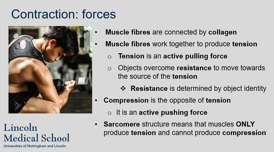
1. Muscle fibers are connected by collagen.
2. Tension is an active pulling force produced by muscle fibers working together.
3. Resistance is determined by object identity in muscle physiology.
4. Compression is the opposite of tension, and it is an active pushing force.
5. Muscles cannot produce compression due to the sarcomere structure, which only allows them to produce tension.
93
New cards
^^Contraction: forces^^
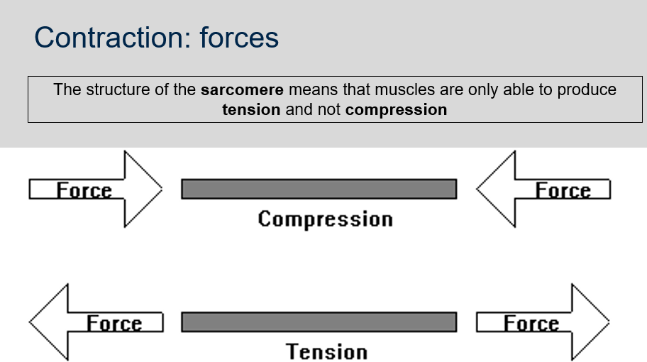
Why can muscles only produce tension and not compression?
Why can muscles only produce tension and not compression?
The structure of the sarcomere, the basic unit of muscle contraction, is such that it can only produce tension and not compression. The sarcomere consists of two types of protein filaments: actin and myosin. During muscle contraction, the myosin filaments pull the actin filaments towards the center of the sarcomere, shortening the sarcomere and producing tension. However, the sarcomere is not designed to push the actin filaments back to their original position, which would be necessary for the muscle to produce compression. Therefore, muscles are limited to producing tension only.
94
New cards
^^Contraction: forces^^
Can you label, describe and explain what this diagram is/shows?
Can you label, describe and explain what this diagram is/shows?

95
New cards
^^Contraction: forces (muscle fibres)^^
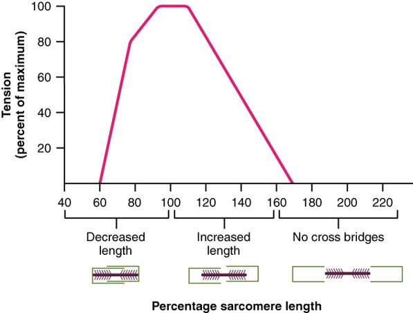
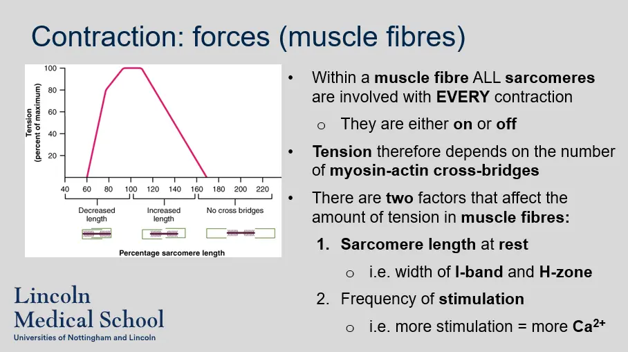
1. Are all sarcomeres within a muscle fibre involved with every contraction?
2. What is tension in muscle fibres dependent on?
3. What are the two factors that affect the amount of tension in muscle fibres?
1. Are all sarcomeres within a muscle fibre involved with every contraction?
2. What is tension in muscle fibres dependent on?
3. What are the two factors that affect the amount of tension in muscle fibres?
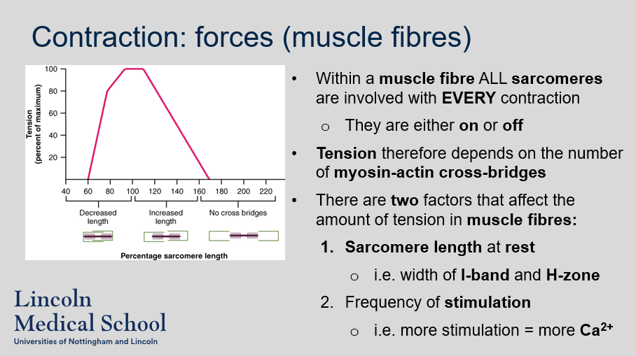
1. Yes, all sarcomeres within a muscle fibre are either on or off during every contraction.
2. Tension in muscle fibres depends on the number of myosin-actin cross-bridges.
3. The two factors that affect the amount of tension in muscle fibres are the sarcomere length at rest, which is determined by the width of the I-band and H-zone, and the frequency of stimulation, which results in more Ca2+ and therefore more cross-bridge formation.
96
New cards
^^Contraction: forces (muscle fibres)^^
Can you label, describe and explain what this diagram is/shows?
Can you label, describe and explain what this diagram is/shows?
97
New cards
^^Contraction: forces (muscles)^^
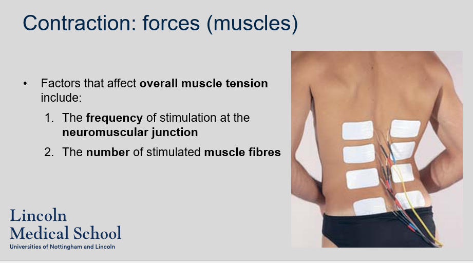
What are the factors that affect overall muscle tension?
What are the factors that affect overall muscle tension?
The factors that affect overall muscle tension include the frequency of stimulation at the neuromuscular junction and the number of stimulated muscle fibers.
98
New cards
^^Contraction: twitches^^
What is a twitch in a muscle fibre?
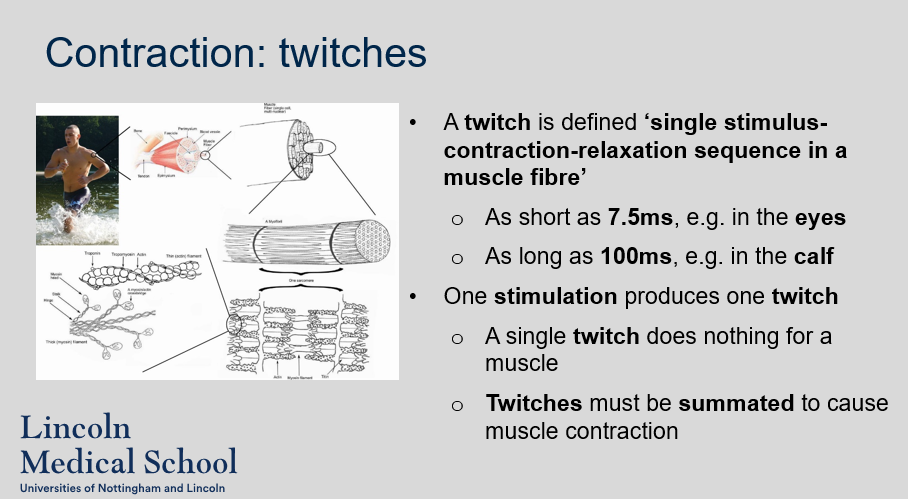
What is a twitch in a muscle fibre?
A twitch is a single stimulus-contraction-relaxation sequence in a muscle fibre, which can be as short as 7.5ms in the eyes or as long as 100ms in the calf. However, a single twitch does not produce any significant effect on a muscle, and multiple twitches need to be summated to cause muscle contraction.

99
New cards
^^Contraction: twitches^^
Can you describe/explain what this diagram is/shows?
Can you describe/explain what this diagram is/shows?
100
New cards
^^Contraction: three stages of a twitch (myograms)^^
1. What is a myogram?
2. What happens during the latent period of a twitch?
3. What happens during the contraction phase of a twitch?
4. What happens during the relaxation period of a twitch?
1. What is a myogram?
2. What happens during the latent period of a twitch?
3. What happens during the contraction phase of a twitch?
4. What happens during the relaxation period of a twitch?
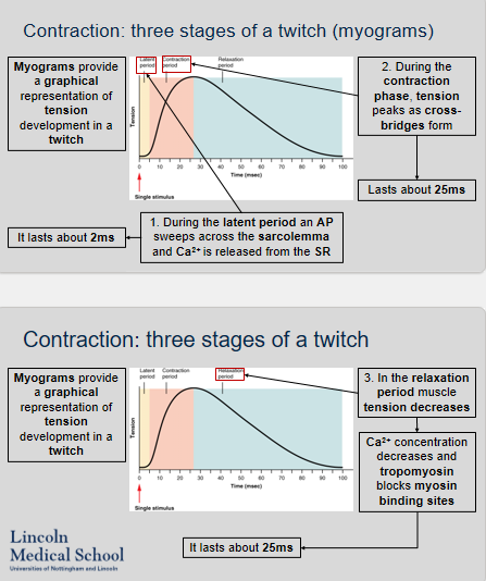
1. A myogram is a graphical representation of tension development in a twitch.
2. During the latent period of a twitch, an action potential (AP) sweeps across the sarcolemma and Ca2+ is released from the SR. This period lasts about 2ms.
3. During the contraction phase of a twitch, tension peaks as cross-bridges form. This phase lasts about 25ms.
4. During the relaxation period of a twitch, muscle tension decreases as Ca2+ concentration decreases and tropomyosin blocks myosin binding sites. This period lasts about 25ms.