The Skeletal System 10/3/25
1/18
There's no tags or description
Looks like no tags are added yet.
Name | Mastery | Learn | Test | Matching | Spaced |
|---|
No study sessions yet.
19 Terms
Pectoral Girdle
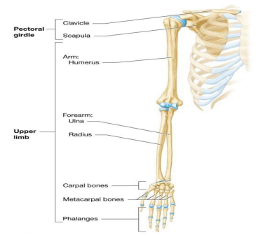
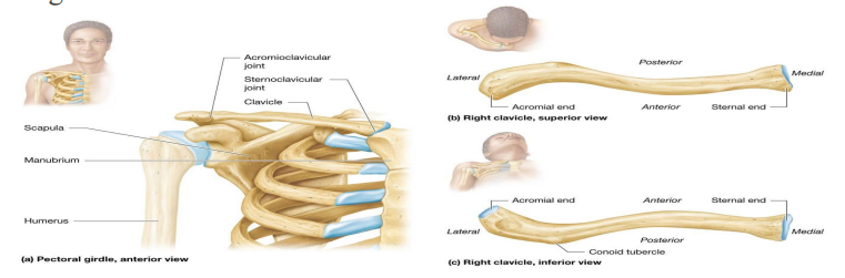
Pectoral girdle – clavicle and scapula; supports 30 bones that make up upper limb; all are components of appendicular skeleton
Each clavicle end is distinct and palpable through skin
Sternal end
articulates medially with manubrium at sternoclavicular joint

Acromial end
articulates laterally with acromion process of scapula at acromioclavicular joint; conoid tubercle provides site for attachment of ligaments near end
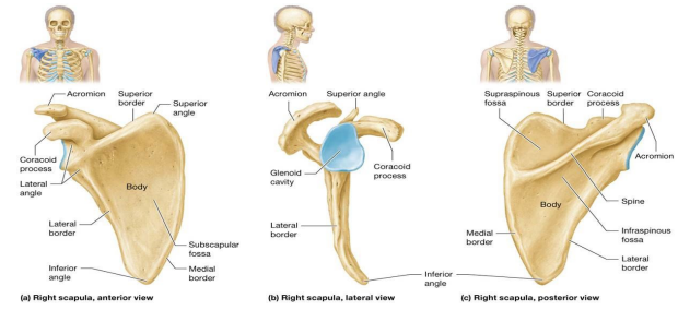
Triangular scapula
Triangular scapula sits on posterosuperior rib cage between second and seventh ribs; body is largest section; has three borders: medial, lateral, and superior
Coracoid process – hook-shaped projection on anterior surface
Subscapular fossa – inferior to coracoid process; provides attachment site for subscapularis muscle
Glenoid cavity – shallow indentation on lateral surface; articulates with humerus in shoulder joint
Spine – posterior ridge of bone; crosses from medial to lateral along superior scapula; terminates as acromion at acromioclavicular joint (AC)
Supraspinous fossa – superior to spine; infraspinous fossa is inferior
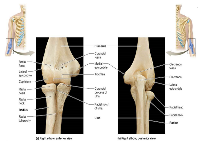
The Humerus
Humerus – largest and strongest bone of upper limb; only bone in arm (brachium); consists of two epiphyses; articulate with other bones and long diaphysis
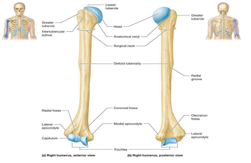
Features associated with humeral articulation with ulna and radius at elbow joint:
Capitulum – spherical knob on anterior and lateral aspect of distal epiphysis
Trochlea – spool-shaped knob on anterior and medial aspect of distal epiphysis
Lateral radial fossa and medial coronoid fossa – small indentations just proximal to capitulum and trochlea
Olecranon fossa – deep indentation on posterior aspect of distal epiphysis; continuation of trochlea

Bones of forearm (antebrachium)
Lateral radius and medial ulna; held to one another by fibrous interosseous membrane
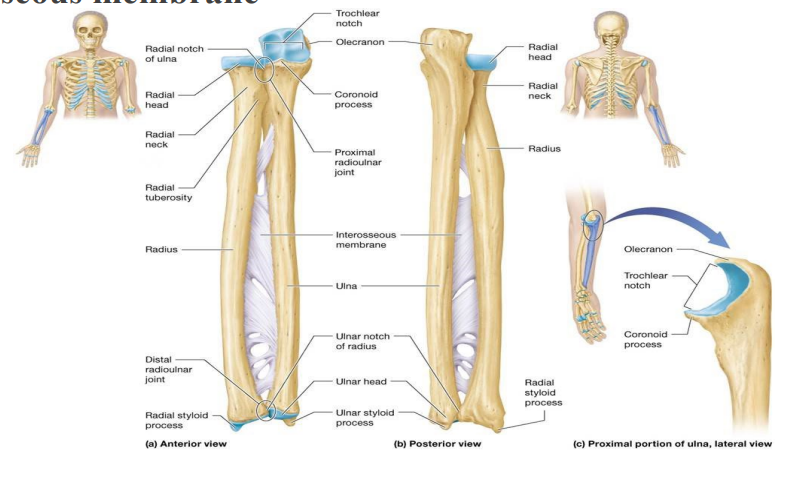
Radius, Ulna
Radius – narrow proximally; progressively enlarges distally
Proximal epiphysis (radial head) – round and flattened structure; articulates with capitulum of humerus at elbow joint and ulna at proximal radioulnar joint
Radial neck – distal to head, ends at radial tuberosity; on medial aspect of radius; attachment site for biceps brachii muscle
Ulna – widest at proximal epiphysis; progressively narrows as it travels distally
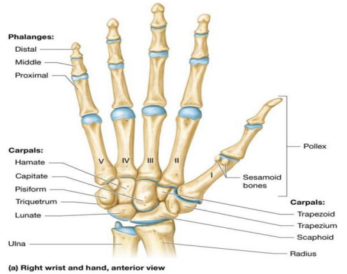
Bones of the Wrist: Carpals
Wrist (carpus) – eight short bones (carpals) arranged in two rows containing four bones each
Hand (manus) – Five long bones (metacarpals)
Bones of the Pelvic Girdle and Lower Limb
Pelvis and bones of pelvic girdle and lower limbs complete appendicular skeleton Hipbones or coxal bones (also known as os coxae) make up pelvic girdle
Articulates with sacrum (component of axial skeleton)
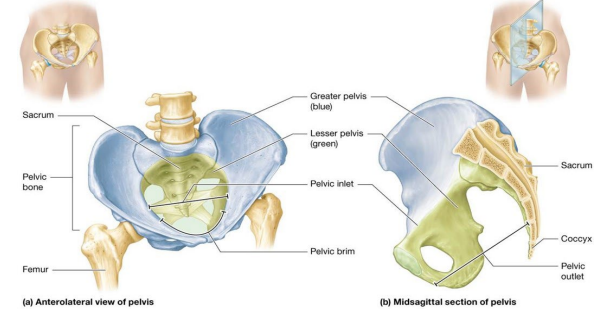
Each coxal bone is three fused bones: ilium, ischium, and pubis
All contribute to acetabulum; deep socket on lateral aspect of coxal bone ; head of femur articulates with acetabulum at hip joint
Ilium and pubis also contribute to obturator foramen; opening in each coxal bone, through which nerves and blood vessels travel
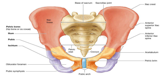
Ilium
forms superior portion of coxal bone
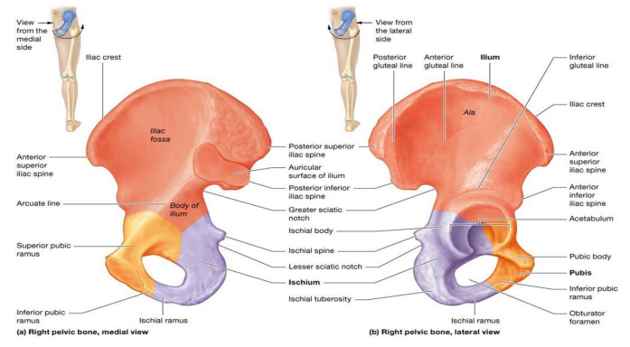
Ischium
ischial body and ramus; forms C-shaped posteroinferior portion of coxal bone

Pubis
smallest coxal bone; three parts that approximate C-shape
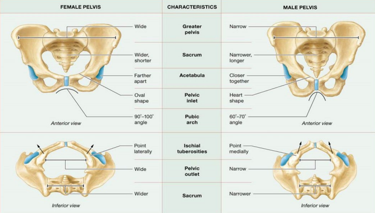
Female vs Male pelvis
Female Pelvis:
Anterior view
Wide greater pelvis
Wider, shorter sacrum
Acetabula farther apart
Pelvic inlet oval shape
Pubic arch 90-100 degree angle
Inferior view
Ischial tuberosities point laterally
Pelvic outlet wide
Sacrum wider
Male Pelvis:
Anterior view
Narrow greater pelvis
narrower, longer sacrum
Acetabula closer together
Pelvic inlet heart shape
Pubic arch 60-70 degree angle
Inferior view
Ischial tuberosities Point medially
Pelvic outlet narrow
Sacrum narrower
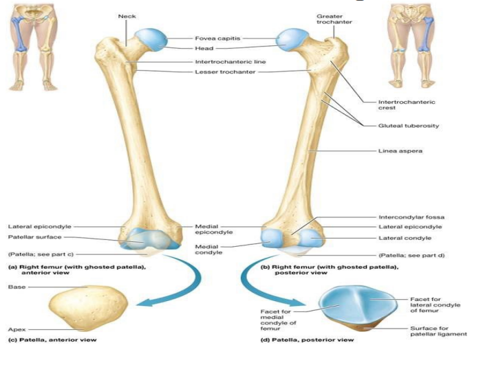
The femur and patella
Femur – largest and strongest bone in body and only bone in thigh
Proximal epiphysis features prominent spherical head; articulates with acetabulum at hip joint
Fovea capitis – small pit in center of head where small ligament attaches to stabilize joint
Triangular patella (kneecap) articulates with patellar surface on femur
Sesamoid bone located within tendon of anterior thigh muscle
Patellar ligament – distal continuation of this tendon; inserts into tibia; secures bone over anterior knee
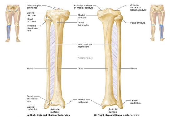
Tibia, Fibula
bones of leg; connected by interosseous membrane; articulate with one another at proximal and distal tibiofibular joints
Fibula – smaller lateral bone of leg, bears only one-sixth weight of tibia
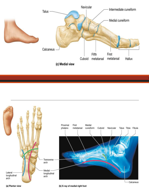
Tarsals, Metatarsals
Tarsals – Seven short bones make up ankle region; connects leg to foot
Metatarsals, numbered I-V from medial to lateral, have proximal base, middle shaft and distal head
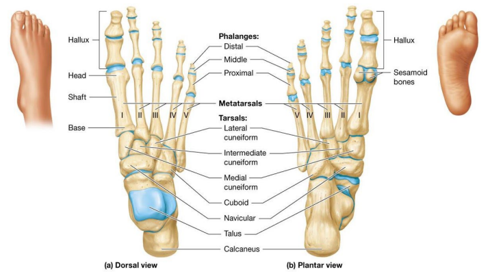
Bones of foot don’t normally lie flat on ground (arched)
three arches supported by ligaments and muscles help support and distribute body weight during movement