Kines 202 Exam 2
1/114
There's no tags or description
Looks like no tags are added yet.
Name | Mastery | Learn | Test | Matching | Spaced | Call with Kai |
|---|
No analytics yet
Send a link to your students to track their progress
115 Terms
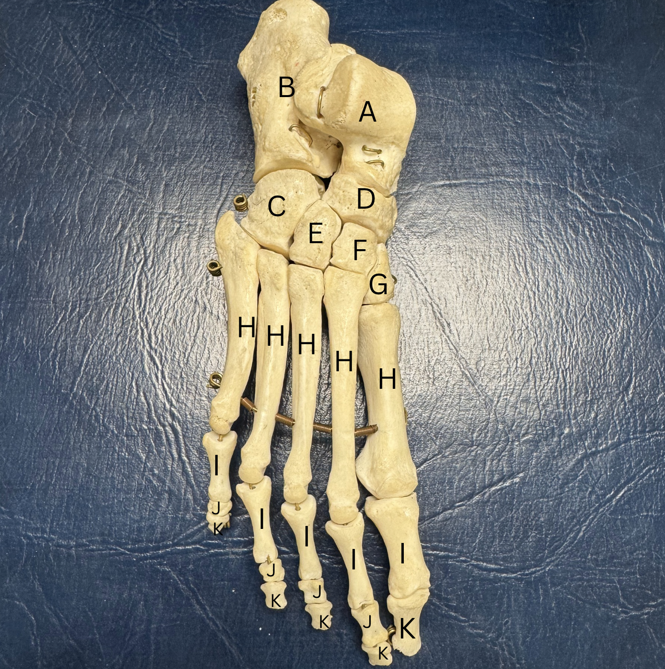
Label the bones of the feet
A. Talus
B. Calcaneus
C.Cuboid
D. Navicular
E.Lateral cuneiform
F. Intermediate cuneiform
G. Medial cuneiform
H. Metatarsals (1-5)
I. Proximal Phalange (1-5)
J. Middle Phalange (2-5)
K. Distal Phalange (1-5)
Label the parts of each of these short bones
Base= proximal
Head=distal
Middle= Shaft
True or False all 14 phalanges have a base, shaft, and head?
True; all 14 phalanges have a base, head, and shaft
What 2 sesamoid bones are in the foot?
2 sesamoid bones are found under the tendon of the flexor hallucis brevis, used to disperse motion for running
What is the purpose of the arch of the foot?
for shock absorption
What 3 arches do we have in our foot?
medial longitudinal arch-determines foot type
lateral longitudinal arch
transverse arch
What are the 3 different types of arches that are determined by the medial longitudinal arch?
Pes rectus- normal arch
Pes cavus- high arch
Pes planus- flat arch
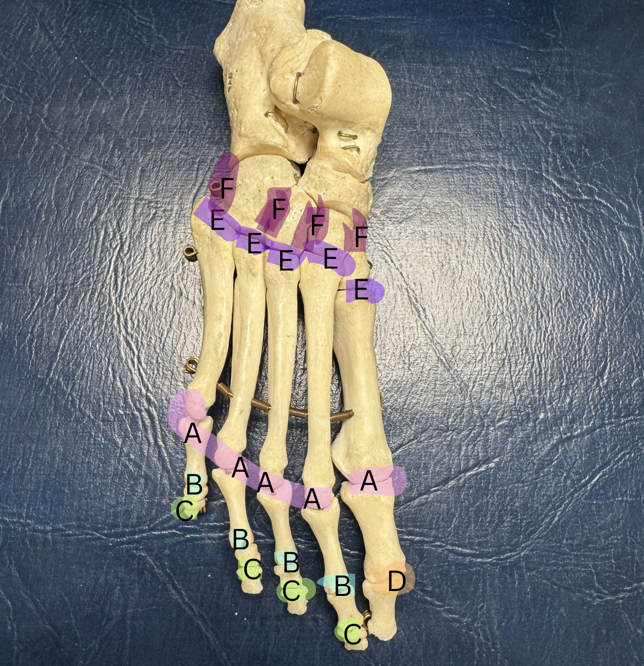
Label these joints A-F
A. Metatarsophalangeal joint
B. Proximal Interphalangeal Joint
C. Distal Interphalangeal Joint
D. Interphalangeal Joint
E. Tarsometatarsal Joint
F. Intermetatarsal Joint
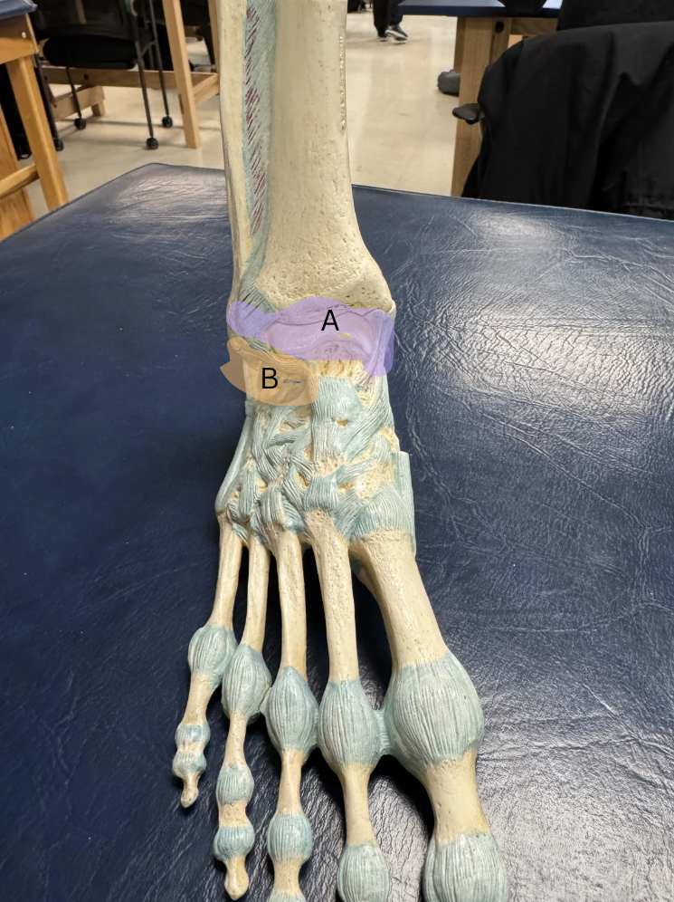
Label the two joints A and B, and what movements they do.
A. Talocrural Joint, Plantar Flexion and Dorsiflexion
B. Subtalar Joint, Ankle Eversion and Inversion
What is the function of the subtalar joint?
tri-planar motion- supination, pronation
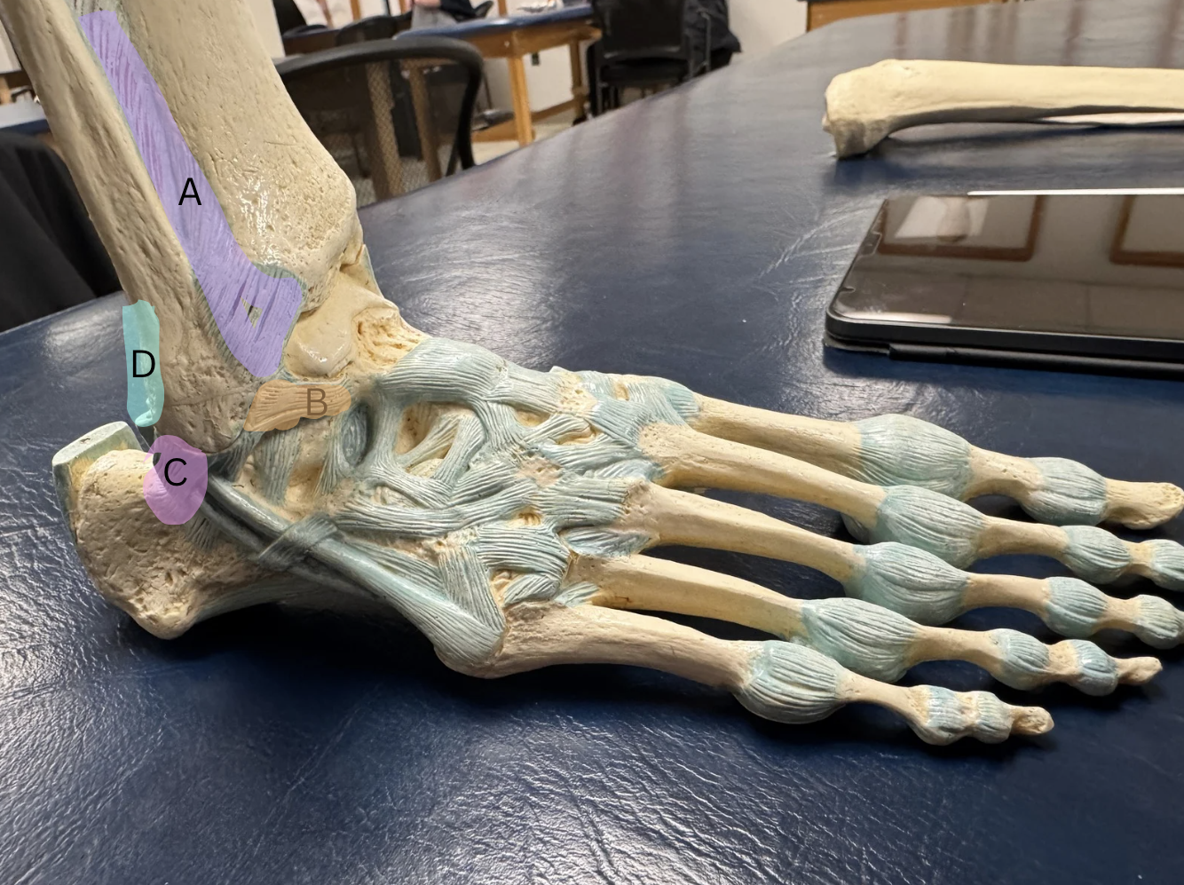
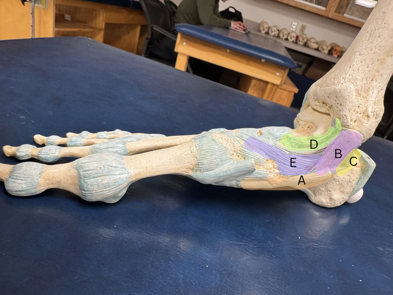
Label the ligaments from A-D
A. Anterior Tibiofibular Ligament
B. Anterior Talofibular Ligament
C. Calcaneofibular Ligament
D. Posterior Tibiofibular Ligament
Label each ligament from A-D
A. Plantar Calcaneonavicular (Spring) Ligament (makes up arch)
B.Tibiocalcaneal Ligament
C. Posterior Tibiotalar Ligament
D. Anterior Tibiotalar Ligament
E. Tibionavicular Ligament
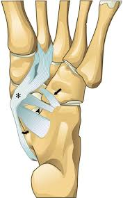
What ligament is on the bottom of the foot?
Long Plantar ligament, from the Calcaneus to the 2nd-5th Metatarsals
What other 3 ligaments make up the plantar fascia?
helps maintain longitudinal arch, includes medial band, central band, and lateral band
What is the difference between intrinsic and extrinsic muscles of the foot?
intrinsic= bone origin and insertion are in the foot
extrinsic= bone origin is out of the foot and insertion is in the foot
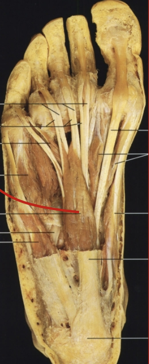
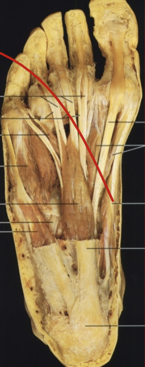
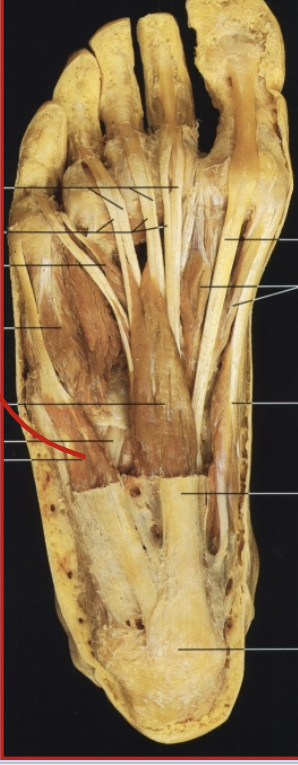
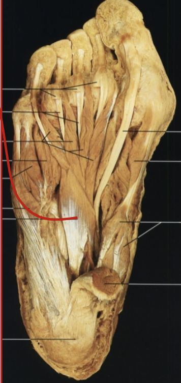
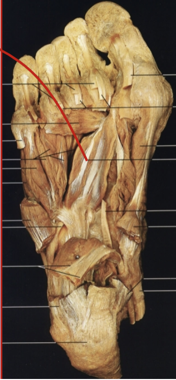
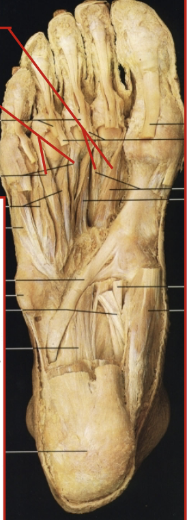
What are the plantar layers and muscles are included in them?
1st Layer- flexor digitorum brevis, abductor hallucis, abductor digiti minimi
2nd Layer- lumbricals and quadratus plantae
3rd LAYER- flexor hallucis brevis, adductor hallucis (2), flexor digiti minimi brevis
4th Layer- plantar interossei, and dorsal interossei
What muscle, layer, action and innervation is this?
muscle= flexor digitorum brevis
layer= 1st layer
action= flexes digits
innervation=medial plantar nerve
What muscle, layer, action and innervation is this?
muscle= abductor hallucis
layer= 1st layer
action= abducts 1st digit
innervation= medial plantar nerve
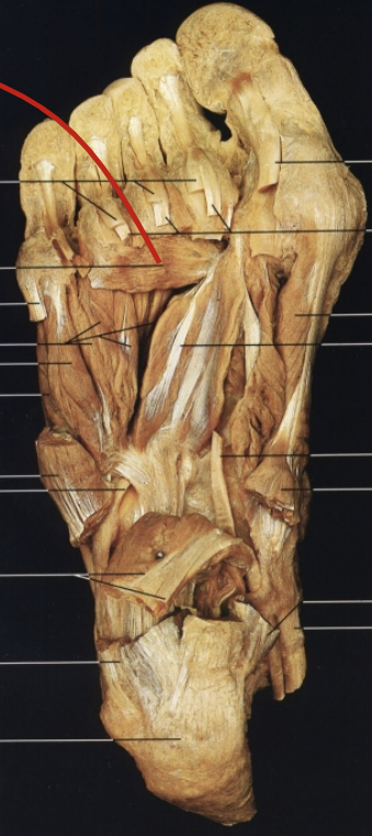
What muscle, layer, action and innervation is this?
muscle= abductor digiti minimi
layer= 1st layer
action= abducts and flexes the 5th metatarsal
innervation= lateral plantar nerve
What muscle, layer, action and innervation is this?
muscle= quadratus plantae
layer= 2nd layer
action= assists in flexor digitorum longus, flexes digits
innervation= lateral plantar nerve
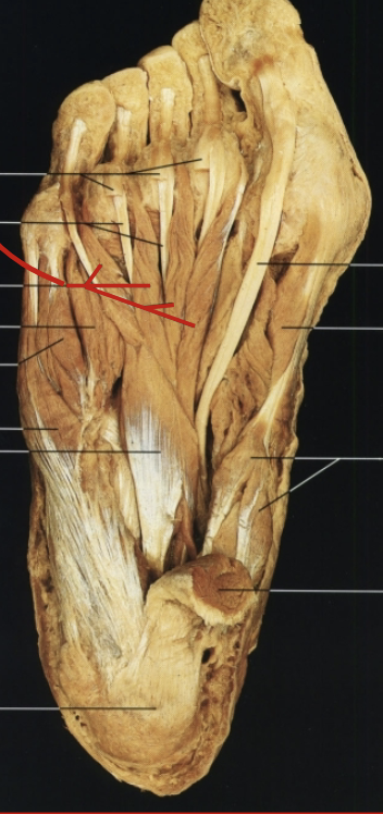
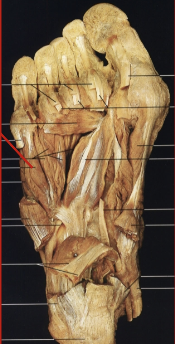
What muscle, layer, action and innervation is this?
muscle= lumbricals
layer= 2nd layer
action= flexes and extends digits 2-5 b/c they attach extrinsically and inserts in foot
innervation= lateral plantar nerve
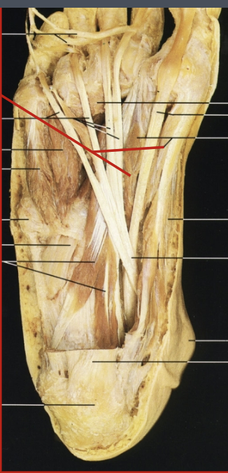
What muscle, layer, action and innervation is this?
muscle= flexor hallucis brevis
layer= 3rd layer
action= flexes 1st digit
innervation= medial plantar nerve
What muscle, layer, action and innervation is this?
muscle= adductor hallucis (oblique head)
layer= 3rd layer
action= adducts the metatarsophalangeal joint of great toe
innervation= lateral plantar nerve
What muscle, layer, action and innervation is this?
muscles= adductor hallucis (transverse head)
layer= 3rd layer
action= adducts metatarsophalangeal joint of great toe
innervation=lateral plantar nerve
What muscle, layer, action and innervation is this?
muscle= flexor digiti brevis
layer= 3rd layer
action= flexes 5th phalange
innervation= lateral plantar nerve
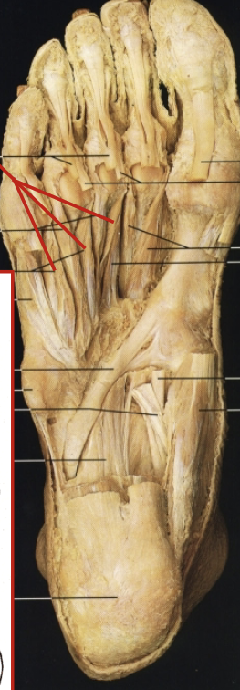
What muscle, layer, action and innervation is this?
muscle= plantar interossei (3)
layer= 4th layer
action= adducts and flexes 3rd, 4th, and 5th toe
innervation= lateral plantar nerve
What muscle, layer, action and innervation is this?
muscle= dorsal interossei (D.A.B)
layer= 4th layer
action= abduction of 2nd, 3rd, and 4th toes
innervation= lateral plantar nerve
What muscles are dorsal intrinsic muscles?
extensor digitorum brevis
extensor hallucis brevis

What muscle, action and innervation is this?
muscle= extensor digitorum brevis
action= helps extensor digitorum longus, extends toes 2-4
innervation= deep peroneal nerve
What muscle, layer, action and innervation is this?
muscle= extensor hallucis brevis
action= helps extensor hallucis longus, extends great toe
innervation= deep peroneal nerve
What two vascular arteries supply the foot?
Anterior tibial artery
Posterior tibial artery
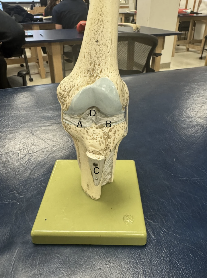
Label the menisci and ligaments
A.Medial Menisci
B.Lateral Menisci
C.Infrapatellar tendon
D. Anterior Cruciate Ligament
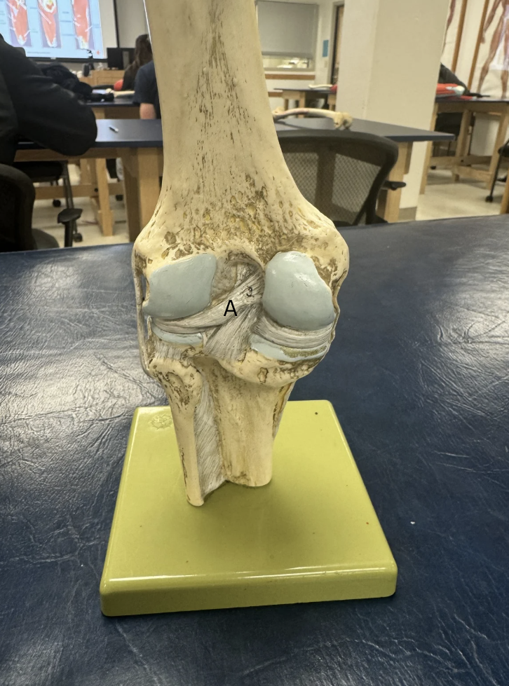
Label this ligament
A. Posterior Cruciate Ligament
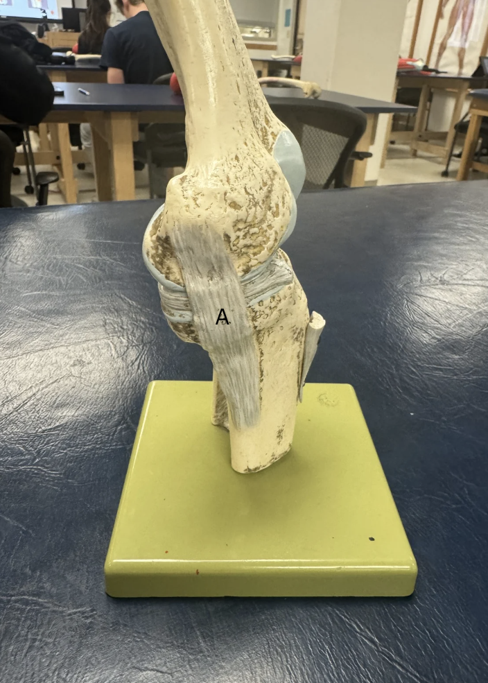
Label this ligament
A. Medial Collateral Ligament

Label this Ligament
A. Lateral Collateral Ligament
What type of joint in the tibiofibular joint?
amphiarthrodial (syndesmosis)
What is membrane is in between the Tibia and Fibula
Interosseous Membrane
Action of Subtalar Joint
eversion and inversion on the frontal plane
Action of Talocrural Joint
plantar flexion and dorsiflexion on the sagittal plane
Action of Tibiofibular Joint
Gliding
Difference between Open and Closed Kinetic Chain
Open Kinetic Chain= both feet off the ground/ in the air
Close Kinestic Chain= some [art of the foot is on the floor, usually leads to sprains
CKC Pronation joints
Subtalar: eversion
Forefront: abduction
Talocrural: plantar flexion
OKC Supination joints
Subtalar: inversion
Forefront: adduction
Talocrural: dorsiflexion
What muscles in the anterior compartment of the lower leg, what nerve innervates them, and what action do they all have in common?
Muscles- Anterior Tibialis, Extensor Digitorum Longus, Extensor Hallucis Longus, and Peroneus Tertius
Nerve- deep peroneal
Action-dorsiflexion
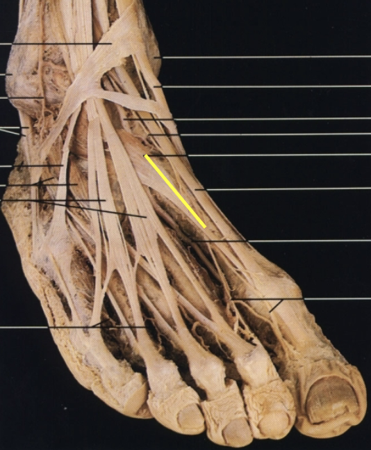
Label this muscle from A-C
A. Tibialis Anterior
B. Extensor Hallucis Longus
C. Extensor Digitorum Longus
Label this muscle
Peroneus Tertius
What kind of joint is the proximal and distal Tibiofibular Joint?
Amphiatrhrodial-syndesmosis
OIF of Anterior Tibialis
O: Lateral Tibial condyle
I: Medial Cuneiform and 1st Metatarsal
F:Dorsiflexion
OIF of Extensor Digitorum Longus and innervation
O: Lateral Tibia and head of Fibula
I: Dorsal (bottom side) of 2-5 Phalanges
F: Dorsiflexion of 4 digits
innervation: deep peroneal nerve
OIF of Extensor Hallucis Longus and innervation
O: Anterior surface of Fibula
I: Dorsal side (bottom side) of 1st Phalange
F: Dorsiflexion of big toes (1st metatarsal)
innervation: deep peroneal nerve
OIF of Peroneus Tertius
O: distal third of fibula
I: base of 5th metatarsal
F: small contribution to ankle dorsiflexion
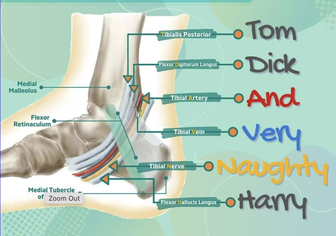
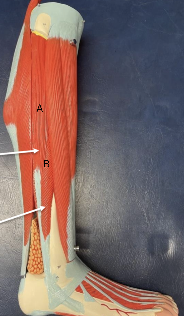
What muscles are in the deep posterior compartment?
Muscles: Tibialis Posterior,Flexor Digitorum Longus, Flexor Hallucis Longus
Nerve: Tibial Nerve
Action: Plantar Flexion
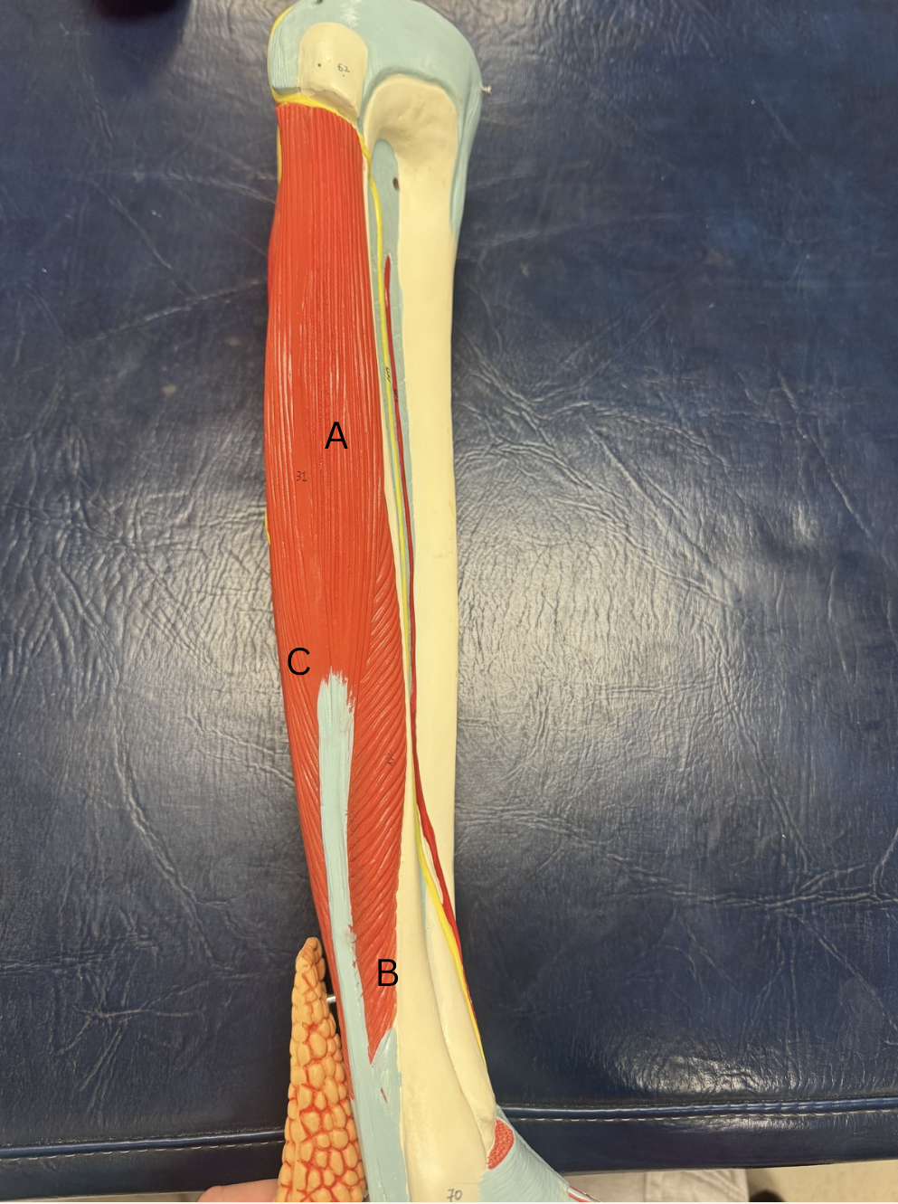
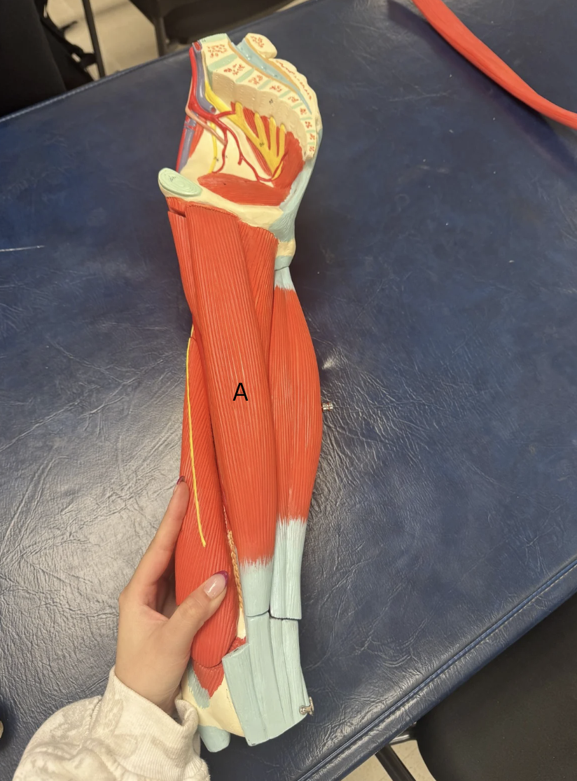
Label the muscles from A-D
A. Popliteus
B. Tibialis Posterior
C. Flexor Digitorum Longus
D. Flexor Hallucis Longus
OIF of Posterior Tibialis
O: Posterior Surface of Upper Intake interosseous membrane
I: Lower surface of navicular and cuneiform
F: Plantar Flexion
OIF of Flexor Digitorum Longus and innervation
O: Middle 1/3rd of Posterior Tibia
I:Base of distal phalanges
F: 4 digit plantar flexion
innervated- tibial nerve
OIF of Flexor Hallucis Longus
O: Posterior Fibula
I: Base of the distal phalanx of the big toe
F: plantar flexion of big toe
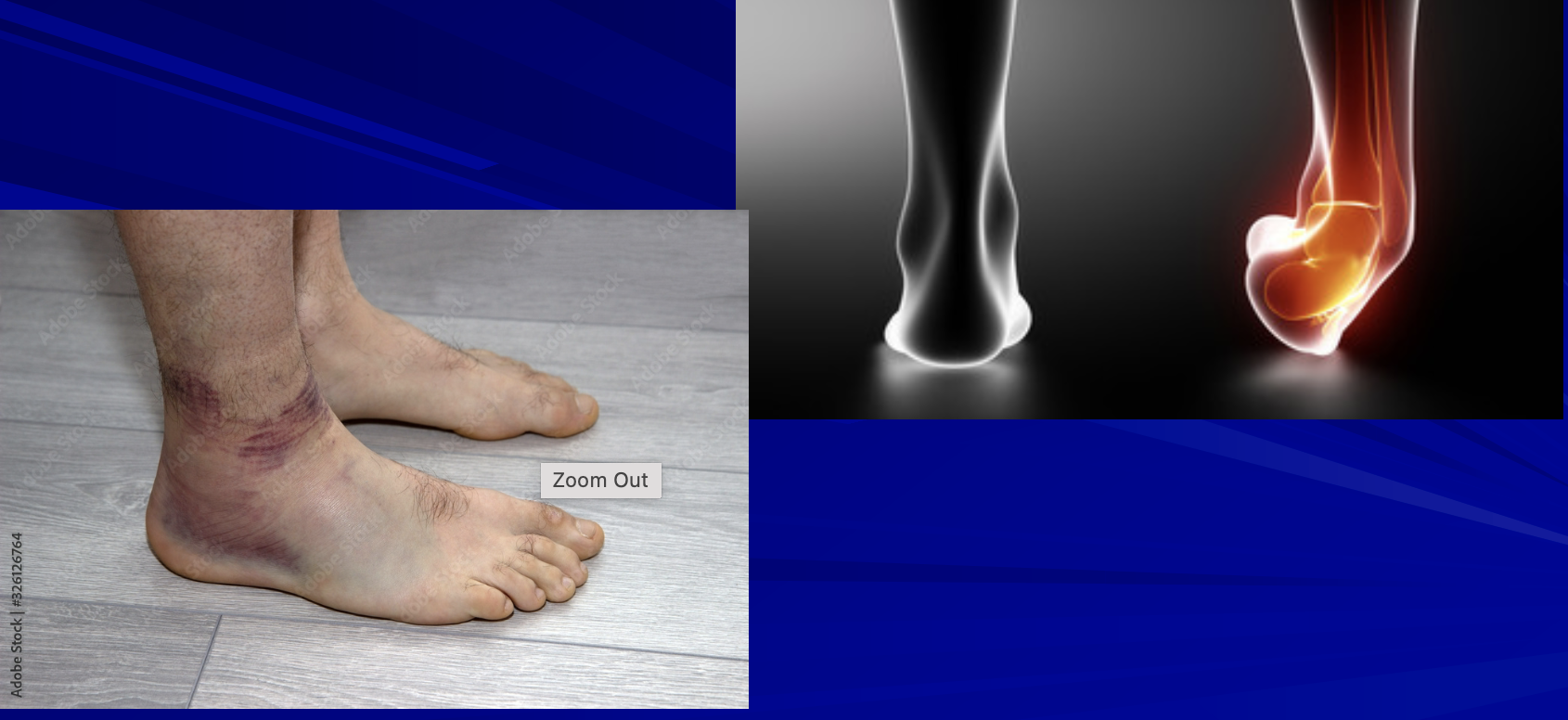
3 types of ankle sprains and which one is the most likely to be injured?
Medial Sprain; MOI- ankle rolls with eversion, damage to medial ligaments
Lateral Sprain: MOI- #1 musculoskeletal injury ankle rolls with inversion, damage to the lateral ligaments
High ankle sprains: MOI- external or dorsiflexion with rotation with damage to tibiofibular joint
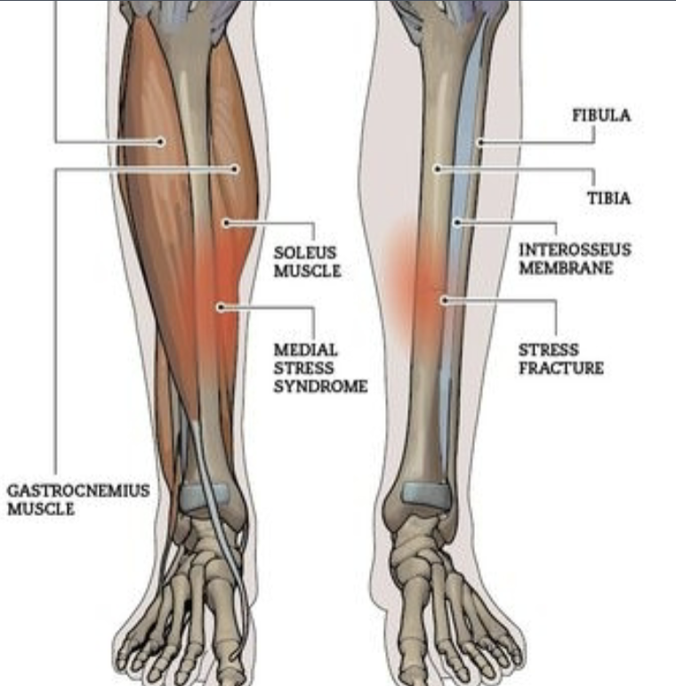
What is the medical term for “shin splints”? What is the MOI and predisposing factors that can lead to shin splints?
Medical term is Medial Tibial Stress Syndrome, due to any muscles attached to Tibia pulling on periosteum from Tibia, is a precursor of tibial stress fractures
MOI: repetitive stress like running and jumping
Predisposing factors: overtraining, flat feet, poor footwear
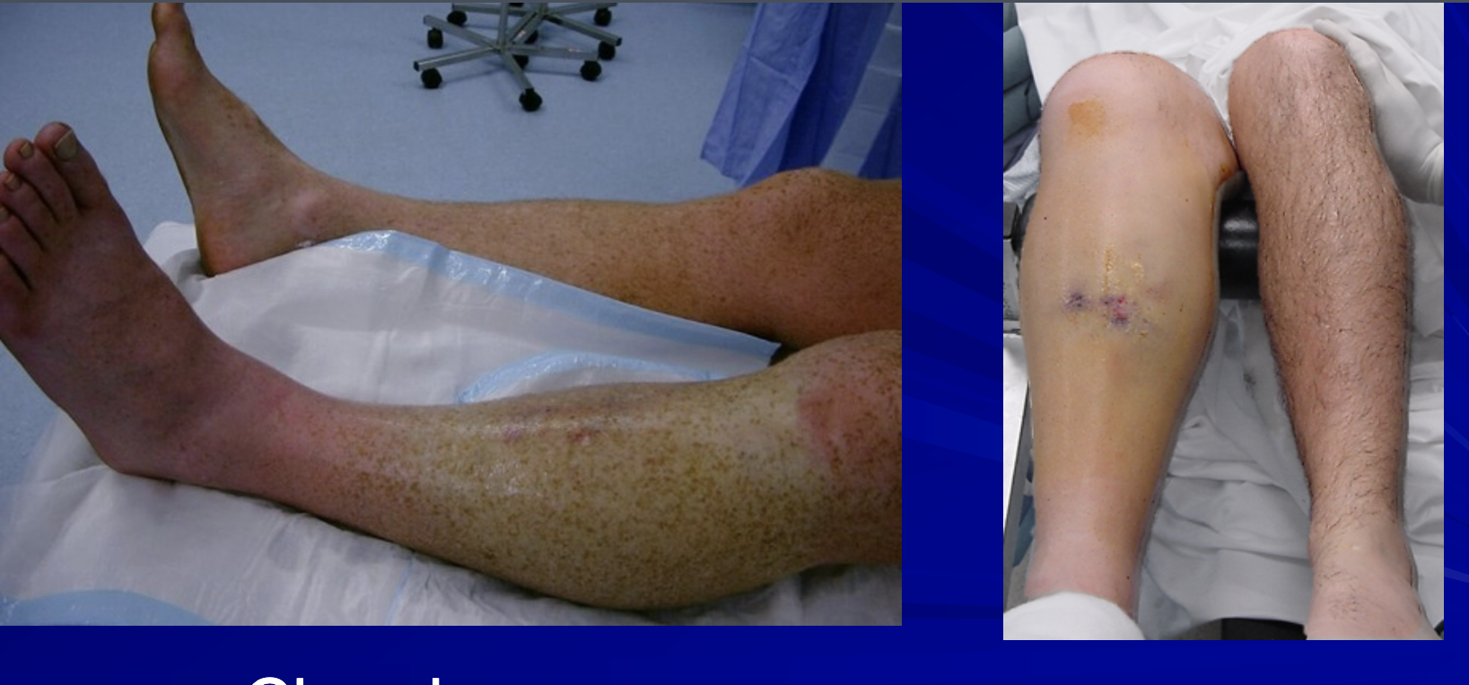
What is compartment syndrome? MOI for acute and chronic? The 6 P’s
The pressure that can build up in a muscle compartment
MOI: acute: trauma, crush injuries, severe swelling chronic: repetitive exercise
Treatment: Fasciotomy
Most common in order is Anterior, Lateral, Posterior, but never Superficial Posterior
6 P’s
Pain
Pressure
Pallor
Pulse
Paresthesia
Passive ROM
What is Tarsal tunnel syndrome? How can we treat it and what muscles are affected?
compression of tibial nerve within tarsal tunnel
muscles: gastrocnemius, tibialis posterior, flexor hallucis longus, flexor digitorum longus, and soleus
treatment: rest, ice, anti-inflammatory meds
What is “turf toe”? MOI?
a sprain on the 1st phalange (big toe) from pushing of ground/turf
MOI: hyperextension of big toe
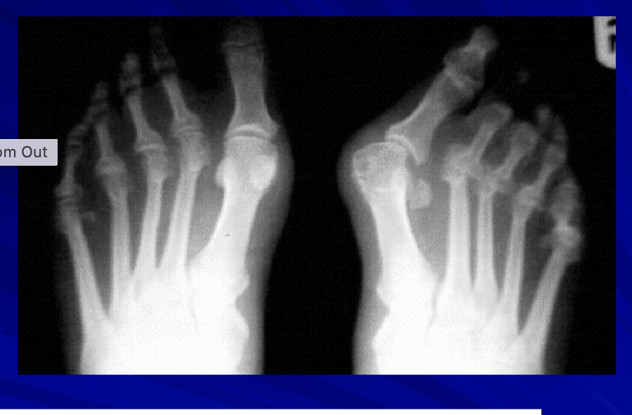
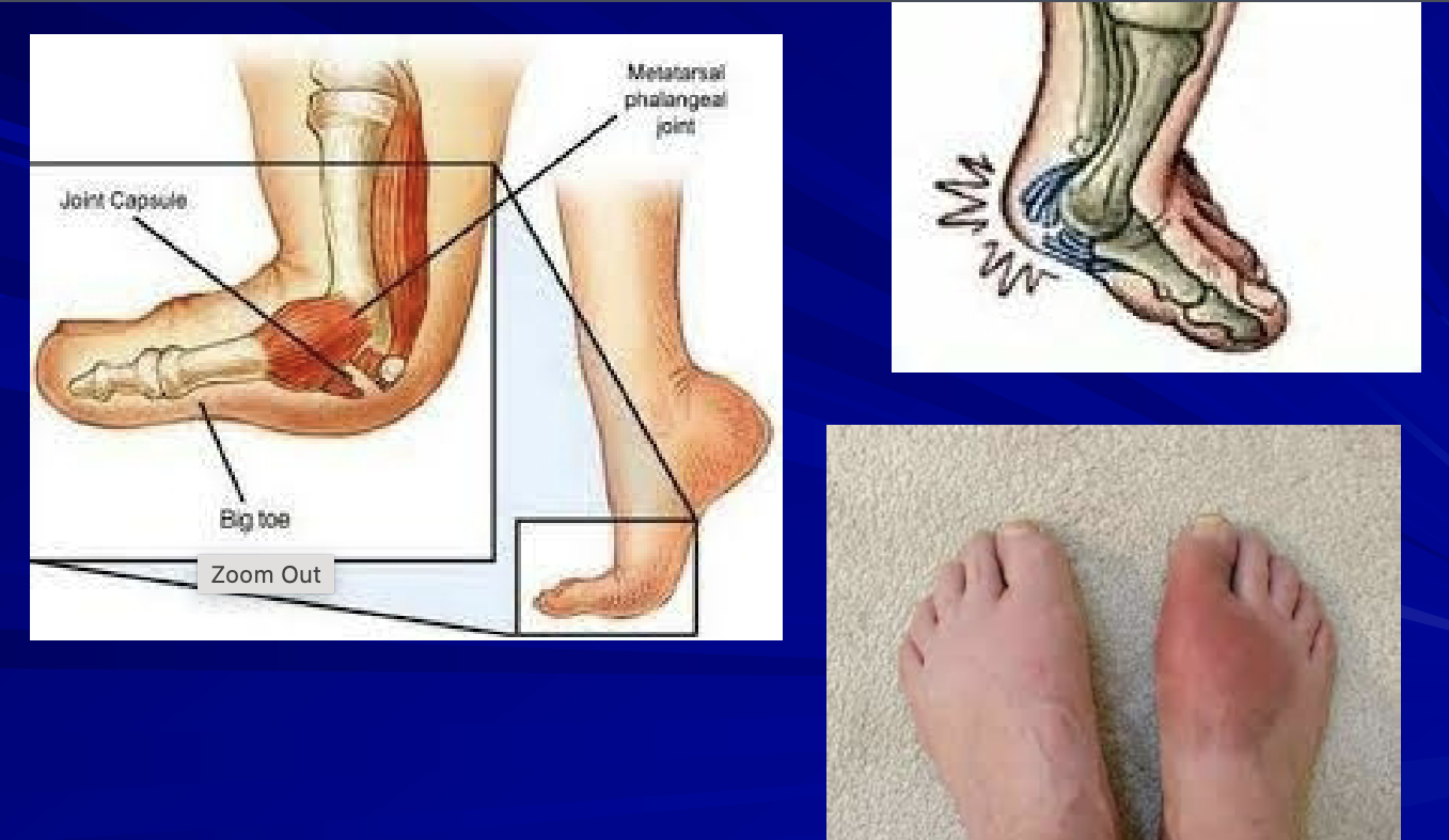
What is “hallux valgus”? MOI?
a deformity where big toe deviates medially than the other phalanges
MOI: genetics, tight shoes, overuse
What is a bunion? MOI?
Excess bony enlargement of the base of big toe
MOI: chronic pressure from tight shoes, genetics
more common in women and passed down by women
What is plantar fasciitis? Symptoms and signs?
inflammation of the plantar fascia
symptoms and signs- heel pain, pain in first steps, tenderness under foot
What is bursitis? List the kinds of bursa happen in the lower leg and ankle?
inflammation in a bursa
Achilles Tendon
Posterior Tibial Tendonitis
Peroneal Tendonitis
Plantar Fasciitis
Sesamoiditis
Bursitis
What is tendonitis? Which tendons would be involved in each compartment?
inflammation in tendons
Anterior Compartment- AT, EDLT, EHLT, Peroneus tertius
Deep Posterior Compartment- PT, FDLT, FHLT
Superficial Posterior Compartment- Gastrocnemius, Soleus
Lateral- Peroneus longus and brevis
OIF of Gastrocnemius and innervation
O: just above superior to the medial and lateral condyles of femur
I: Calcaneal tuberosity via Achilles Tendon
F: two joint muscle, knee flexion and plantar flexion
Innervation: Tibial nerve
OIF of Soleus and innervation
O: proximal tibia and fibular head
I: inserts into calcaneal tuberosity via Achilles Tendon
F: plantar flexion
Nerve: Tibial nerve
OIF of Plantaris and innervation
O: superior lateral condyle of femur
I: calcaneal tuberosity via Achilles Tendon
F: weak plantar flexion
Nerve: Tibial nerve
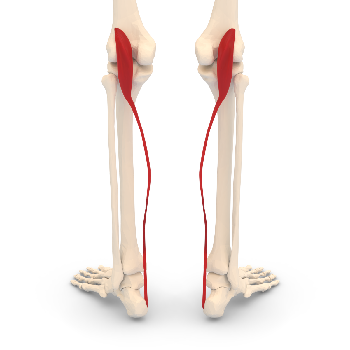
OIF of Popliteus and innervation
O: lateral surface of femoral condyle
I: posterior proximal surface of tibia
F: knee flexion; screw-home method
Nerve: Tibial nerve
OIF of Peroneus Longus and innervation
O: proximal fibular shaft/head
I: plantar aspect of 1st metatarsal
F: ankle eversion
Nerve: superficial peroneal nerve
OIF of Peroneus Brevis and innervation
O: Distal fibular shaft
I: plantar aspect of 5th metatarsal
F: ankle eversion
Nerve: Superficial peroneal nerve
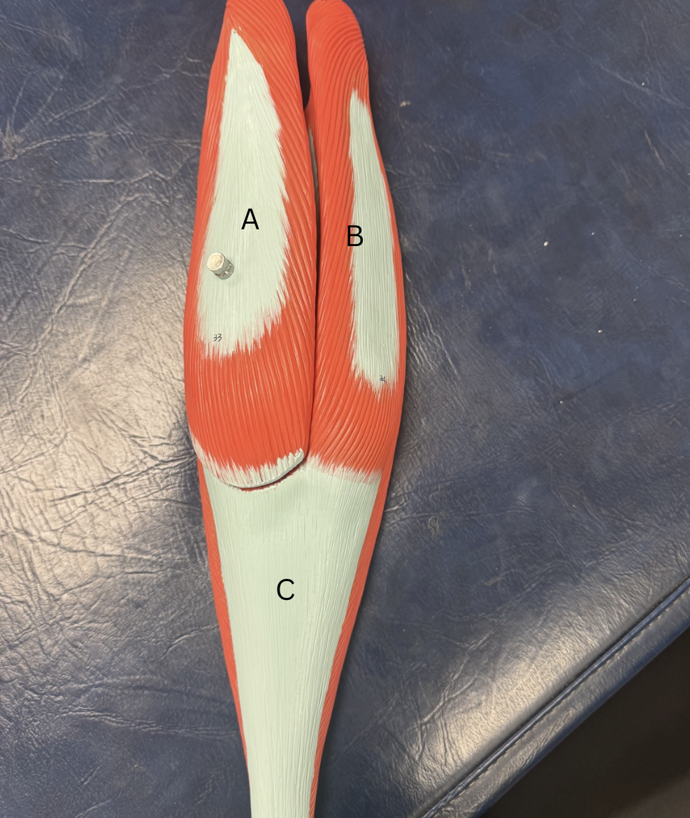
Label these muscles
A. Peroneus Longus
B. Peroneus Brevis

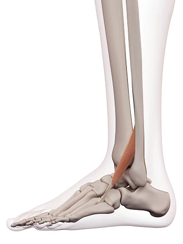
Label this muscle
Peroneus tertius
Label this muscle
Plantaris
Label these muscles
A. Medial head of gastrocnemius
B. Lateral head of gastrocnemius
C. Achilles Tendon
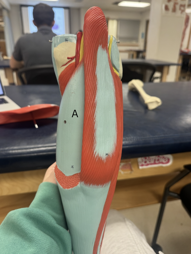
Label this muscle
Soleus
What is Retinaculum, Synovial Sheaths, and Bursa?
Medial and Lateral Retinacula- connective tissue that are non-contractile used to hold tendons in place
Synovial Sheaths-reduce friction and allow smooth movements of tendons
Bursa- fluid filled sacs to reduce friction of tissues
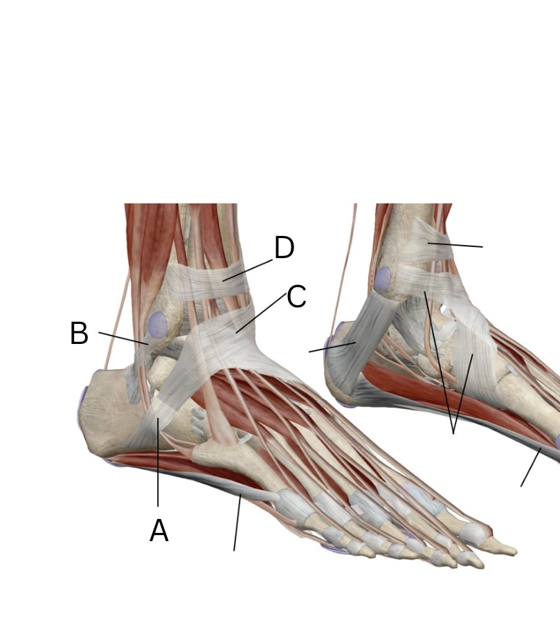
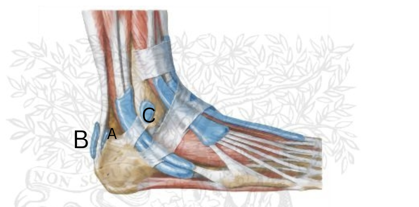
Label these retinaculum? medial or lateral?
Lateral Retinaculums:
A. Inferior Peroneal Retinaculum
B. Superior Peroneal Retinaculum
C. Inferior Extensor Retinaculum
D. Superior Extensor Retinaculum
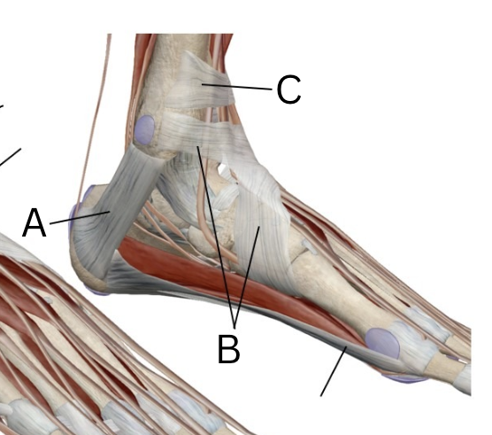
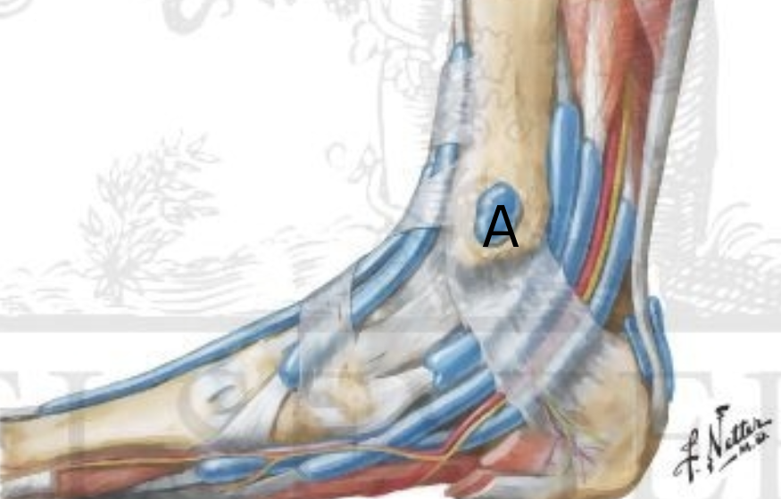
Label these retinaculum? medial or lateral?
Medial Retinaculum:
A. Flexor Retinaculum
B Inferior Extensor Retinaculum
C. Superior Extensor Retinaculum
Label the bursa, medial or lateral?
Lateral Bursa
A. subcutaneous calcaneal bursa
B. subtendinous calcaneal bursa
C. lateral malleolus and subcutaneous bursa
Label this bursa, medial or lateral?
Medial bursa
A. medial malleolus and subcutaneous bursa
What is the flow of vascular supply for the lower leg started at the iliac hip?
Iliac Artery
Femoral Artery
Popliteal Artery
Anterior Tibial Artery-splits into 4 anterior and dorsal arteries in foot
Posterior Tibial Artery-splits into 2 posterior arteries, caudal, in foot
Peroneal Artery
Medial and Lateral Plantar Artery
After the Anterior Artery is split off from the Popliteal Artery what 4 main arteries does it goe into?
On the dorsal side of the foot it goes into
Dorsi pedis artery
Lateral Tarsal Artery
Dorsal metatarsal artery
Digital artery
After the Posterior Artery is split off from the Popliteal Artery what 2 main arteries does it goe into?
On the caudal side of the foot
medial plantar artery
lateral plantar artery
What 3 joints are important in the knee? What type of joint is the knee joint?
Patellofemoral joint
Proximal Tibiofibular joint
Tibiofemoral joint
Biaxial diarthrodial joint for flexion and extension and internal and external rotation
What kinds of bone is the Patella?
Sesamoid bone and increases moment arm (leverage) of knee extension
What is the screw-home method?
The popliteus muscle origin is lateral femoral condyle to insert into medial posterior side of Tibia that helps lock the knee for extension and flexion stability
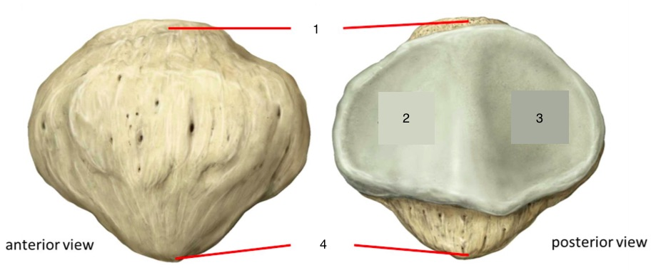
Label this bone
Patella bone
Base
Lateral Facet
Medial Facet
Apex of Patella
What is the terrible triad?
a sprain ACL, MCL, and medial meniscus tear all at the same time
Where are most injuries to menisci in the knee if they tear?
On the Posterior horns because the hamstrings and popliteus are attached posteriorly
OIF of MCL (medial collateral ligament)
O: Medial condyle of Femur
I: Medial condyle of Tibia
F: prevents valgus deformity
OIF of LCL (lateral collateral ligament)
O: Lateral condyle of Femur
I: Lateral head of Fibula
F: prevents varus deformity
What are static restraints of the Patellofemoral joint
Static restraint provides joint stability without actively contracting
Medial Retinaculum
Lateral Retinaculum
Patellotibial Ligaments
Patellofemoral Ligaments
Iliotibial Tract (easy to tear)
What are dynamic restraints of the Patellofemoral Joint
muscular structures that help stabilize joints through active contraction
Vastus Lateralis
Vastus Medialis
Vastus Intermedius
Rectus Femoris
Tensor Vastus Intermedius
Suprapatellar tendon
Infrapatellar tendon
What type of joint is the proximal Anterior and Posterior Tibiofibular joint and Interosseous membrane?
Both anterior and posterior proximal tibfib joints are diarthrodial more specifically synovial
The interosseous membrane is syndesmosis
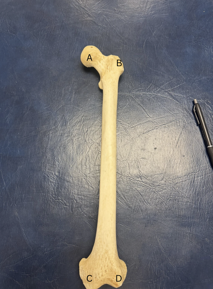
Label the Femur
inside head of Femur is Fovea Capitis
A.Head of Femur
B. Greater Trochanter
C. Medial Condyle (above is epicondyle)
D. Lateral Condyle ( above is epicondyle)
E. (Not labeled) lesser Trochanter
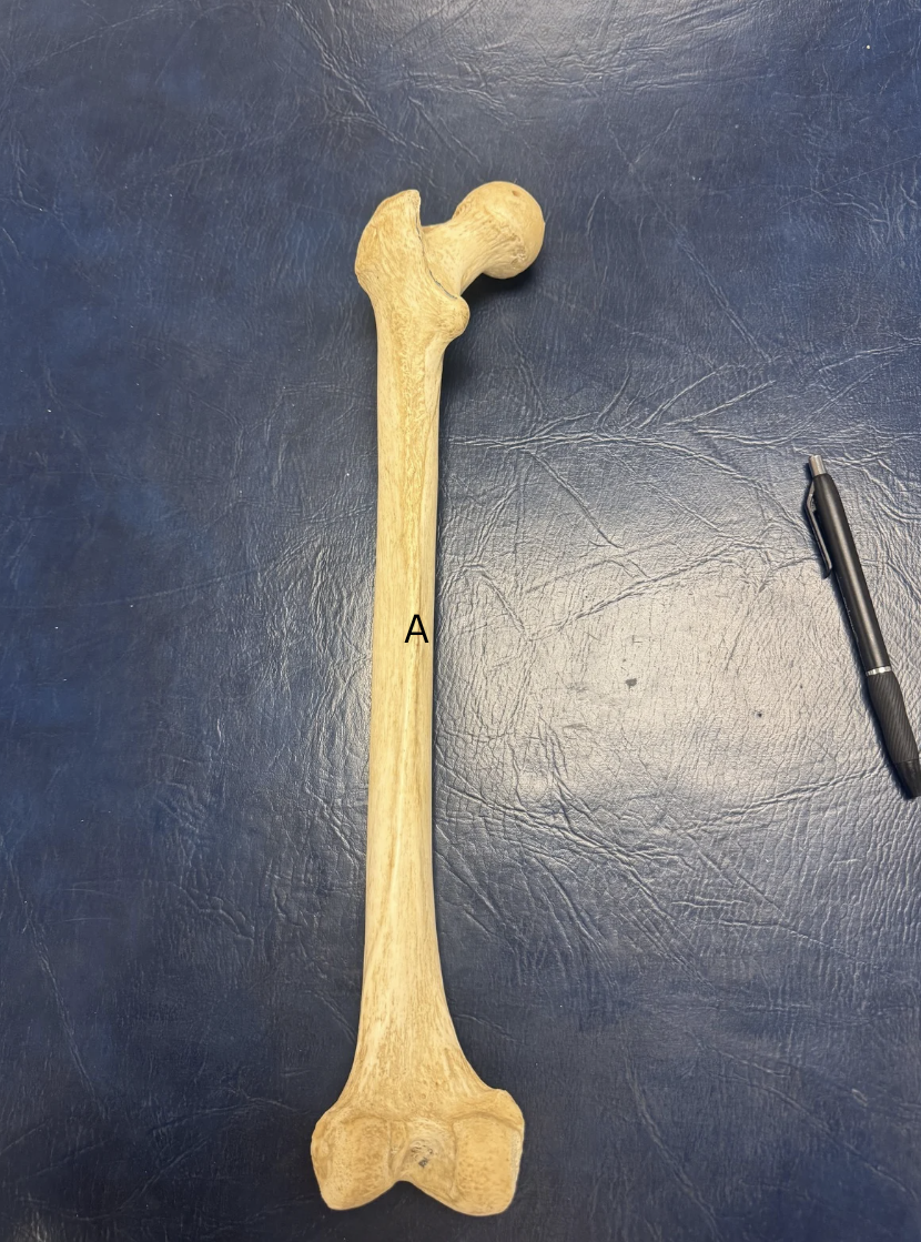
Label A
A. Linea Aspera
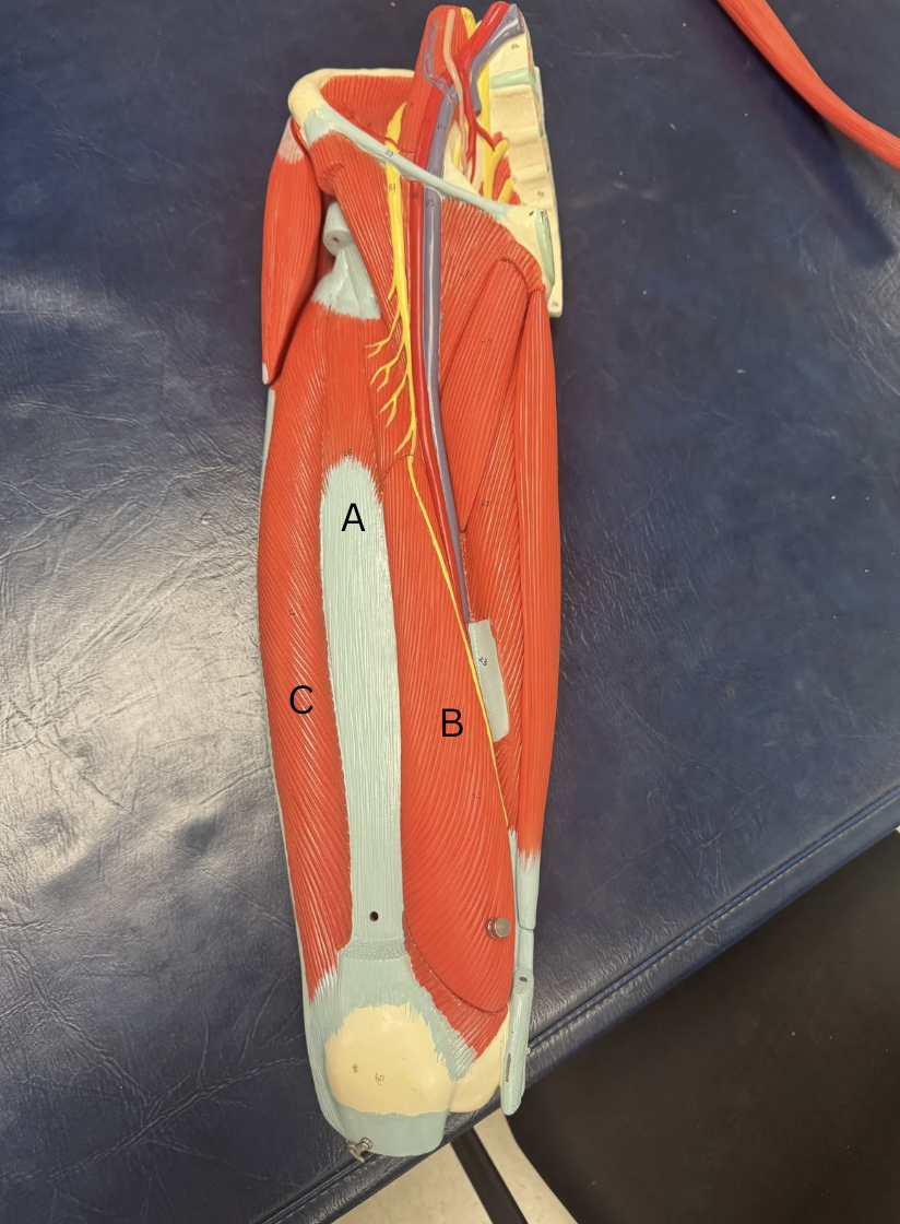
Label the quad muscles
A. Vastus Intermedius ( Rectus Femoris taken out)
B. Vastus Medialis
C. Vastus Lateralis
Label this muscle
Gracilis