psyc 211 - research methods
1/31
There's no tags or description
Looks like no tags are added yet.
Name | Mastery | Learn | Test | Matching | Spaced | Call with Kai |
|---|
No analytics yet
Send a link to your students to track their progress
32 Terms
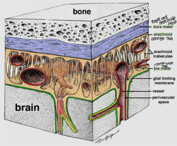
What is the meninges and what are its 3 layers
the meninges are the three protective layers that are wrapped around the spinal cord and brain
The outer layer is dura mater. It is thick, though, unstretchable tissue
the middle layer is arachnoid mater. Its web-like extensions create a soft spongy layer that is filled with cerebrospinal fluid
The third layer is Lia mater. This layer sits closets to the brain and is a bit like Saran-wrap
What is cerebrospinal fluid
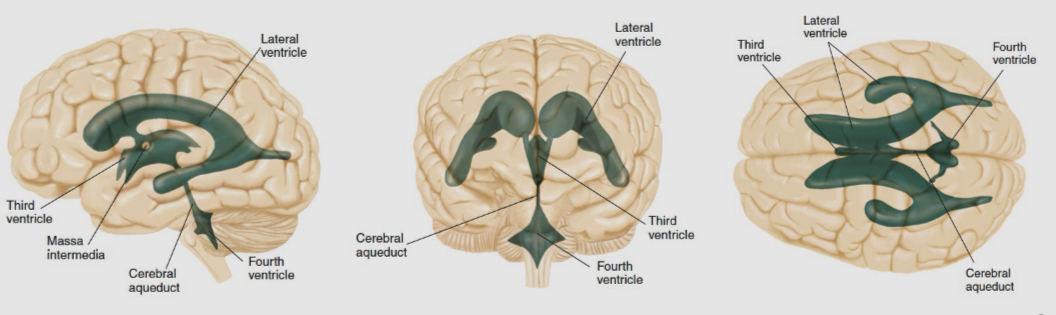
It is made by it is a distinct type of tissue that is located in each of the brain’s ventricles: choriod plexus.
The two lateral ventricles are the largest. They are underneath the cerebral cortex
The third ventricle lies between the two thalamic nuclei at the center of the brain
The fourth ventricle is in the hindbrain, between the pins and cerebellum
Small canals interconnect each ventricle and bring CSF to the spinal cord
What are the characteristics of CSF
there are immune system cells in the dura mater that analyze the CSF before it gets returned to the blood supply
It is made continuously by chronic plexus
It circulates around and hints the brain providing nutrients and removing waste
It exits the CNS by passing through holes in the dura mater, where it is absorbed into the blood supply
What are the four primary areas
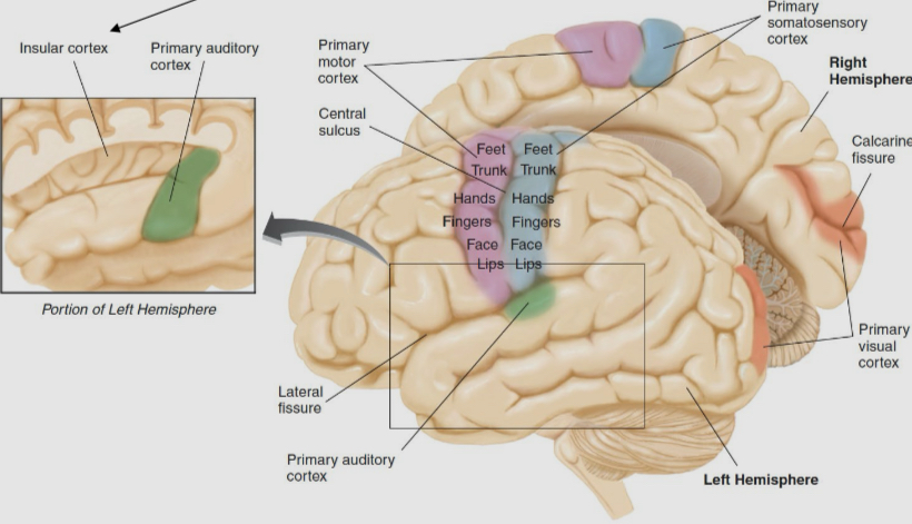
Primary motor cortex (frontal lobe): contains motor neurons that Synapse in the spinal cord. Different regions of the primary motor cortex control different parts of the body.
Somatosensory cortex (parietal lobe): is where touch information enters the cerebral cortex. Different regions of somatosensory cortex receive information from I different parts of the body
Primary auditory cortex (temporal lobe): is where auditory information enters the cortex
Primary visual cortex (occipital lobe): is where visual information enters the cerebral cortex
There is also the insular cortex, where gustatory information enters the cerebral cortex
What is the sensory association cortex
it is where perception takes place and memories are formed
A sensory association complex is adjacent to each primary sensory area and receive information only from the nearest one
What is the limbic system
It is a collection of subcortical brain areas that regulate emotions and the formation of episodic memories
Its principle areas include the hippocampus, amygdala, and cingulate cortex
Its its
What is the cingulate cortex
It is a large area that overlies the corpus callosum. This region interconnects many limbic areas of the brain
How does the brain form
it starts of as a neural tube during the first month of human development in the womb
The first cells in the tube a neural progenitor cells
In the 8th week, the cells undergo symmetrical cell division, so each neural progenitor cell becomes two neural progenitor cells
Over the next 3 months, when a neural progenitor cell divides one of the daughter cells detaches from the center of the neural tube.
When that cell divides, it will produce two neurons or two glial cells.
What is neurogenesis
it is the production of new neurons
Neural progenitor cells produce neurons and glia after they undergo asymmetrical division. Huam neurgofensis largely stops five months after conception when neural progenitor cells start to undergo apoptosis
There is little neurones is in adult mammals
What is apoptosis
it is programmed cell death that occurs in multicellular organism
Human neural progenitor cells undergo apoptosis around t eh fights month of development in the womb. This is when neurogenesis stops
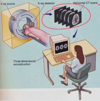
What is computerised Tomography (CT scan)
It is a machine that takes pictures of the brain
The resolution is not great for soft brain tissue
An X-ray beam is projected through the head to an X-ray detector. The X-ray beam is delivered from all angles
Cheap and fast
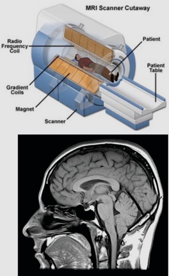
What is magnetic resonance imaging (MRI)
It uses a strong magnetic field and low energy radio waves
The magnet causes every hydrogen proton in the body to spin in a particular direction
A pulse of low energy radio waves changes the spin of these protons. Then, each proton flips back to the spin direction determined by the magnet. During this, every proton emits a new radio waves changes, which is detected by the scanner
The scanner provides an estimate of the relative density of hydrogen atoms
MRI identifies the density of water molecules and fat molecules
High resolution three dimensional image
Why is diffusion tensor imaging (DTI)
it is used to measure the direction and speed of water molecules
It creates images that show the location and direction of every axon
It is basically and MRI that is more sensitive to water molecule than to lipid ones
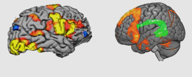
What is functional magnetic resonance imaging (fMRI)
it is a rapid series of MRI scans that reveal the movement of oxygenated blood
Ex: when a brain area is active, blood flow to that region increases within 5 seconds. We can then identify changes in neural activity by tracking oxygenated blood
Doesn’t involve needles, surgery, or radioactivity
Decent spatial resolution and temporal resolution
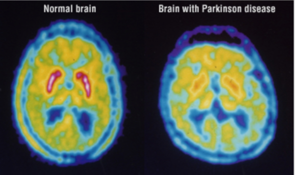
what is positron emission tomography (PET)
it uses 2-DG as a measure of neural activity, but has been replaced by the fMRI
It is used with other radioactive tracers
It is uses to medausde changes in the expression levels of neurotransmitter receptors across weeks
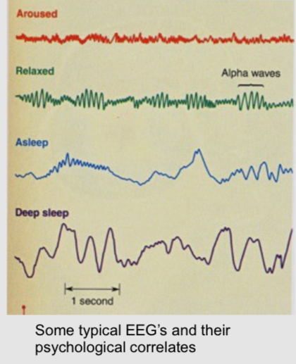
What is electroencephalogram (EEG)
it is a measure of electrical activity in the brain that uses macro electrodes (metal discs) attached to the scalp
It records the summed up population level activity of millions of neurons
It can be used as a diagnostic stool
Specific patterns are associated with consciousness, sleep stages, etc
What is experimental ablation
it involves the removal or destruction of a portion of the brain. The functions that can no longer be performed following the surgery are probably controlled by that brain area
Used to know the function of a brain region
What are radio frequency lesions
they are small lesions made by passing radiofrequency current through a metal wire that is insulated everywhere but the tip
the electric current produces heat that burns cells around the tip of the wire
The size and shape of the lesion is determined by the duration and intensity of the current
Axons that are passing through will also be burned
What is an excitoxic lesion
it is a brain lesion technique that is selective for cell bodies. It involves micro injections of a glutamate receptor agonist, which causes neurons to spike so much they undergo apoptosis
Since there are profs on axons, any that are passing thought the area are spared
What is a sham lesion
it is a placebo procedure that duplicates all steps of producing a brain lesion except die the step that causes extensive brain damage
What is a reversible lesion
it can be achieved by injecting drugs that temporarily inhibit neuronal spiking, like voltage-gated sodium channel blockers that stop all action potentials
What is chemical stimulation
it is the infusion of anasthetics which will shut down neural activity (stop all actin potentials)
The infusion of receptor agonists or antagonists will selectively affect cell bodies and synapses. There are no neurotransmitter receptors on axons, so fiber of passage are not directly affected by drug infusions
What is electrical stimulation
it is the delivering of electrical current through an implanted metal wire which will stimulate everything in the area (cell bodies and axons passing through)
Low frequency stimulation → increases spiking activity. Stimulating faster than 10p times per second can produce the same behavioural efffect as lesioning in the brain area
What are micro electrodes
they are thin metal wires with a fine tip that can record the electrical activity of individual neurons
They are used during behavioral tests to record every action potential from a given neuron
It is possible to simultaneously records from hundreds of individual neurons
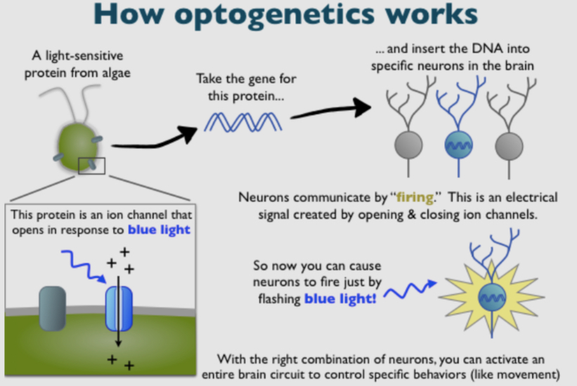
What are optogenetics
it is a way to make neurons sensitive to light to depolarize or hyperpolarize collections of them at any moment during a behavioral test
this foreign DNA provides instructions to make light-sensitive proteins
What is a virus
it is a DNA delivery system
they replicate by injecting viral DNA in a host organism
Its DNA contains instructions on how to make more vireus
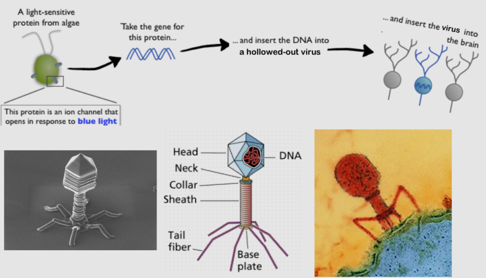
What are replication deficient viruses
they are viruses in which the viral DNA is edited so that they won’t be able to replicate
They can infect cells, but the DNA they deliver is created by the researchers
What is GFP
it is a fluorescent protein that is used to shows infected cells in a lab setting
What are mad-made fluorescent receptors
they are fluorescent proteins that a designed by scientists
It allows to visualize neurotransmitter release in living brains in real time
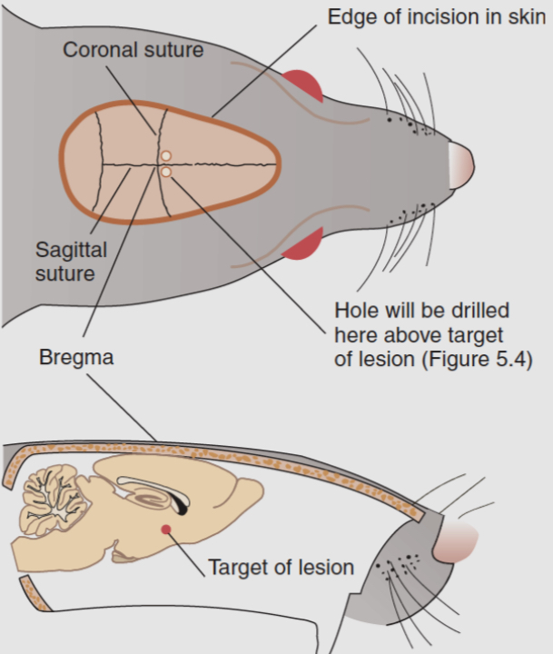
What is stereotaxic surgery
it is a surgical intervention that uses a stereotaxic apparatus
It is used to infuse drugs in specific parts of the brain
It is also used to permanently implant metal straws, metal electrodes and fibre optic cables
What is Bregma
it is the junction where pieces of the skull fuse together.
Bregma is often used as a reference point for stereotaxic brain surgery
what are the reasons for stereotaxic surgery
it is commonly used for one time injections of a drug or virus to:
lesion a brain area
Lesion a specific type of cell in a particular brain area
To alter gene expression, which typically involves viral-mediated gene delivery