exam 3 day 2
1/51
There's no tags or description
Looks like no tags are added yet.
Name | Mastery | Learn | Test | Matching | Spaced | Call with Kai |
|---|
No analytics yet
Send a link to your students to track their progress
52 Terms
what gives white matter its color
high proportion of myelinated nerve fibers
what does white matter consist of
nerve fibers, (axons), neuroglia, and blood vessels
how is white matter orgnanized
into columns (funiculli); anterior, posterior, lateral
what is a tract (fassiculus)
group of axons with same origins, termination, and function
white matter tracts are divided into what 2 types
ascending (sensory) and descending (motor)
ascending tracts carry info in what direction
to the brain
descending tracts carry info in what direction
from the brain
blue regions are what
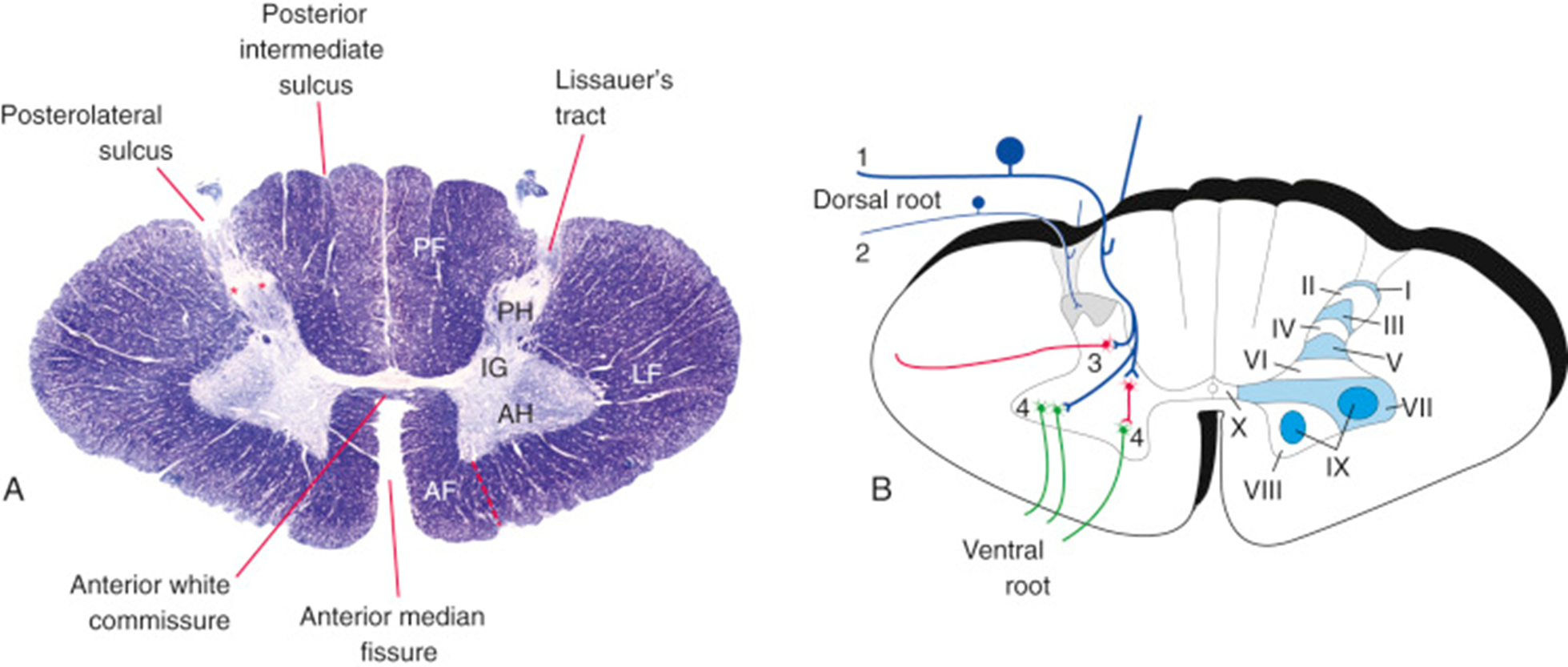
ascending (sensory/afferent) pathways

red regions represent what
descending (motor/efferent) pathways
dorsal columns carry what type of info
touch and position (proprioception)
spinothalamic tract carries what
pain and temp
corticospinal tract function
voluntary motor control
what does gray matter consist of
cell bodies, dendrites, neuroglia, blood vessels
shape of gray matter in spinal cord
H-shaped
gray matter is divided into what
horns (posterior, anterior, lateral)
posterior (dorsal) horn function
sensory processing
anterior (ventral) horn function
motor output (skeletal muscle)
lateral horn contains what
preganglionic autonomic neurons
where is the lateral horn found
T1-L2 sympathetic
what connects the two halves of gray matter
gray commisure
what is in the center of gray matter
central canal (CSF)
sensory neuron are located where
dorsal horn
lower motor neurons are located where
ventral horn
interneurons function
connect sensory + motor → reflex arcs
preganglionic neurons are found where
lateral horn
how many Rexed laminae
10
lamina II =
substantia gelatinosa
function of substanta gelatinosa
modulates pain and temp
clarke’s nucleus function
posterior spinocerebellar tracts (proprioception)
clarke’s nucleus location
T1-L3
white matter increases in what direction
caudal→ cranial
why does white matter increase upward
more ascending fibers added
where is gray matter enlarged
cervical and lumbosacral enlargements
why are enlargements present
innervation of limbs
thoracic spinal cord
has lateral horns (ANS)
cervical level
large ventral horns (upper limb)
lumbar level
large ventral horns (upper limb)
thoracic level
small horns + lateral horns present
fasciculus gracilis carries info from
lower limb
fasciculus cuneatus carries info from
upper limb
posterior intermediate sulcus is present where
above T6
lissauer’s tract function
pain fibers enter and ascend / descend slightly
white commissure function
crossing of fibers
what are ventricles
CSF-filled spaces in CNS
lateral ventricles connect to 3rd via
foramen of monro
3rd→ 4th ventricle via
cerebral aqueduct
4th ventricle drains into
subarachnoid space
CSF is produced by what
choroid plexus
CSF composition
low cells, high Na+/Cl-, low K+/Ca2+
csf function
protection
waste removal
ionic balance
largest subarachnoid cistern
cisterna magna
CSF exits 4th ventricle via
foramen of Magendie and Luschka