Principles of homeostasis
1/9
Earn XP
Description and Tags
» Define the terms: • homeostasis • compensation • decompensation • positive and negative feedback » Explain that homeostatic mechanisms are responsible for maintaining optimal tissue function » Explain that loss of homeostasis occurring as a result of external insults can lead to disease
Name | Mastery | Learn | Test | Matching | Spaced |
|---|
No study sessions yet.
10 Terms
what is homeostasis?
» …the relative constancy of the body
» …the maintenance of constant conditions in the body’s environment
» Greek
• Homoios = unchanging/similar to
• stasis = standing still/stability
» It is how an organism regulates it’s internal environment to maintain stability
» Compensation
The return to homeostasis after being challenged
» Decompensation
The failure to compensate, adapt, heal, etc
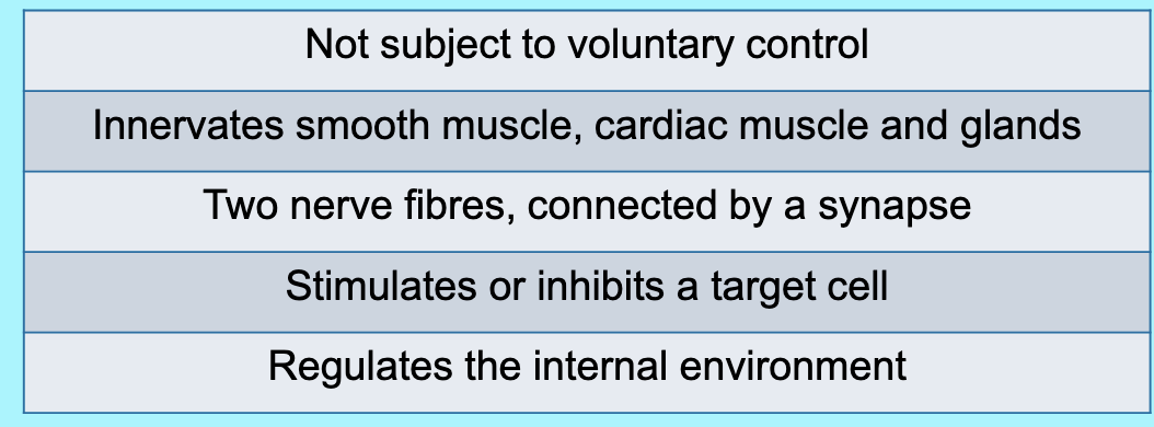
what is the autonomous nervous system
» Primary function
» Involuntary control
» Collaboration between ANS and the endocrine system
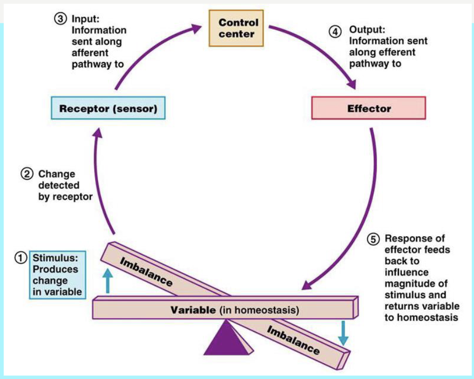
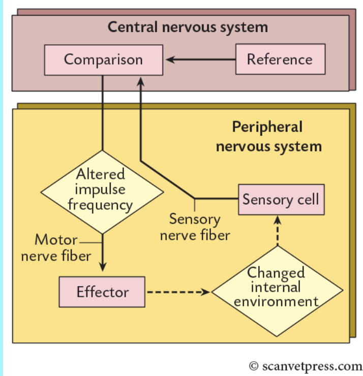
what is negative feedback
examples are temperature control, water balance, pH of the blood, blood glucose
a control mechanism that reverses a change and restores a system to a stable state
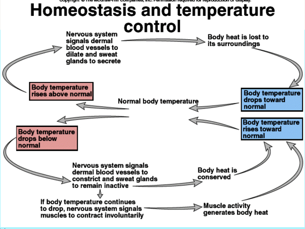
homeostasis and temperature control
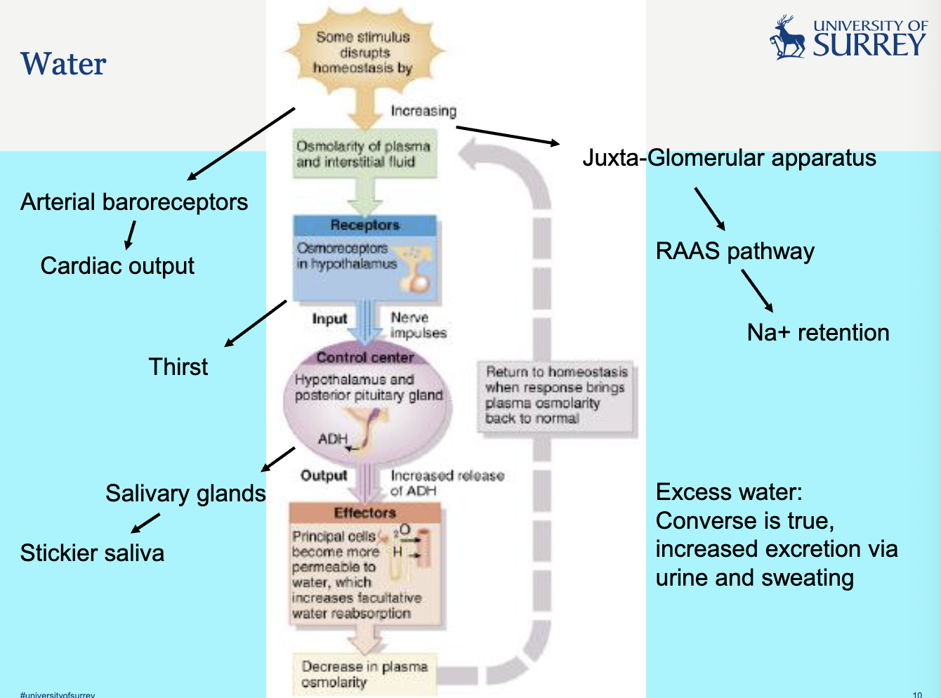
describe water control homeostasis
review this when you are revising - check if panapto is finally up
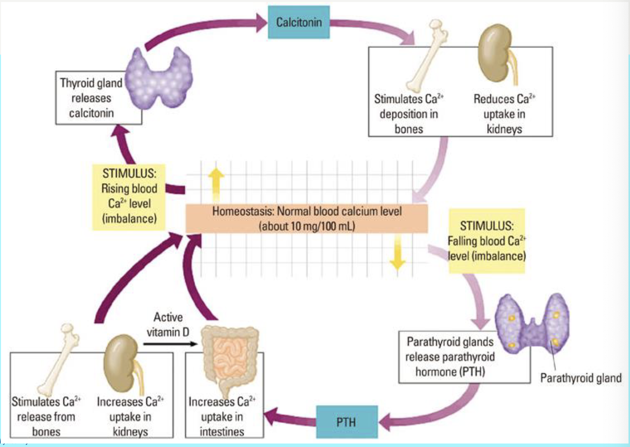
describe calcium blood level homeostasis
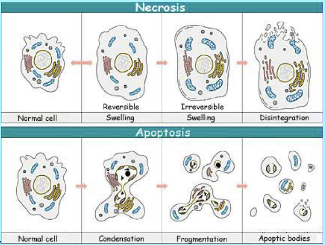
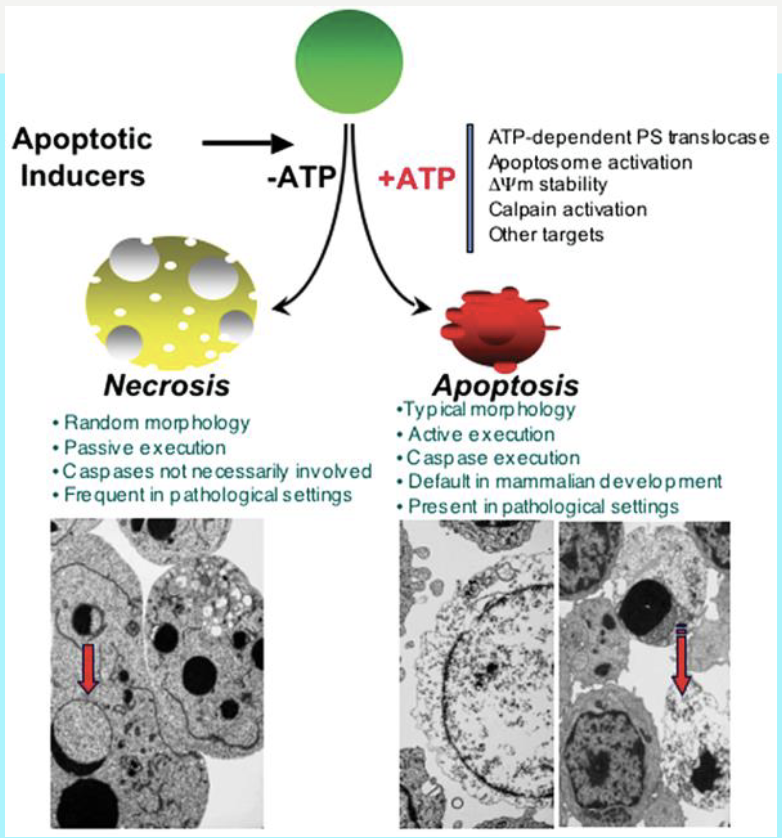
what are the basic concepts of cell death?
we have necrosis and apoptosis
necrosis is is an accidental cell death caused by injury or infection
whereas
apoptosis is a natural, programmed cell death essential for development
again review this when revising
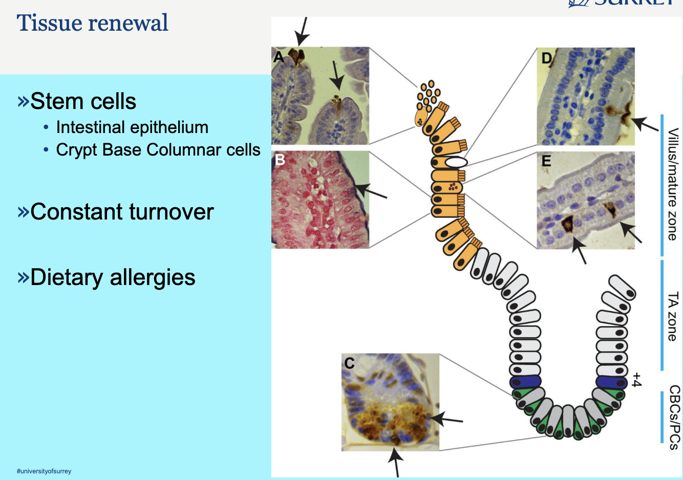
describe tissue renewal in the intestine
What You Are Looking At: The Intestinal Crypt-Villus Axis
The diagram illustrates the cellular organization of the lining of the small intestine, which is one of the most rapidly self-renewing tissues in the body. The entire lining is replaced every 4 to 5 days. The structure is divided into two main functional compartments:
The Crypt (The Factory/Stem Cell Niche): The invaginated, gland-like structure at the bottom, where all new cells are generated.
The Villus (The Functional/Differentiated Zone): The finger-like projection extending upward into the gut lumen, where mature cells perform absorption and are eventually shed.
Explanation of the Notes and Zones
The notes and zones describe the different cell populations responsible for this constant renewal:
1. The Stem Cell Niche (The Crypt Base)
This is the area at the very bottom of the crypt, which houses the true stem cells and their supporting cells.
CBCs (Crypt Base Columnar Cells):
Role: These are the Intestinal Stem Cells (ISCs). They are the engine of the entire system.
Function: They are responsible for self-renewal (making more stem cells) and multipotency (generating all the different cell types needed for the intestine).
Location: They are interspersed between the Paneth cells at the very bottom of the crypt.
PCs (Paneth Cells):
Role: These are specialized secretory cells that form the Stem Cell Niche.
Function: They do not renew the tissue themselves. Instead, they secrete essential signaling molecules (like Wnt ligands) that are critical for maintaining the CBCs in their stem cell state and promoting their proliferation. They are the support staff for the stem cells.
Location: They are located at the base of the crypt, intermingled with the CBCs.
2. The Proliferative Zone
This zone is located just above the stem cell niche.
TA Zone (Transit-Amplifying Zone):
Role: This is the zone of rapid cell division.
Function: Cells generated by the CBCs enter the TA zone. They are called progenitor cells. They undergo a limited number of rapid cell divisions (amplification) to quickly generate a large pool of new cells before they stop dividing and begin to differentiate. They are the production line of the tissue.
Location: The middle and upper parts of the crypt.
3. The Differentiated Zone
This is the area where cells mature and perform their final function.
The "Other Zone" (The Villus):
Role: This is the functional surface of the intestine.
Function: Cells that leave the TA zone migrate upward onto the villus. As they migrate, they differentiate into mature cell types (e.g., enterocytes for absorption, goblet cells for mucus secretion).
Tissue Renewal: Once they reach the tip of the villus, they are shed into the gut lumen, completing the renewal cycle.
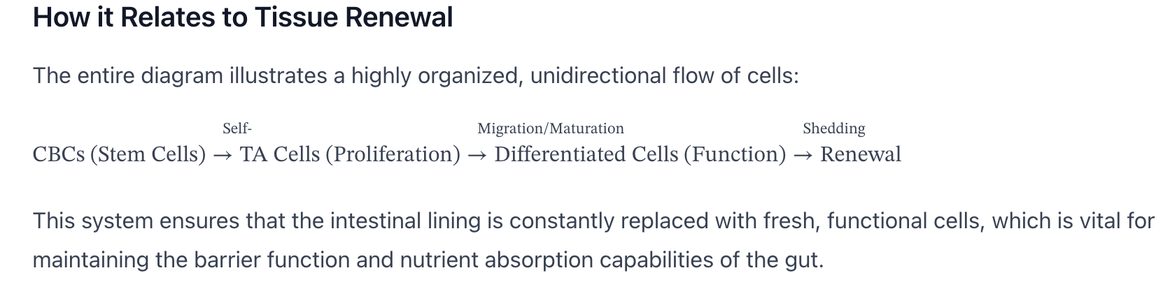
How it Relates to Tissue Renewal
The entire diagram illustrates a highly organized, unidirectional flow of cells:
CBCs (Stem Cells)→Self-Renewal/DifferentiationTA Cells (Proliferation)→Migration/MaturationDifferentiated Cells (Function)→SheddingRenewal
This system ensures that the intestinal lining is constantly replaced with fresh, functional cells, which is vital for maintaining the barrier function and nutrient absorption capabilities of the gut.
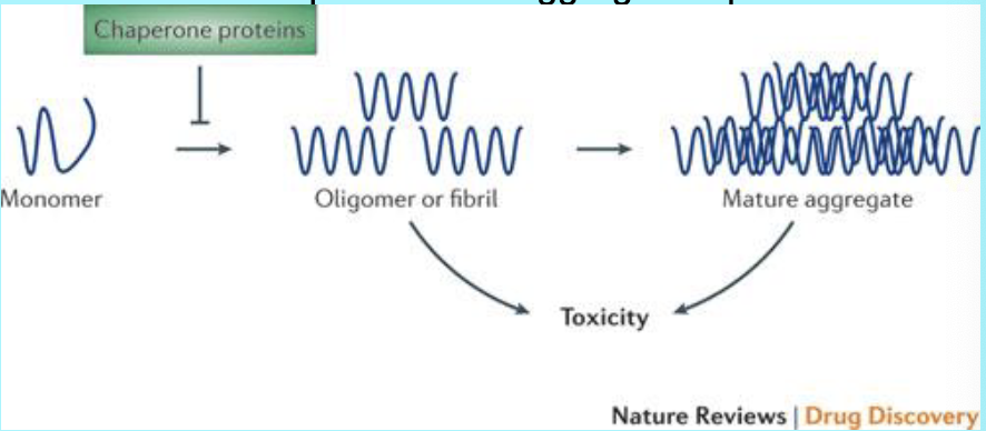
what is the impact of misfolding of proteins?
» Misfolding of disease-causing proteins results in the disruption of protein homeostasis when misfolded monomers accumulate and begin to form
intermediate soluble oligomers or fibrils, and eventually form mature
insoluble aggregates
• Chaperone proteins assist in the correct folding of proteins and prevent the
formation of toxic oligomeric species
• Increasing the expression of chaperone proteins enhances the ability of cells to
maintain protein homeostasis even in the presence of aggregation-prone
proteins
Examples of protein-misfolding diseases are
• BSE/Scrapie/CWD
• Parkinson’s
• Huntingdon’s
• Alzheimers
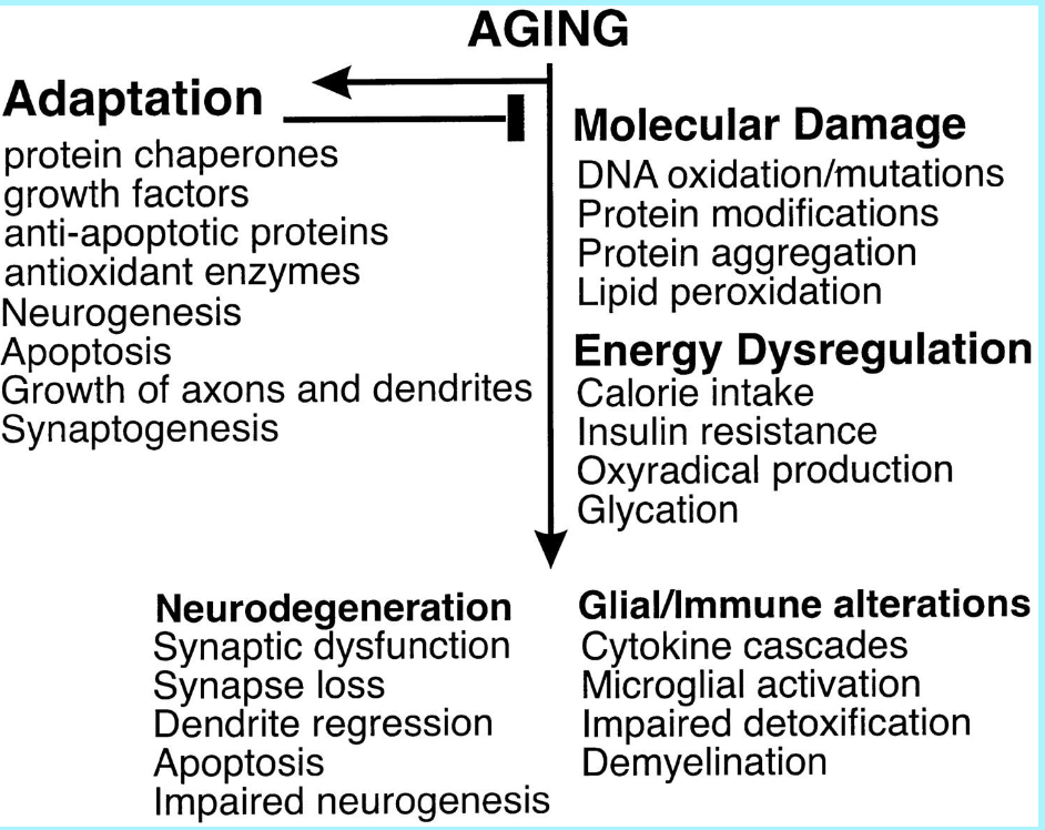
how does aging work?
this image shows the biological changes in the brain associated with aging, and the adaptation side shows what natural processes the brain uses to slow down aging and counteract the damage
so the arrow is showing for example as we age we have an increase in molecular damage, as we have an adaptation like protein chaperones, but as we age it would be harder to regulate it wont be as efficient
this is how disease develops