3.8 transport in animals
 Call Kai
 Learn
Learn Practice Test
 Spaced Repetition
 Match
Match Flashcards
 Knowt Play
1/120

There's no tags or description
Looks like no tags are added yet.
Name | Mastery | Learn | Test | Matching | Spaced | Call with Kai | Chat |
|---|
No analytics yet
Send a link to your students to track their progress
121 Terms
what is the need for transport systems?
as organisms get bigger, the distance between the cells get greater
as diffusion is only effective over very short diffusion distance, the organism won’t survive
organisms therefore need a specialised transport system to move substance around the body
how are exchange systems adapted to maximise effectiveness?
thin for short diffusion distance
large surface area so lots of a substance can diffuse at once
good blood supply to maintain a concentration gradient
what are the features of a transport system?
transport medium
vessels to transport
pump to transport medium
medium which flows in one direction (valves)
ability to control flow of medium suit the changing needs of the organism
what are the 4 types of circulatory systems?
single closed
double closed
open circulatory system
closed circulatory system
what is the single closed circulatory system?
only passes through the heart once for each complete circuit of the body
the heart had 2 chambers
because it goes through 2 sets of narrow vessels, the pressure is low which limits exchange process
what is the double closed circulatory system?
the blood travels twice through he heart each circuit
this ensures relatively high pressure and fast flow of blood
what are advantages of double circulation?
the blood only travels through 1 capillary network before retuning to heart
this maintains high pressure and steep concentration gradient
what is the close circulatory system?
the blood is enclosed in blood vessels and doesn't come in direct contact with cells
the blood is pumped under pressure and things leave through walls of the blood vessels
the volume of blood flowing can be adjusted by widening or narrowing blood vessels
what is the open circulatory system?
the blood (haemolymph) flows freely from the heart into body cavities (haemocoel) and comes into direct contact with the cells
this is where change takes place before returning to heart through open-ended vessels
no transport of O2 or CO2 as directly exchanged from tracheoles
what is the function of an artery?
carry blood away from the heart
what is the structure of an artery? (outside to inside)
tunica externa (adventita)
tunica media
tunica intima
narrow lumen
what is the structure/function of tunica externa in artery?
mostly made of collagen and elastic fibres
strong protein that protects the blood vessel from over-stretching
what is the structure/function of tunica media in artery?
thick layer of smooth muscle and elastic tissue
muscle which withstands high pressure, can contract to narrow the lumen and reduce blood flow
elastic tissue which maintains high blood pressure by stretching and recoiling
what is the structure/function of tunica intima in artery?
made up of:
endothelium which is one cell thick, smooth, reduces friction for free blood flow
connective tissue
elastic fibers
what is the function of the narrow lumen in artery?
helps maintain high blood pressure
what is the function of the arterioles?
carry blood from an artery to the capillaries
what is the structure of the arterioles and why?
contains rings of smooth muscle which contract and constrict and the diameter of the arteriole
this increases resistance to flow and reduces the rate of flow of blood which diverts the flow to parts of the body that are demanding more oxygen
what is the structure of capillaries and why?
very narrow diameter which reduces blood flow to allow exchange between blood and surrounding cells more efficiently
thin walls only one cell thick, reduces the diffusion distance
walls are leaky to allow plasma and dissolved substances to leave the blood
what are capillary beds?
network of capillaries running though tissues
what is precapillary sphincters?
capillary beds which regulate the flow of blood to tissues
what is the purpose of venule?
transport blood from capillaries to veins
what does the venula walls consist of?
muscle
elastic
collagen
what is the structure of a vein? (outside to inside)
tunica externa (adventita)
tunica media
tunica intima
large lumen and valve
what is the structure/function of the tunica externa in veins?
mostly made of collagen and elastic fibres
strong protein that maintains the shape of the vein, prevents collapse under low pressure
what is the structure/function of the tunica media in veins?
thin layer of muscle and elastic fibres
much thinner in veins
no need for thick muscle as don’t need to withstand high pressure
what is the structure/function of the tunica intima in veins?
made up of:
endothelium-one cell thick, smooth and reduces friction for free blood flow
connective tissue
elastic fibres
why do veins have a large lumen?
low blood pressure
why do veins have valves?
prevents the backflow of blood
blood flows in 1 direction
how do valves work in veins?
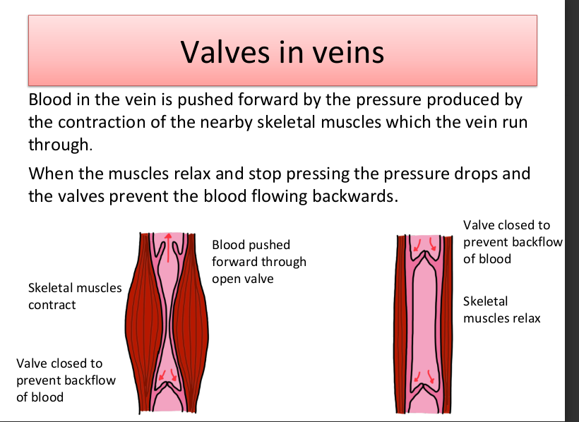
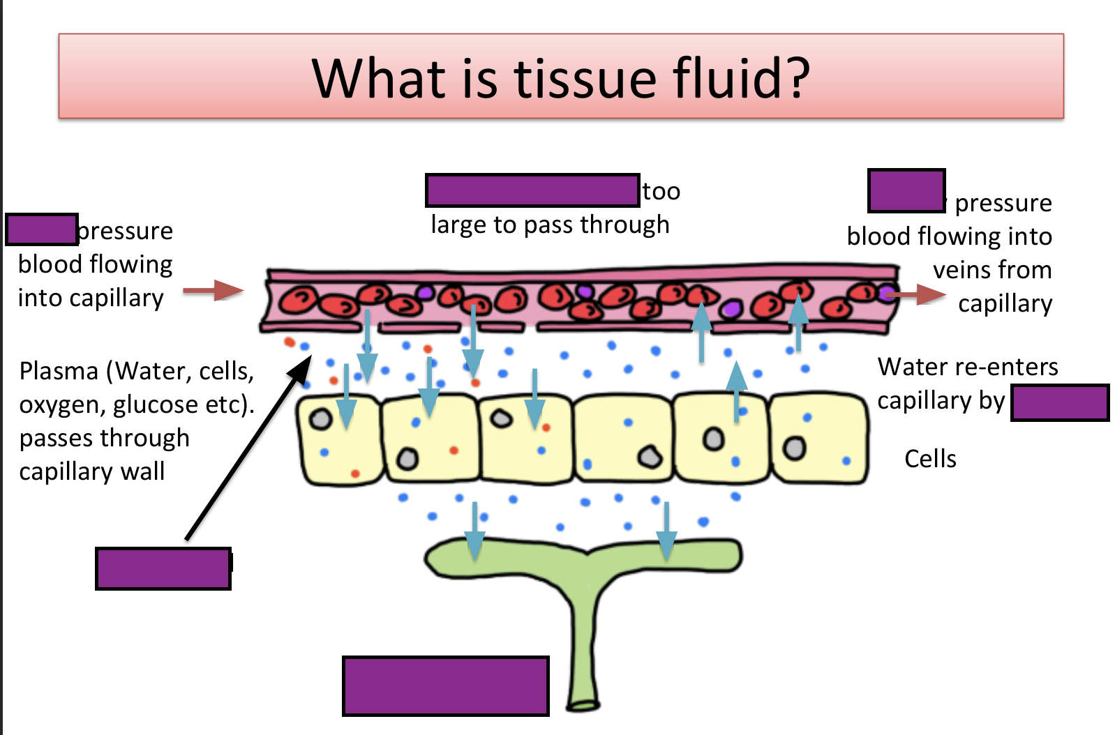
fill in labels
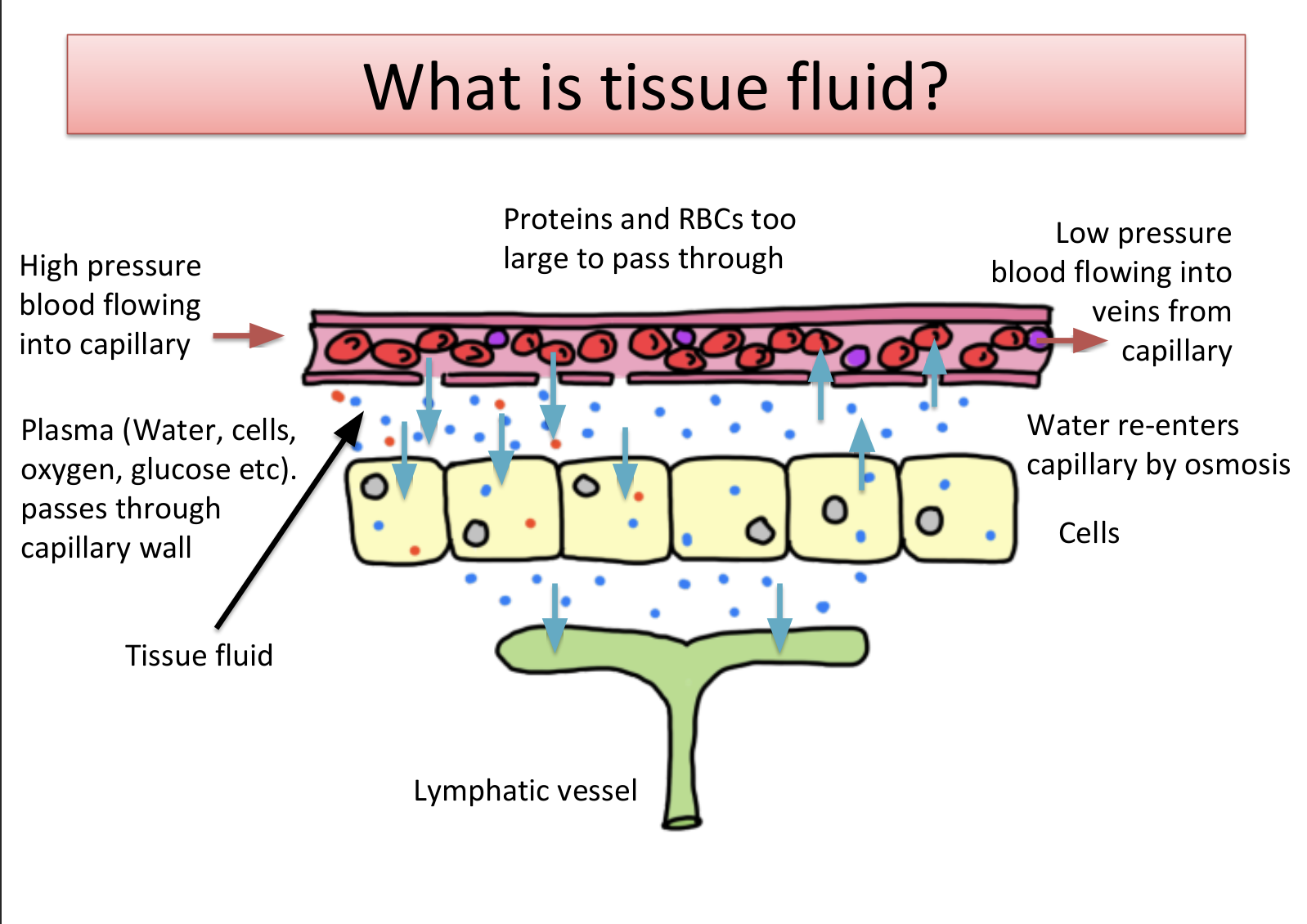
what is tissue fluid?
formed by plasma leaking from capillaries
it surrounds cells and supplies them with oxygen and nutrients
formed at the arteriole end of capillary beds
drained from the cells by lymph vessels and venules
what is hydrostatic pressure?
the pressure caused by a fluid pushing against the container that its’s within
what is oncotic pressure?
the pressure created by the osmotic effects of the solutes in the solution
at the arterial ends which pressure is bigger?
hydrostatic pressure > oncotic pressure
at the venue ends which pressure is bigger?
oncotic pressure > hydrostatic pressure
why is there oncotic pressure?
in blood plasma we have proteins such as albumin
these are highly hydrophilic and lower water potential of the blood plasma in capillary
so tendency of water to move back to blood by osmosis
what is the purpose of the lymphatic system?
drain surplus tissue fluid away from the cells
supports immune system by producing and containing lymphocytes (type of WBC)
helps fat and fat soluble vitamin absorption
how much tissue cells drain into lymphatic system instead of back into the venule?
10% drains into lymphatic system, where it forms a lymph
the lymphatic system drains into the circulatory system near the vena cava vie the thoracic duct
why does blood travel through the heart twice for each circuit?
to ensure relatively high pressure, fast flow of blood
what do the coronary arteries do?
supply the heart muscles itself with oxygen and glucose needed for respiration
what happens if coronary arteries become constricted?
arteries become more narrow and restricts the blood flow to the heart muscles
less oxygen and less glucose pass through
less respiration to release energy for the heart to contract
how can you treat cardiovascular disease?
using a stent
a tube is inserted into the artery to keep it open to make sure the blood can pass through
what are the 2 branches of the vena cava?
superior
inferior
what does the superior vena cava do?
brings in blood from head and upper parts of the body
what does the inferior vena cava do?
brings in blood from lower parts of the body
why is the left ventricle thicker than the right?
left ventricle pumps blood around the whole body whereas right only pumps to the lung
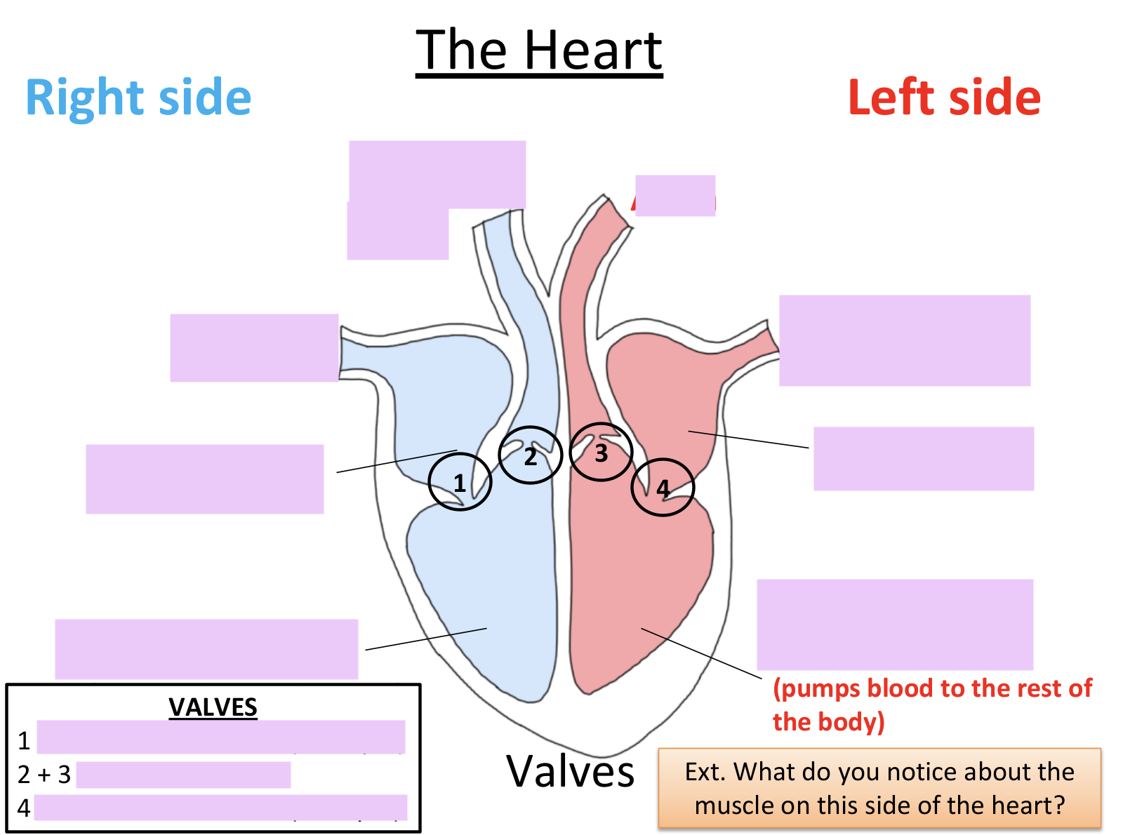
label this
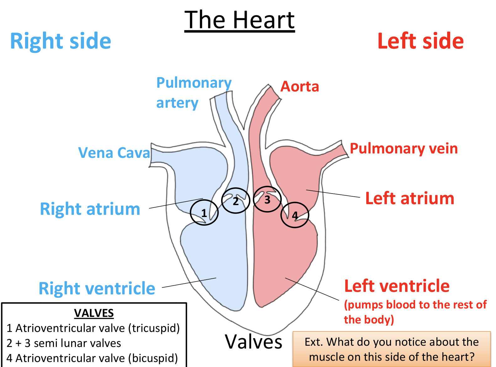
what is the tissue separating the left and right side of the heart called?
septum
what does systole mean?
contracting
what does diastole mean?
relaxing
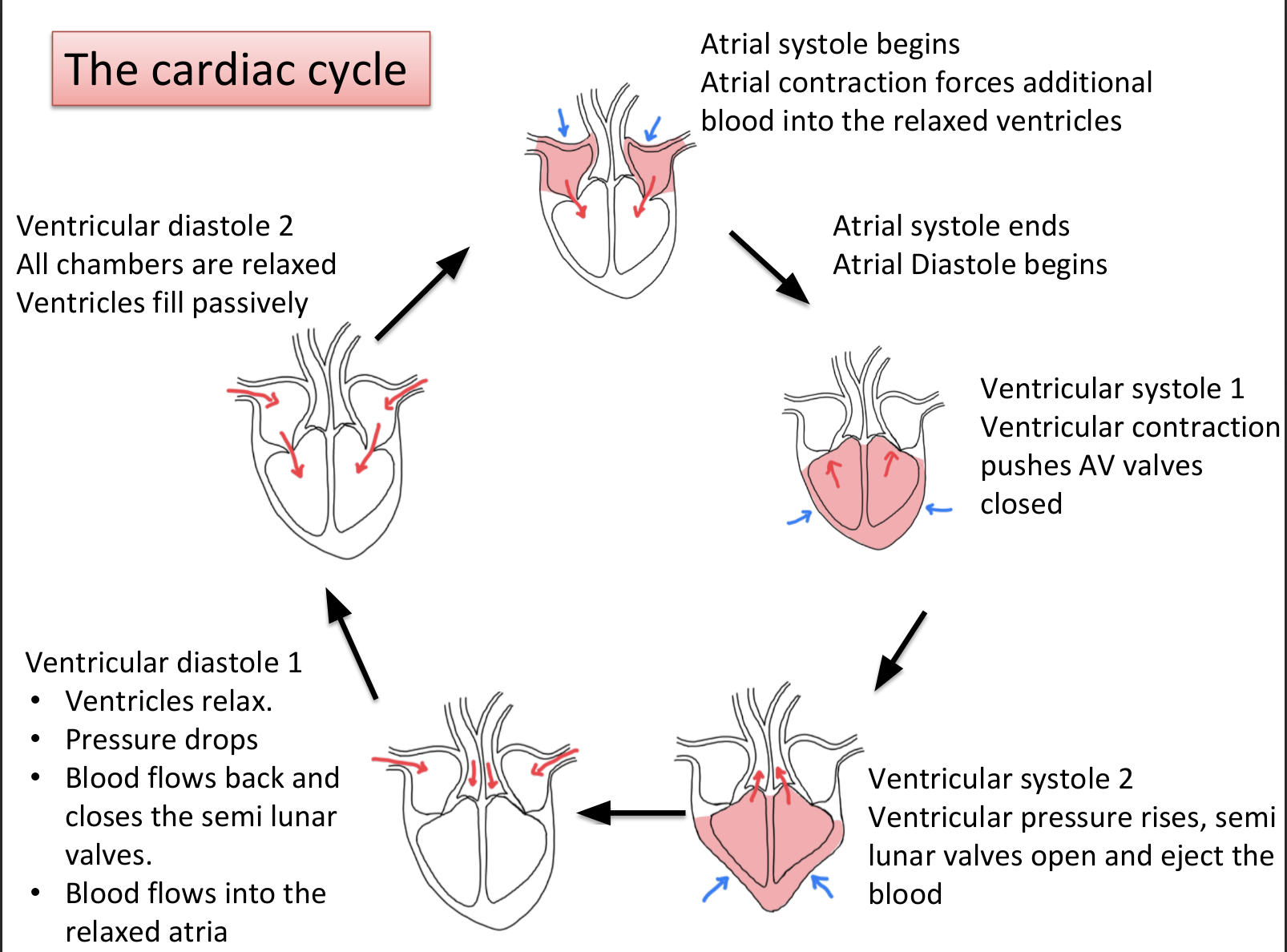
explain the cardiac cycle?
both atria and ventricle are in diastole
blood flows into atria through vena cava and pulmonary vein, causes pressure in atria to rise
pressure in atria > pressure in ventricle
AV valves open so blood flows from atria to ventricle
atria systole takes place, pushes remaining blood into ventricle, stretching ventricle
ventricular systole takes place, pressure increases
ventricular pressure > atrial pressure
AV valves close, prevents movement of blood back into atria
semi-lunar valves in pulmonary artery and aorta also open, blood pumped out of heart
ventricle diastole, ventricular pressure is lower than pulmonary artery, semi-lunar valves close
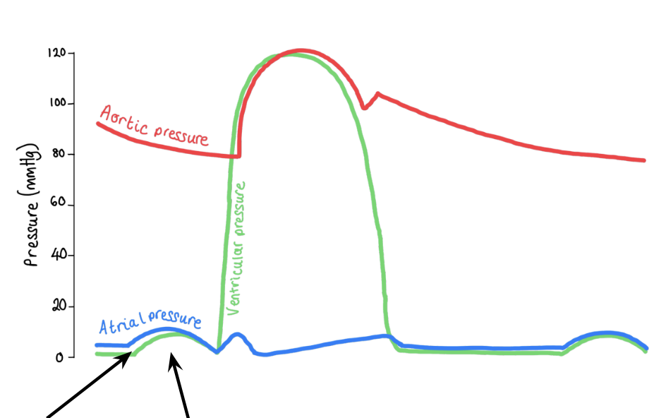
explain what happens on this part of the graph
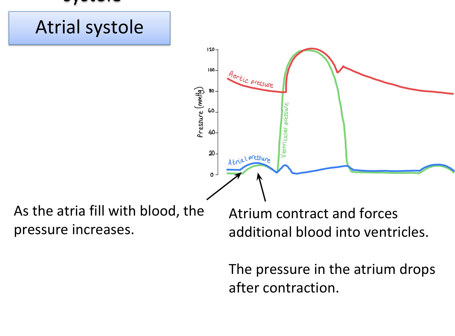
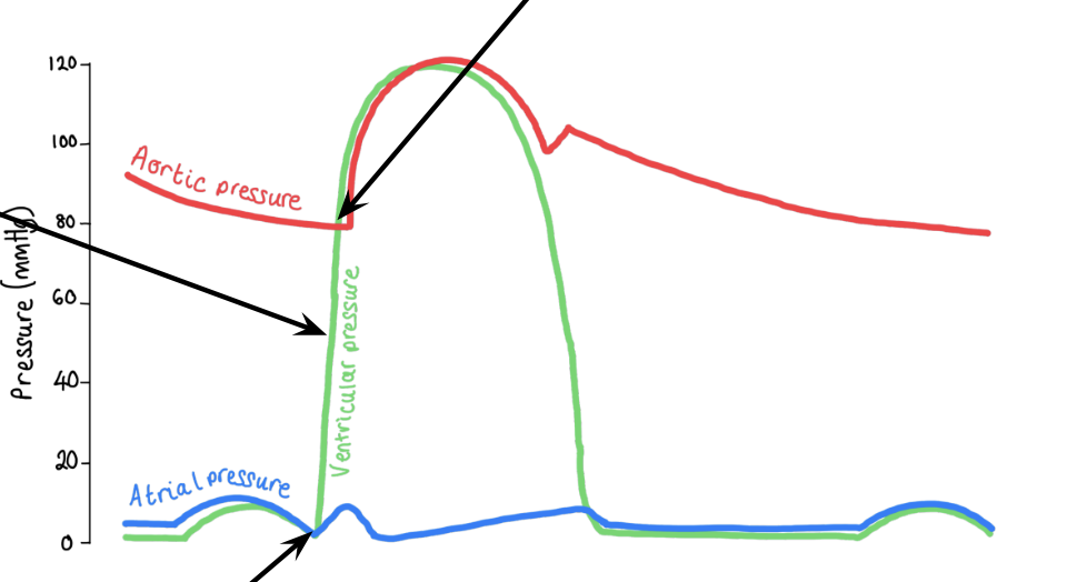
explain what happens on this part of the graph
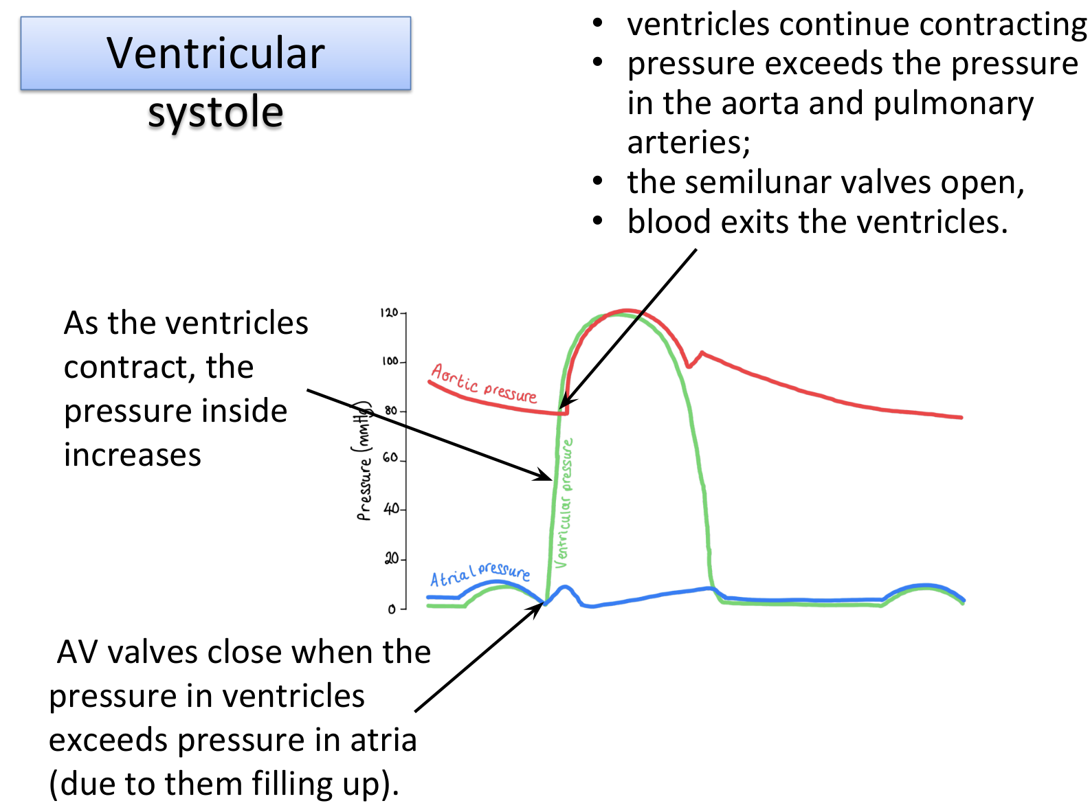
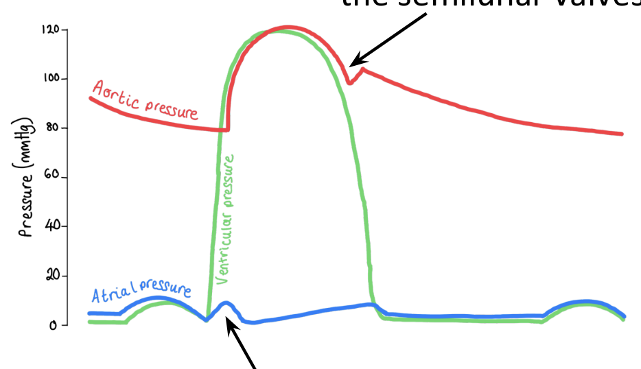
explain what happens on this part of the graph
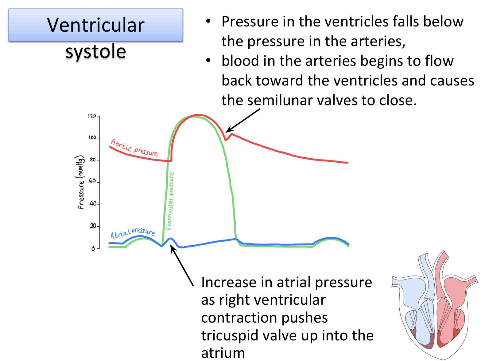
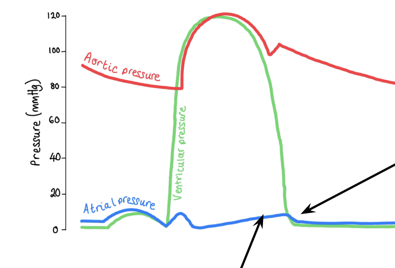
explain what happens on this part of the graph
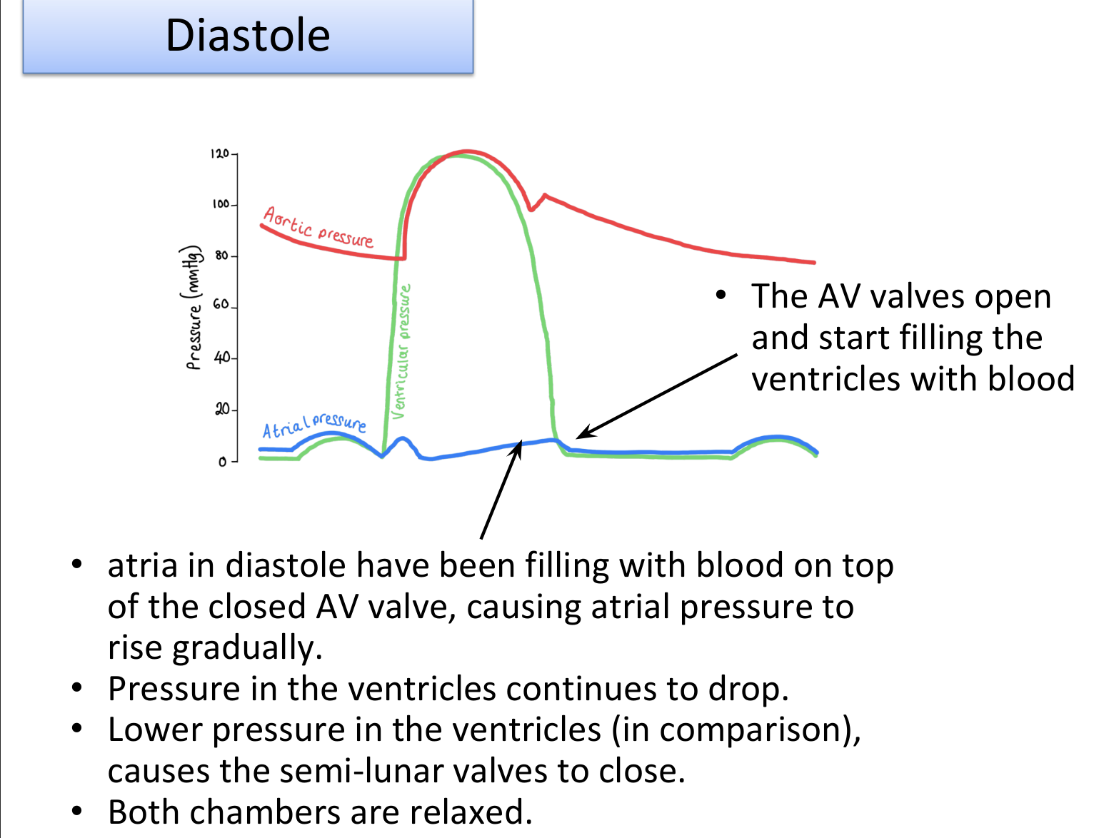
what is the sound the heart makes described as?
lub-dub
what happens during the lub sound of the heart?
AV valves closing due to contraction of ventricles
what happens during the dub sound of the heart?
semi-lunar valves closing as a result of the back flow from the aorta and pulmonary artery as the ventricles relax and pressure is lower
what is systolic blood pressure?
maximum blood pressure
occurs when ventricles are contracting (at the end of the cardiac cycle)
what is diastolic blood pressure?
minimum blood pressure
occurs when ventricles are relaxed and filled with blood (at the beginning of cardiac cycle)
how do you record blood pressure, layout wise?
systolic blood pressure / diastolic blood pressure
e.g. 120/80 mm Hg
what does diastolic pressure give the clearest indication of?
resistance to flow in blood vessels
what is blood pressure?
measured in arteries
determined by cardiac output and resistance to flow of blood in vessels
resistance to flow is affected by diameter of blood vessels
narrow vessels (vasoconstriction)
wider vessels (vasodilation)
what factors affect blood pressure?
cardiovascular centre
diameter of blood vessels controlled by stimulation of sympathetic and parasympathetic nerves
smoking
nicotine causes vasoconstriction
build up of fatty deposits in vessels
diet
high fat diet leads to build up of fatty deposits in blood vessels
adrenaline
causes selective vasoconstriction and vasodilation
increases in blood viscosity
excess water loss (sweating / excessive urination)
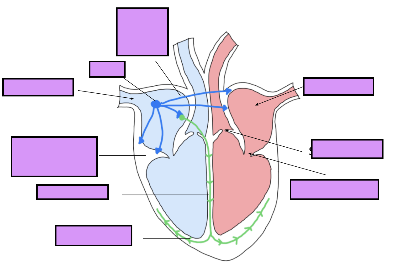
label this
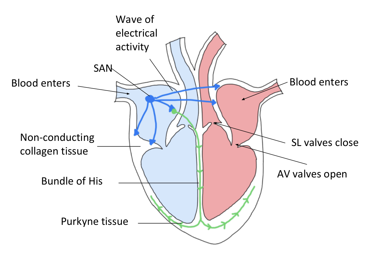
what is the mass of specialised cells in the right atrium called?
Sino-atrial node (SAN)
In gsce this was called a pacemaker
why are pacemaker cells myogenic?
initiates its own heart beat
how is a heartbeat established? Step by step
The SAN produces electrical impulses which cause both atria to contract (systole)
This forces open the AV valves, blood flows into ventricles. There is a band of non-conducting fibres which prevents the atria sending impulses to the ventricles
A patch of muscle fibres,AVN, conducts electrical impulses through the Bundle of His to the tip of the ventricles. There is a short delay in this node to allow ventricles to fill up with blood before contracting the Purkyne tissue
The 2 ventricles contract, base upwards, the Av valves shut and blood passes through semilunar valves into the arteries
as the ventricles are contracting, the atria relax and fill up with blood
what is heart rate?
number of beats per minute
how would you calculate heart rate?
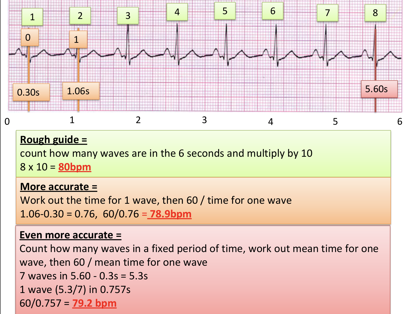
beats per minute = heart rate
what is stroke volume?
volume of blood (cm3) pumped by heart in 1 beat
how would you calculate stroke volume?
stroke volume = blood cm3 per beat
what is cardiac output?
stroke volume multiplied by the heart rate gives the amount of blood (cm3) pumped by heart 1 min
how would you calculate cardiac output?
cardiac output (cm3 min) = heart rate x stroke volume
what do cardiac muscle consist of?
fibres that branch, producing cross-bridges
why is it good that cardiac muscles consists of fibres that branch?
help the stimulus to spread around the heart, producing a squeezing effect
why do cardiac muscles have a lot of mitochondria?
to supply energy for contraction and are separated by intercalated discs which helps to synchronise the contraction
what are features of involuntary cardiac muscle?
myogenic
intermediate contraction speed
cells branch and interconnect for simultaneous contraction
uninucleated
has striation (bands)
connected by intercalated discs (low electrical resistance so impulses pass through easily)
what are electrocardiograms (EEGs) used for?
to monitor electrical activity of the heart
what is the process for getting a EEG?
EEG leads are attached to the body and a small amount of gel is applied which allows the electrical impulses of the heart to be more easily transmitted to the heads
how does a healthy persons EEG looks like?
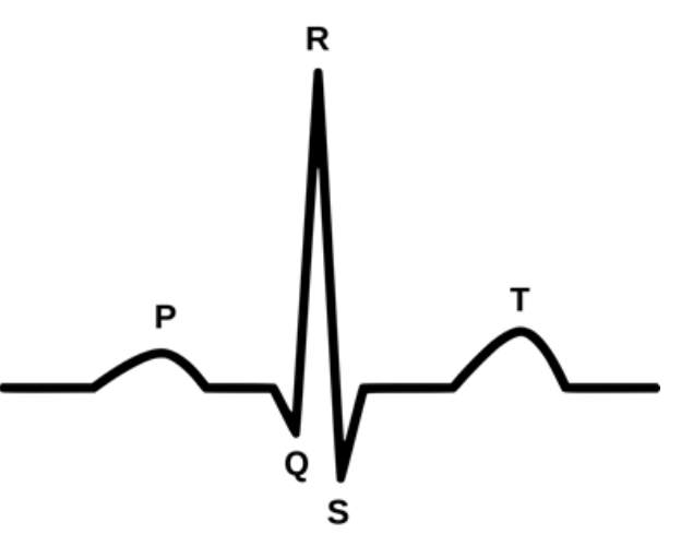
what does each letter mean?
P = atria contraction (only very small)
Q = electrical impulses going down the Bundle of His
QRS = ventricular contraction
T = ventricular repolarisation
how would you calculate heart rate?
what is bradycardia?
heart rate of <60bpm
what is normal heart rate?
60-100bpm
what is tachycardia?
heart rate of >100bpm
normally 100-160bpm
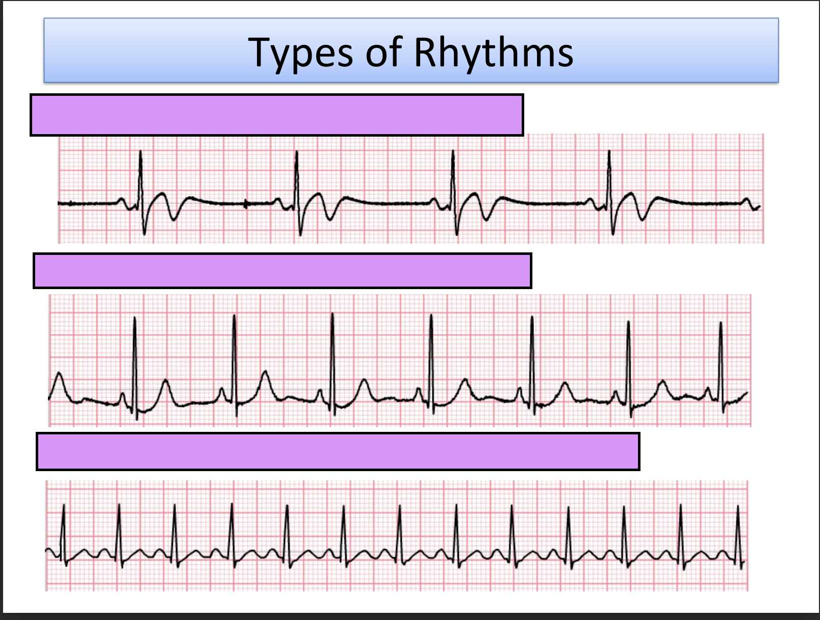
name these types of rhythms
bradycardia
normal rate
tachycardia
what are the two ventricular conditions?
ventricular fibrillation
ventricular tachycardia
what is ventricular fibrillation and how does it look like?
impulses originating from several places in ventricles at a high rate

what is ventricular tachycardia and how does it look like?
rapid heart rhythm originating from ventricles

what is atrial fibrillation and how does it look like?
Atria beating more frequently than ventricles so no clear P wave is seen
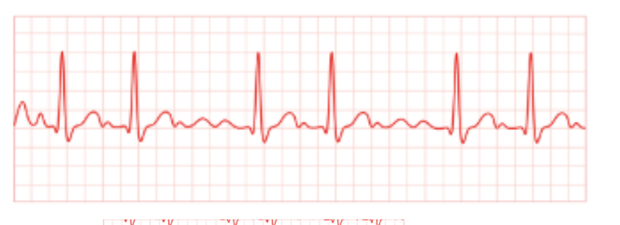
what is ectopic heartbeat?
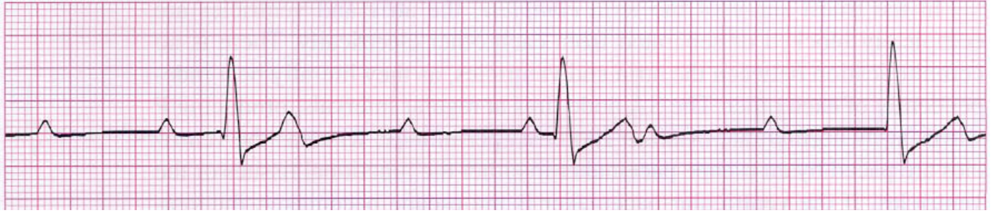
when either the atria or ventricle has an extra beat
how would ectopic heartbeat of ventricles having an extra beat look like?
what are key facts about oxygen?
low solubility in water
only about 3ml of O2 are dissolved in each litre of plasma
If we have a total plasma volume of 3-5 litres only about 9-15ml of O2 can be carried in the dissolved states
resting body requires 250ml of O2 per minute
what percent is oxygen transported for both ways?
98.5% combines with haemoglobin
1.5% is dissolved in the plasma
what are advantages of haemoglobin in red blood cells?
Hb is close to the enzymes that maintain or vary the binding property
Within RBCs, Hb doesn’t affect osmotic pressure of plasma
RBC’s clear stagnant plasma near capillary walls making exchange of O2, CO2, nutrients and waste more efficient
what is the structure of Hb?
2 alpha-globin polypeptide chain, 2 beta-globin polypeptide chain
each polypeptide chain has it’s own tertiary structure
haem group is a ‘prosthetic group’ since it contains Fe+
so haemoglobin is a conjugated protein as has a non-protein attached
what is the short hand way of saying partial pressure of oxygen?
pO2
what is partial pressure?
the individual pressure exerted independently by a particular gas within a mixture of gases
(basically conc in a gas)
how many oxygen molecules can Hb transport?
4 oxygen molecules