MCB 244: Nervous System III
1/143
There's no tags or description
Looks like no tags are added yet.
Name | Mastery | Learn | Test | Matching | Spaced |
|---|
No study sessions yet.
144 Terms
Brain Ventricles:
cavities within the brain; lined with ependymal cells; contain cerebrospinal fluid; connect with each other and with the spinal cord's central canal. Four ventricles within the brain:
There are _____ ventricles in the brain
four
Two lateral ventricles:
large cavities in cerebrum, separated by a medial partition called septum pellucidum
Third ventricle:
a narrow space in middle of diencephalon; connected to eachlateral ventricle by an interventricular foramen
Fourth ventricle:
a sickle-shaped space between pons and cerebellum; connected to third ventricle by cerebral aqueduct; opens to subarachnoid space medially and laterally; narrows before merging with central canal of spinal cord
Cerebrospinal fluid (CSF):
a clear, colorless liquid surrounding CNS thatcirculates in ventricles and subarachnoid space
Functions of the CSF
-Buoyancy: reduces the brain's apparent weight by 95%
- Protection: provides a liquid cushion
- Environmental stability: transport of nutrients/ wastes and protects against fluctuations
CSF formation:
- Formed by choroid plexus in each ventricle: a layer of ependymal cells and blood capillaries (within pia mater)
- Blood plasma is filtered through the capillary and modified by ependymal cells (compared to plasma, CSF has more Na+, Cl-; less K+, Ca2+, glucose)
- In addition, ependymal cell secretions and interstitial fluid from subarachnoidspace help make up CSF
CSF is continually formed and reabsorbed..
Excess CSF flows into arachnoid villi and drains into dural venous sinuses; anarachnoid granulation is a collection of these villi
Step 1: circulation of cerebrospinal fluid
CSF is secreted by chloroid plexus in each lateral ventricle
Step 2: circulation of cerebrospinal fluid
CSF flows through interventricular foramina into third ventricle
Step 3: circulation of cerebrospinal fluid
Choroid plexus in third ventricle adds more CSF.
Step 4: circulation of cerebrospinal fluid
CSF flows down cerebral aqueduct to fourth ventricle
Step 5: circulation of cerebrospinal fluid
Chlorid plexus in fourth ventricle adds more CSF
Step 6: circulation of cerebrospinal fluid
CSF flows out through two lateral aperatures and one median aperture
Step 7: circulation of cerebrospinal fluid
CSF fills subarachnoid space ad bathes external surfaces of brain and spinal cord
Step 8: circulation of cerebrospinal fluid
At arachoid will, CSF is reabsorbed into venous blood of dural venous sinuses
Clinical View: Hydrocephalus
- Pathologic condition of excessive CSF
- Often leads to brain distortion -May result from obstruction in CSF restricting reabsorption
- May result from intrinsic problem with arachnoid villi
-In a young child, head enlarged with possible neurological damage
- May be treated surgically
- Implant shunts that drain CSF to other body regions
Blood-brain barrier (BBB)
➢ Regulates which substances enter brain's interstitial fluid
➢ Helps prevent neuron exposure to harmful substances [e.g., drugs, wastes,abnormal solute concentrations; note: some drugs can pass and affect the brain(e.g., alcohol)]
BBB composed of specialized capillaries
Endothelial cells are connected by many tight junctions; walls have a thick basement membrane; wrapped by perivascular feet of astrocytes
BBB reduced in certain locations for functional reasons
-Choroid plexus needs to produce CSF
- The hypothalamus and pineal gland need to secrete hormones
Cerebrum
➢ Origin of all complex intellectual functions
➢ Two large hemispheres on superior aspect of brain
➢ Center of: Intelligence and reasoning; thought, memory, and judgment; voluntary control of skeletal muscle; conscious perception of senses
➢ Cerebrum is composed of left and right cerebral hemispheres
cerebrum: Longitudinal fissure:
deep cleft separating hemispheres
crebrum: Corpus callosum
- At a few locations, white matter tracts connect the hemispheres.
- Corpus callosum is the largest tract providing connection between them
Regions of cerebrum may exhibit ______ functions, some not easily assigned to one region
multiple
Cerebrum: Each hemisphere interacts with the opposite side of the body; example:
the left hemisphere receives sensory signals from the right side of the body and sends motor signals to the right side of the body
Some higher-order functions exhibit ___________ ___________. They are primarily controlled by one side of the brain; for e.g., speech is frequently located in left cerebral hemisphere
cerebral lateralization
Lobes of the cerebrum
Five lobes in each hemisphere: 4 are named for overlying cranial bones: frontal,parietal, temporal, occipital; insular lobe not visible at surface
Frontal lobe:
-frontal lobe is the anterior part of cerebrum
- Posterior border is deep central sulcus; precentral gyrus controls voluntary movement
- Lateral sulcus separates inferior frontal lobe from temporal lobe
- Frontal lobe has varied functions: motor control, concentration, verbal communication, decision making, planning, personality
Parietal lobe (supero-posterior part of cerebrum)
- Anterior border is central sulcus; postcentral gyrus is a ridge just posterior to central sulcus
- Posterior border is parieto-occipital sulcus
- Lateral border is lateral sulcus
- Serves general sensory functions; e.g., evaluating shape and texture of objects
Temporal lobe (internal to temporal bone)
- Located inferior to the lateral sulcus
- Functions include hearing and smell
Occipital lobe (posterior part of cerebrum)
Functions in vision and visual memories
Insula (deep to lateral sulcus)
- Small lobe that can be observed by pulling away the temporal lobe
- Functions in memory and sense of taste
Frontal lobe key functions
- Abstract thought
-Explicit memory
- Mood
- Motivation
-Foresight and planning
-Decision making
-Emotional control
-Social judgment
-Voluntary motor control
- Speech production
Insula key functions
-Taste
-Pain
-Visceral sensation
-Consciousness
-Emotion and empathy
-Cardiovascular homeostasis
Parietal lobe key functions
-Taste
-Somatic sensation
-Sensory integration
-Visual processing
-Spatial perception
-Language processing
-Numerical awareness
Occipital lobe
- visual awareness
-visual processing
Temporal lobe
-Hearing
-Smell
-Emotion
-Learning
-Language
-comprehension
-Memory consolidation
-Verbal memory
- Visual and auditory memory
-Language
Functional Area of the Cerebrum: Motor area
is housed within frontal lobes
Functional Area of the Cerebrum: Primary motor cortex (also called somatic motor area)
located in precentral gyrus; controls skeletal muscle activity on opposite side of body (i.e., projects contralaterally (opposite side) within brainstem or spinal cord)
Functional Area of the Cerebrum: motor homunculus:--> The controlled body regions map Functional Area of the Cerebrum:
distorted proportions of the body reflect amount of cortex dedicated to each part (e.g., hands are large on homunculus because large area of brain controls their precise movements)
Functional Area of the Cerebrum: Motor speech area (Broca area)
located in inferolateral portion of left frontallobe (in most people); controls movements for vocalization
Functional Area of the Cerebrum: Frontal eye field:
on superior surface of middle frontal gyrus; regulates eyemovements needed for reading and binocular vision
Functional Area of the Cerebrum: Premotor cortex
(somatic motor association area): located anterior to premotor cortex; coordinates learned, skilled activities
Sensory areas: Primary somatosensory cortex
located in postcentral gyrus of parietal lobes;receives somatic sensory informationfrom proprioceptors, touch, pressure,pain, temperature receptors
sensory homunculus:
- Areas of the body sending input can be mapped
- distorted proportions reflect the amount of sensory information collected from that region; large regions for lips, fingers, genital regions
Somatosensory association area:
immediately posterior to the postcentral gyrus (in the parietal lobe); integrates touch information allowing us to identify objects by feel
Primary visual cortex:
located within the occipital lobe; receives, processes, stores visual information
Visual association area:
surrounds primary visual cortex; integrates and interpretscolor, form, to allow identification/recognition of things (for example, faces)
Primary auditory cortex:
located within the temporal lobe; receives, processes, stores auditory information
Auditory association area:
located in temporal lobe; integrates and interprets sounds
Primary olfactory cortex:
located within temporal lobe; receives, processes, stores odor information
Primary gustatory cortex:
located within insula; receives, processes, stores tasteinformation
Functional brain regions
Integrate information from multiple association areas
Prefrontal cortex:
located rostral to premotor cortex (in frontal lobe); complex thought, judgment, personality, planning, deciding; still developing in adolescence
Wernicke area:
typically located in left hemisphere; involved in languagecomprehension (recognizing and understanding spoken or written words; 'Wernicke areahelps us comprehend words')
Clinical View: Autism Spectrum Disorder
- Autism affects 1 in 88 U.S. children
- Characterized by social and communication difficulties
- Severity varies across autism spectrum
-Best predictors of independent adulthood are intelligence and communication ability
- Specific causes unknown
➢ Genetic, environmental, and biochemical factors have been explored
➢ Males have four times higher incidence than females
➢ Vaccines found not to be a factor
Central white matter
- lies deep to gray cerebral cortex
- Composed of myelinated axons grouped into tracts
Association tracts
- Connect regions of cerebral cortex within the same hemisphere
➢ Arcuate ('bowed') fibers: short tracts connecting neighboring gyri
➢ Longitudinal fasciculi: longer tracts connecting gyri in different lobes
Commissural tracts
➢ Commissures connect regions in different hemispheres; include corpus callosum(C-shaped), anterior and posterior commissures
Projection tracts
➢ Link cerebral cortex to inferior brain regions and spinal cord
➢ For e.g., corticospinal tracts carry signal from cerebral cortex to spinal cord: as this projection tract passes between thalamus and cerebral nuclei it is called the internal capsule
Cerebral Lateralization: Anatomical asymmetries
Petalias: protrusion of a lobe on one side compared to other side
➢ Right-handed individuals tend to have right frontal petalias and left occipital petalias
➢ Left-handed individual tend to show the opposite Cerebral Lateralization
Cerebral lateralization
➢ Two sides of cerebrum exhibit differences in higher-order functions
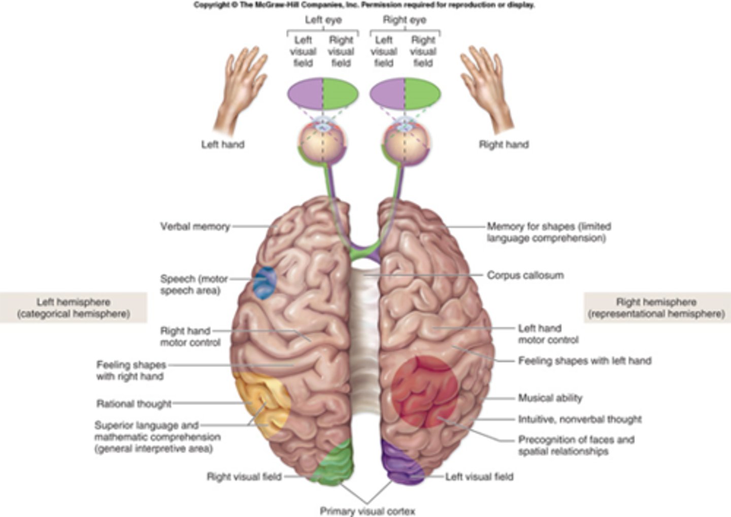
Cerebral lateralization: Categorical hemisphere
is usually the left hemisphere- specialized for languageabilities, functions in categorization and analysis; contains Wernicke area andmotor speech area; sequential, analytical and reasoning tasks (science andMathematics)
Representational hemisphere
is usually the right hemisphere; concerned withvisuospatial relationships, imagination, comparison of senses (musical and artisticskills, patterns and spatial relationships, comparison of sight, sound, smell and taste)
➢ The two hemispheres communicate through the corpus callosum and othercommissures
Lateralization develops in early childhood- seen prior to ____ to ____ years of age
5 to 6
Lateralization differs between sexes:
women's posterior corpus callosum is thicker(more connections); males suffer more functional loss when one hemisphere damaged
Lateralization is correlated with handedness:
in right handers, the left hemisphere is almost always categorical, speech-dominant; left-handed individuals may have either hemisphere be categorical
Clinical View: Epilepsy and Cerebral Lateralization
- Epilepsy is a neurological disorder
- Neurons transmitting APs too frequently and rapidly
- Usually controlled by medications, but may require surgical removal of part of brain
➢ In most severe cases, may require hemispherectomy: removal of side of brain responsible for seizure activity
➢ Remaining hemisphere able to take over some functions of missing hemisphere
Cerebral nuclei (Basal nuclei)
gray matter deep in cerebrum; help regulate motoroutput; diseases of these nuclei associated with involuntary movements
Cerebral nuclei : Caudate nucleus
enlarged head and slender tail paralleling lateral ventricle; helps produce pattern and rhythm of walking movements
Cerebral nuclei : Lentiform ('lentil-shaped) nucleus
rounded mass between insula anddiencephalon; composed of putamen and globus pallidus
Cerebral nuclei : Putamen ('shell): helps control movements at subconscious level
Globus pallidus ('pale ball'): influences thalamus to adjust muscle tone
Cerebral nuclei :Globus pallidus ('pale ball')
influences thalamus to adjust muscle tone
Cerebral nuclei: Claustrum ('barrier'):
thin sliver of gray matter immediately internal to insulacortex: extensive connections to cerebral areas suggest involvement inconsciousness
Cerebral nuclei : Amygdaloid body (amygdala; 'almond')
expanded region at tail of caudatenucleus; functions in mood, emotions
Cerebral nuclei : Corpus striatum
striated or striped appearance of the internal capsule. consist of the caudate nucleus, putamen, glbous pallidus
Nuclei'
denotes cellbodies within the CNS
ganglia
denotes cell bodies external to the CNS
in a coronal section you see:
- cerebral cortex
-corpus callosum
-lateral ventricle
-septum pellucidum
-thalamus
-internal capsule
-lateral sulcus
-insula
-third ventricle
-optic tract
-hypothalamus
Clinical View: Cerebrovascular accident (CVA, or stroke)
-Reduced blood supply to part of brain
- Due to blocked arterial blood vessele or hemorrhage
-May cause brain tissue death if prolonged for several minutes
-Symptoms of blurred vision, weakness, headache, dizziness, walking difficulty
-Affects opposite side of body
-Brief episode is a transient ischemic attack (TIA)
Clinical View: Brain Disorders; Headache
➢ Due to dilated blood vessels in skull or muscle contraction
➢ Migraine headaches: severe, recurring, often unilateral
➢ Not true brain disorder, but may accompany them
Cerebral palsy
➢ Group of neuromuscular disorders
➢ Result from damage to infant brain before, during, or right after birth
➢ Impairment of skeletal muscle, sometimes mental retardation
Huntington disease
➢ Hereditary disease affecting cerebral nuclei
➢ Rapid, jerky, involuntary movements
➢ Intellectual deterioration
➢ Fatal within 10 to 20 years after onset
Parkinson disease
➢ Affects muscle movement and balance
➢ Stiff posture, slow voluntary movements, resting tremor
➢ Caused by decreased dopamine production in substantia nigra
Diencephalon ('in-between brain'):
Includes the epithalamus, thalamus,and hypothalamus
Epithalamus
Forms posterior part of roof of diencephalon, covers third ventricle
Pineal gland:
endocrine gland secreting melatonin; helps regulate day-night cycles,circadian rhythm
Habenular nuclei
help relay signals from limbic system to midbrain; involved in visceral andemotional responses to odors
Thalamus
➢ Oval masses of gray matter on lateral sides of
Interthalamic adhesion:
- midline mass of gray matter connecting left and right thalamus;composed of about a dozen thalamic nuclei; axons from a given nucleus project to aparticular region of cortex
- Receives signals from all conscious senses except olfaction; relays some signals to appropriate part of cortex and filters out other signals distracting from subject of attention (for example, background noise in crowded room)
Hypothalamus: ('CEO of the ANS and endocrine system corporations!')
Anteroinferior region of the diencephalon
Infundibulum
stalk of pituitary that extends from hypothalamus
hypothalamus: Master control of autonomic nervous system
influences heart rate, blood pressure,digestive activities, respiration
hypothalamus: Master control of endocrine system:
secretes hormones that control activities in anteriorpituitary gland; produces antidiuretic hormone and oxytocin
Hypothalamus: Regulation of body temperature:
neurons in preoptic area detect altered temperature;signal other hypothalamic nuclei to heat or cool the body
Hypothalamus: Food intake:
ventromedial nucleus monitors nutrient levels, regulates hunger
Hypothalamus: Water intake:
anterior nucleus monitors concentration of dissolved substances in blood,regulates thirst
Hypothalamus: Sleep-wake rhythms:
suprachiasmatic nucleus directs pineal gland to secrete melatonin,regulates circadian rhythms
Emotional behavior:
part of the limbic system; controls emotional responses (pleasure, fear, etc.)