NEUR1020 Module 3 Content
1/15
There's no tags or description
Looks like no tags are added yet.
Name | Mastery | Learn | Test | Matching | Spaced | Call with Kai |
|---|
No study sessions yet.
16 Terms
Detectors of stimuli become less sensitive after prolonged periods of exposure. This frees our attention to make new stimuli/inputs more prominent.
this encourages opposite experiences relative to the adaptor.
Reduced responding in the brain to an adapted input enhances the prominence of new inputs relative to the old ones
eg. If you stare at a spiral a sensory adaptation occurs where after the spiral stops a visual after-effect of the spiral spinning in the opposite direction occurs. This is a motion-after effect
eg. Troxler fading: inputs slowly fade because of adaptation as we stare, whenever we stare at a static scene. This occurs so slowly we usually don’t notice. If a new, identical input were to appear, it would look bolder than the original input.
these adaptation effects occur in all sensory modalities
We do not have veridical experiences of sensory input: our experiences are a product of activity in our brains, and that activity can promote experiences that differ from sensory inputs from sensory organs.
McGurk effect= when you hear something different when a voice is accompanied by visual lip movements.
we can experience an ‘averaging’ of audio and visual information, which is different to the audio/visual experience we would have had if those inputs were experienced separately
eg. if you see someone mouthing ‘ga’ but the noise they make is ‘ba’, the brain can perceive it as a ‘da’ sound.
This shows people don’t have a veridical sense of input from any single sensory modality. Instead, our brains tend to create perceptual experiences that sum together all the sensory information that is available to us.
Perception= the brain’s hypothesis about what is out in the world based on analysis of evidence provided by sensory organs and guided by past experience
sense organs are needed for the important first step that leads to perception: transducing environmental energy into electrochemical signals that can be sent to the brain.
The ability to hear relies on the transformation of sound waves into signals that can be sent to your brain
sound waves are generated by vibrations that alternate in exerting increased and decreased force on air molecules (creating a wave). These alternations in pressure propagate through the air until they reach the ear.
the frequency of the wave determines the pitch of the sound=how often air pressure is increased then decreased over time. High pitch=low frequency
the amplitude of a wave controls the intensity/loudness of a sound=the magnitude of changes in air pressure. Big changes create louder sounds The mechanisms in the ear:
Hair cells in the cochlea of our inner ears allow us to transduce soundwave signals to the brain
The cell’s hair-like stereocilia extend into the surrounding liquid and move back and forth when the liquid is vibrated by sound waves
When the stereocilia bends, ions rush to the top of the hair cells and release chemicals at the base. These chemicals bind to auditory nerve cells, creating an electrical signal that propagates along the auditory nerve to the brain.
1. Photoreceptors, which form the retinae of human eyes, transduce light into signals that can be sent to the brain
photoreceptors contain light-sensitive chemicals called pigments that absorb light photons. This begins a process that changes the photoreceptor’s membrane potential, causing a wave of depolarisation.
2. Ganglion cells gather signals from the back of photoreceptors via bipolar and amacrine cells and converge to form the optic nerve
ganglion cells have circular receptive fields (regions of retina they respond to) and respond positively to inptu from a central region and negatively to input in a surrounding region OR vice versa: you can change your focus. If you try to stimulate the entire receptive field the on/off regions balance and no signal is produced (blur)
Retinal ganglion are like little adding machines that detect circular blobs of contrast. At fixation=0 brightness contrast, away from fixation (in between intersections)= +4 brightness contrast, at intersection= +2 brightness contrast
each receives input from many photoreceptors
3.the optic nerve carries visual signals from our eye to the brain
Some limits to vision:
the pigments are contained in the outer segments at the back of the retina: away from the light. This limits the eye’s ability as all the cellular layers that refract light blur the image before it can even reach photoreceptors.
The physiological blind spot stops us from having veridical impressions of the images that reach the retinae. This mistaken notion is called naive realism.
the blind spot= where the optic nerve has to pass through the surface of the retina to carry signals to our brain. There are no photoreceptors at this spot so you are blind to any image that falls on this region of the retina. The brain goes through a process called ‘perceptual filling in’ to assume that what is on either side of the blind spot is also within it and fills it in.
Some people have different experiences and sensitivities to certain colours and it is possible that we are all having idiosyncratic experiences. The colours we experience are associated with different wavelengths of light
The range of colours an animal can see are determined by the range of wavelengths of light their photoreceptors can absorb (Humans=400nm (blue)→700nm(Red), birds include UV, goldfish= UV + Infrared)
This also depends on the different types of photoreceptors we have in our retina:
Different types of photoreceptors are called ‘cones’. Each cone maximally absorbs (MA) a different wavelength (colour).
Humans are trichromats= we rely on 3 types of cones: Short cones (MA 430nm, blue), Medium cones (MA 530nm, green) and Long cones (MA 517nm, red). Birds are tetrachromats (+UV range). Dogs/cats are dichromates (only yellow and blue cones).
Colour vision comes from the ability to detect the ratio of activation across these 3 cones.
blue lights maximally activate short cones, minimally activate medium and don’t activate long at all
red lights maximally active long cones, minimally activate medium and don’t active short at all
grey= equal activation across all 3 * white= all maximally activated
black=all minimally activated
even people with normal colour vision have different ratios of the 3 receptors in the retina; supporting the theory that we are all having idiosyncratic experiences Red-green colour blindness=don’t have long or medium cones.
this is less common in females as the defective gene is carried along teh X chromosome: as females have 2 a non-defective chromosome can compensate, but males only have one=no opportunity for compensation
some females even have a 4th cone, allowing them to distinguish many more colours
We can tell if people have different experience of colour when they have different patterns of sensitivity to physical inputs
Colour perception is also a product of more complex processes that discount the impact of illumination
lighting affects the different wavelengths objects refract.
eg. a banana refracts more red light at sunset than at midday, but we want to see it as yellow despite the lighitng.
So, our visual system strives to subtract that impact of lighting away from our impressions of colour in a process called colour constancy
eg. the meme dress. If your visual system perceives the dress as lit by the sun, it will subtract yellow from your impressions of colour and you will see the dress as blue and black. If it perceives it as artficial lighting, it will subtract more blue lighting and the dress will appear white and gold.
How are signals carried from eyes to brain?
The primary visual pathway
signals leave the eye via the optic nerve, travelling to the optic chiasm
at the chiasm, signals cross so information from right side of visual space travel to the left side of the brain and vice versa
these signals don’t relate to eyes: signals from both eyes can relate to one side of visual space
if there is a problem specific to information from one eye, it must be the eye itself or in the optic nerve prior to the optic chiasm.
If there is a problem specific to information from one side of visual space, the problem is due to damage beyond the chiasm
From the chiasm, signals travel to the lateral geniculate nucleus/LGN= the subcortical brain structures, then to the primary visual cortex (V1) at the back of the brain. V1 is the first cortical region to receive visual signals.
End-stopped V1 cells are responsive to oriented stripes of contrast of specific, limited length
complex V1 cells are responsive to oriented stripes of contrast anywhere within its receptive field
complex direction-selective V1 cells are responsive to oriented stripes of contrast located anywhere within its RF that move in a particular direction
How does the primary visual cortex process visual signals?
There are two primary visual cortexes, HV1 and HV2, one in each hemisphere
HV1 responds to all visual inputs from one side of visual space, HV2 the other (as per the left/right split at the chiasm)
Humans do not see all V1 activity
eg. flicker fusion threshold for colour describes how many times in a second a light can switch between colours before we stop seeing flickering and start seeing a constant, averaged colour. For humans this is 30hz=30 times a second. Even though our vision stops, V1 cells are responsive to the flickering lights at rates far beyond flicker threshold.
we can do retinotopic mapping of V1 responses with fMRI
=adjacent neurons in visual brain regions encode information from adjacent regions on the retinal surfaces
as inputs become more and more peripheral, they are mapped onto progressively anterior regions of V1=more forward on the surface of the brain
as inputs become more to the top of the visual field they are mapped toward the bottom of V1 (and vice versa)
this is most apparent in lower level visual brain structures with small receptive fields (LGN, V1, V2, V3) but holds in all visual brain regions
There are many other cortical brain regions that receive input directly/indirectly from V1 that all have retina topically mapped responses
What is blindsight? How is it measured? What was discovered about it?
A phenomenon where people think they are blind but they aren’t. It occurs when you suffer damage to the primary visual cortex
people report blindness for a proportion of the visual field encoded by damaged VI regions known as cortical scotoma
Polarimetry quantifies the extent of this blindness
patients fixate a point and we flush spots of light at various speed in their visual field. After each flash they are asked if they saw it
experiments would ask a patient to guess, even when they couldn’t see an input, details about the flash (eg. colour). Patients who were blind, however, performed well above chance
Blind sight shows that some visual operations can be carried out without activity in V1. Humans can still navigate and avoid obstacles despite insisting they can’t see.
How can we get a true measure of visual sensitivity despite blind sight?
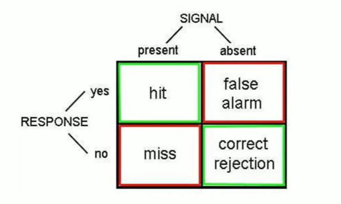
Signal detection theory
a light is flashed or not flashed and a participant guesses if it was or wasnt
we measure the hit rate (proportion of times a participant correctly reports an input) and false alarm rate (proportion of times a participant reports an input when it isn’t present)
d prime statistics (the ratio between hit rates and false alarm rates) reflects sensitivity
1:1 ratio=0 sensitivity
if the hit rate is greater than the false alarm rate the participant has sensitivity
What are the fields of all visual neurons?
Receptive field: position on the retina that images must fall upon to make that visual neuron respond
Response selectivity: the type of input to which a cell will respond to
The architecture of human vision. Higher order examples and what would happen if they were damaged.
Cells at progressive stages of processing respond to increasingly complex features
visual cells (V1) respond to simple things like bars of light, coloured bars of light, bars of light moving in a particular direction etc.
These cells then project to other cortical brain regions (cortical cells) and cells in these regions respond to increasingly complex properties
eg. particular patterns of motion, not just any direction, or particular objects like faces
the regions of different cells that respond to different specific inputs can be detected with fMRI
eg. V5= 2 regions that respond selectively to complex patterns of motion (directional)
cerebral atinetopsia=no motion vision, the world is viewed as frames jumping from one level to the next. Only experienced if both V5 are damaged (Rare)
eg. V4= 2 regions where large numbers of cells are attuned to specific colours (very few are direction tuned).
cerebral icromatopsia= absense of colour vision caused by bilateral damage of both V4s (only one side=only one side of visual space colourblind)
Face vision
Humans have a whole network of cortical brain regions responding to faces in the temporal lobe, including:
superior temporal sulcus (STS)
fusiform face area (FFA)
the occipital face area (OFA)
adjacent to both V1 and V4
Presopagnosia=face blindness that correlates to damage to the OFA
What is functional modularity? The binding problem
=The fact that our brains contain multiple regions that are specialised for processing different visual properties (eg. colour, faces, motion)
The binding problem= how can the brain tell when activity spread across the brain is related to a common object?
miss binding occurs due to the brain incorrectly binding features in the complex combined activity from different brain regions to create coherent perceptions
eg. steady state miss binding happens because visual bindings of colour and motion are disproportionally shaped by central vision. The brain assumes bindings that prevail in central vision also do in the periphery
we don’t know how the brain solves the binding problem
Dual visual stream theory
Humans have 2 visual systems: one that promotes perception (ventral stream) and one that guides our actions (dorsal stream)
ventral important for generating conscious visual sensations. Prohects into the temporal love for semantic memory.
dorsal is where the secondary visual pathway projects, important for vision for action