PS2021 (Perception, Attention and Action)
1/279
There's no tags or description
Looks like no tags are added yet.
Name | Mastery | Learn | Test | Matching | Spaced | Call with Kai |
|---|
No analytics yet
Send a link to your students to track their progress
280 Terms
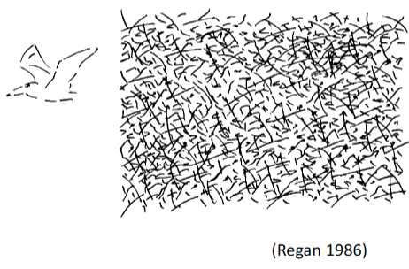
How do we know vision is not simple
Machines struggle with replicating human vision
How does a camera record images
Records pixel values. Records light intensity as numbers assigned to sensor locations
How does the human eye record images
Records light intensity as neural activation of photo receptors
How is the visual system like a camera
At best it is similar to the earliest stages of visual system
What is the purpose of the visual system and perception
Perception = generate meaningful and adaptive representations of the environment
Visual system = meaning and adaptive representations explicitly encode relevant aspect of the world and help guide behaviour
What is the Craik-O’Brien-Cornsweet Effect
Seeing the colours as 2 different colours when they are the same. This is due to the edge boundaries and our brain trying to make a 2D image, 3D

What does the two-tone image effect show
Initially, it looks like meaningless black and white splotches but if given prior knowledge it influences how we see it
What is the information processing paradigm
Input (stimulus)→ Brain/mind (information processing)→Output (perception)
What other disciplines is vision science important for
Psychology
Neuroscience
Evolutionary biology and sensory ecology
Engineering and machine vision
Applied mathematics and physics
What does psychophysics try to determine
The relationship between stimulus and perception quantitatively

What is the absolute threshold
The smallest amount of stimulation that can be reliably detected
What is the difference threshold
The smallest difference between two stimuli that can be reliably detected
What is just noticeable difference (JND)
Difference between guessing (getting 50% right) and knowing (e.g. 75% right)
What do thresholds typically measure
Limits of the perceptual system
Studies perception at the level of the whole organism
What is the Weber Fraction
Ratio between JND and reference intensity
The ration is always constant
What is electrophysiology
Microelectrode located near (or inside) the neuron will pick up action potentials
Provides information about processing in individual neurons or a small number of neurons
Typically done in animals (due to ethics)
What are the pros and cons of EEG/ MEG
Good temporal resolution but bad spatial resolution
What are the pros and cons of fMRI
Good spatial resolution but bad temporal resolution
Why would we use neuroimagining techniques
They can pick up consequences of electrical signals in the brain without the need for direct access
They can provide information about processing in the brain at the level of whole brain areas
What are computational methods
Build mathematical models of information processing at different levels
Provide very precise explanations (mechanistic and functional) of biological vision
Important implications for machine vision
How does vision link to neuropsychology and neuropsychiatry
Study individuals with brain lesions or with neurological/ psychiatric conditions
Important for understanding healthy vision
Allow for the creation of treatment and interventions
Two types of photoreceptors
Rods and cones
Rods
Approx 100 million
One type of photopigment
Very sensitive to light; used in dim light (Scotopic vision)
Poor spatial resolution
High temporal resolution
Cones
Approx 6 million
S-, M-, L- with peak sensitivities at different wavelengths (colour vision)
Less sensitive to light; used in bright light (photopic vision)
High spatial resolution
Poor temporal resolution
What is the receptive field
A specific region of sensory space in which an appropriate stimulus leads to a response in a sensory neuron
Structure of a ganglion cell
positive centre with negative surrounding or vice versa (equal number of pos and neg cells)
On-centre bipolar cells
Positive centre.
Response to light in the centre = depolarisation (become more positive)
Response to light in the surround = hyperpolarisation (become more negative)
Overall = excited by light shined in their centre and inhibited by light shining in their surround
Off-centre bipolar cells
Negative centre
Response to light in the centre = hyperpolarisation (become more negative)
Response to light in the surround = depolarisation (become more positive)
Overall = inhibited by light shined in their centre and excited by light shining in their surround
Eye structure
Nasal part projects contralaterally
Lateral part projects ipsilaterally
Let visual field is represented in right LGN and vice versa
Lateral Geniculate Nucleus (LGN)
Each layer gets inputs from only one eye (cells are monocular)
Arranged in a retinotopic map
The neighbouring relationships that exist in the retina, continue
Physiology of LGN
Sub cortical
Each layer contains monocular cells that form a retinotopic map of half a visual field
Layers 1 and 2 = Large cell bodies (Magnocellular layers), receive input from M ganglion cells (rod input), high contrast sensitivity and low spatial resolution, process course features and motion
Layers 3 to 6 = Small cell bodies (parvocellular layers), receive input from p ganglion cells (cone input), low contrast sensitivity and high spatial resolution, process fine features and colours
Function of LGN
First relay station between eyes and cortex (axons link to primary visual cortex)
Magno- and parvo cellular layers respond in similar ways as their input ganglion cells
Unknown the specific function
Richly connected to many other parts of the brain
A locus at which retinal information can be modulated by brain areas
More than just a relay station
primary visual cortex (V1)
LGN projects to this. Striate cortex
Left hemisphere process right visual field and vice versa
Most neurons are binocular (recieves input from both eyes)
Organised in a retinotopic map
Map is distorted (cortical magnification)
How does binocular vision help us
Helps us see depth
What is cortical magnification and why does it happen
More neural ‘real estate’ in the cortex for the centre of the visual field compared to peripheral
Causes peripheral vision to be less clear
Clarity increases the more central the object is
What does cortical magnification show us the importance of
Eye-movements
Why do we not use the same amount of neurons for the whole visual field?
Tradeoff between having a large visual field and having (some areas of) high acuity
If we would have the same acuity as in fovea in the whole visual field, we would need eyes and brains multiple times the size of our skull
Fovea
Centre of our visual field
Which receptive field do we have in ganglion cells/ LGN neurons
Centre-surround receptive field
What type of light do centre-surround receptive fields respond to
Spots of light
What receptive field do we have in V1
Elongated receptive fields
What type of light do elongated receptive fields respond to
Lines, bars and edges
If more negative cells are activated in comparisson to positive cells what happens
Inhibits. Deactivation. Do not see the light
If more positive cells are activated in comparisson to negative cells what happens
Excitatory. Activation. Sees light
What does receptor activation depend on
Orientation and size
Feature detectors
Cell responds with same firing rate to an optimally oriented but faint stimulus as to a suboptimal intense stimulus (response of single cell is ambiguous)
Bright light but not optimal orientation = faint light with optimal orientation (firing rate level)
Population codes
Takes all cells into account to make an accurate representation of light
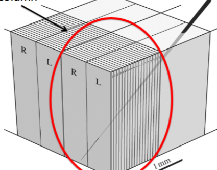
Ocular dominance bands
Certain brain cells prefer information from the left eye over the right and vice versa.
Occurs in the V1
Stellate cells and pyramidal cells create bands of neurons that prefer information specifically from one eye
These bands are alternating (e.g. left, right, left)
Stellate cells
Receives direct input from the thalamus - specfically LGN
First cortical neurons to get visual information
Respond more to one eye than the other (ocular dominance)
Pyramidal neurons
Receive signals from stellate cells and pass them on to other brain areas
Show ocular dominance but it’s more about combining and interpreting the information
Hypercolumns
Each layer responds to a different orientation and size. Takes information from both eyes. Contains ocular dominance columns
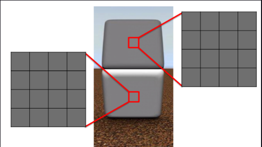
Visual angle
Tells us the size of the retinal image (given a certain object size at a certain distance)
Fourier analysis
A way of breaking down complex patterns into its simple parts. E.g. into simple sine waves.
Early vision as local Fourier analysis
Hypercolumns contain neurons tuned to different orientations and spatial frequencies
All of these neurons analyse the same patch of visual space
Together, they extract spatial frequencies and orientations contained in 'their' local patch
Adaption in vision
method = stare at the same stimulus for a long time
Process = as a consequence of long exposure, those neurons that are tuned to the stimulus property decrease their sensitivity
Low level vision
Extracts local information about lines, bars and edges
Mid-level vision
joins isolated features into larger groups
Forms basis for high-level vision
High level vision
Object recognition
Ventral stream
‘What’ pathway = object identification
Starts in V1 and goes to temporal lobe
Receptive field size increases the further along the stream you go
Dorsal stream
‘Where’ pathway = visuo-spatial information processing
Starts in V1 and goes into parietal lobe
How does mid-level vision overcome ambiguity
Assumptions and prior knowledge
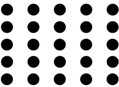
Principle of proximity
View dots as columns rather than rows due to their closeness
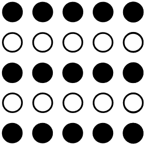
Principle of similarity
View it as rows due to them being separated by colour
Principle of common fate
Things that move together are bound together
Principle of good continuation
Assumes smooth (good) continuation = same object
Brain areas responsible for face perception
Inferior occipital gyri
Superior temporal sulcus
Lateral fusiform gyrus
Domain specificity account for facial processing
Mechanisms operate independently of general object perception
Expertise account for facial processing
Mechanism derive from general object perception but become finely tuned due to extensive experience
Part-whole effect
Features are easier to identify when presentedd as part of a face (e.g. eyes)
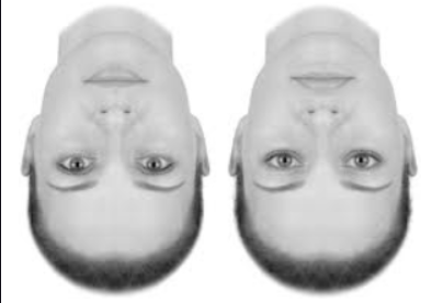
Face inversion effect (thatcher illusion)
Inversion disrupts the processing of fine details and relationship between features
As faces are upside down it is not the same process as normal facial processing
Prosopagnosia
Failure to identify or distinguish between faces, despite (otherwise) normal visual and cognitive ability
Special case of visual agnosia
Familiarity but no identification (opposite of delusional misidentification)
Norm-based code in facial processing
Facial features are represented as deviations from the average face
Makes explicit what is distinctive about a face
Emphasizes subtle variations that define individuals
if norm changes then all faces perceptions should change
Aftereffect in high-level vision
Neurons that code for facial features adapt to specific characteristics of adaptor face
As a consequence, perception of subsequent face is biased away from adaptor characteristics
Gamma rays
High energy and short wace
Radio rays
Low energy andn long waves
Visual sprectrum for humans
Between ultraviolet and infrared (approx. 400 - 800)
White light
Combination of all wavelengths
Why can’t you see colour at night
Although wavelengths stay the same, our brains ability to see differences in wavelength changes. Due to cones needing more light to activate
Rods
Very sensitive., Can react to a single photon of light
Cones
Not very sensitive and requires thousands of photons to work
What colours are we most sensitive to in lower light
Blue and green as it is when rods peak
Short cones
Blue cones
Medium cones
Green cones
Long cones
Red cones (technically most sensitive to yellow)
Principle of univariance
One cone is not sufficient to determine colour as it has nothing to compare it to. Therefore the brain compiles and compares information from all the cones to determine colour
Two receptor system
Each stimulus activates both receptors but in different ratios, and this ratio does not change with intensity. So colour can be calculated by taking the ratio of activity in the two channels
Primordial colour system
Most mammals are dichromatic (a few S cones and lots of M cones). A blue/yellow system differs to humans as our M cones are green. A purely chromatic channel with little spatial resolution
The second subsystem (colour vision)
Approx. 10 million years ago the L cones split into two. Red/green system. Only old world primates. Co-evolved with certain fruits so we could forage them
Monochromats
Only have 1 cone or just rods. Very rare. Don’t see colour at all
Dichromats
Have 2 cones
Protanopes
Deutranopes
Tritranopes
Protanopes
Lack long wavelength cone (red)
1% of males
0.02% females
Deutranopes
Lack medium wavelength cone (green)
1.2% males
0.1% females
Tritanopes
Lack short wavelength cone (blue)
Very rare
Why is colour blindness more common in males
Gene responsible is on the X chromosome. As men only have one X chromosome they only need one chromosome to have the fault whereas women need a fault on both chromosomes
Anomalous trichromacies
Cone does not peak in the ‘right’ place
Protanomaly
Deutranomaly
Protanomaly
Abnormal long wavelength cone
1% males
0.02% females
Deutranomaly
Abnormal medium wavelength cone
4.9% males
0.04% females
Cerebral achromatopsia
Colour blindness that occurs due to brain damage in the area dedicated to colour vision
What is required to see moving dots on a kinematrogram
Small displacement
Time intervals
Present pattern to same eye
Why is it surprising that we can determine depth
No dedicated part of the brain but is more intrinsic
Nothing in a photon that describes distance
Retina is basically flat so receives a flat image of the world