6.0 Afferent pathways: Somatosensory system
1/46
There's no tags or description
Looks like no tags are added yet.
Name | Mastery | Learn | Test | Matching | Spaced | Call with Kai |
|---|
No analytics yet
Send a link to your students to track their progress
47 Terms
What is the pathway from stimulus to response in the nervous system?
Stimulus (input)
Sensory division (PNS) → carries signals from receptors to CNS
CNS (brain & spinal cord) → integration and processing
Motor division (PNS) → sends signals to effectors
Response (output)
👉 Key idea:
Input → Integration → Output
What are the main divisions of the nervous system and their functions?
Central Nervous System (CNS)
Peripheral Nervous System (PNS)
Motor (efferent)
Motor (efferent) division
Carries signals from CNS to body
Somatic nervous system:
→ Controls skeletal muscle (voluntary)Autonomic nervous system:
→ Controls smooth muscle, cardiac muscle, glands (involuntary)
Peripheral Nervous System (PNS)
Carries information to CNS from receptors
Central Nervous System (CNS)
Brain + spinal cord
Function: processing, integration, decision-making
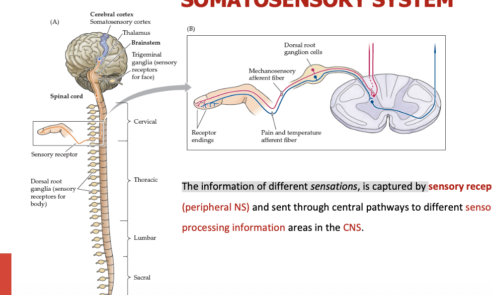
The information of different sensations, is captured by
Sensory receptors (PNS) → sent to diffeerent processing information areas in CNS
Sensory processing begins when
information reaches CNS & sensory information is encoded as electrical signals
The CNS must be able to identify four characteristics of sensory information:
Type/nature of the stimulus
Location
Intensity
Duration
Type/nature of the stimulus:
There is a receptor-stimulus association (e.g., photoreceptor, mechanoreceptor, etc) determines the interpretation of sensory information, regardless of the type of stimulus that caused the stimulation of the receptor.
Example: “seing stars” when rubbing eyes.
Location
encoded by the receptive fields
Intensity
is determined by the frequency of action potentials
Duration
is encoded by how long the receptor’s is activated, tonic receptors keep firing as long as the stimulus persists.
SENSORY RECEPTORS (types)
Mechanoreceptors
Thermoreceptors
Nociceptors
Photoreceptors
Chemoreceptors
Osmoreceptors
Osmoreceptors
changes in the blood’s osmolarity
Chemoreceptors
changes in the chemical molecules (mouth, nose, & internal fluids).
Photoreceptors
light (in the retina)
Nociceptors
painful stimuli (tissue damage)
Thermoreceptors
changes in temperature
Mechanoreceptors
mechanical stimuli: touch, pressure, hearing, balance.
Location of the stimulus
Exteroceptive
Proprioceptive
Interoceptive
Interoceptive receptors
They are in visceral organs, blood vessels
Provide information regarding the internal body environment (O2, osmotic pressure...)
Proprioceptive receptors
They generate conscious information about body position & movement (kinesthesia)
They are in the muscles, tendons & joints
They respond to stretch, tension & movement in these structures.
Exteroceptive receptors
Located in the body surface, sensitive to stimuli from the external environment
Mechanoreceptors, nociceptors, thermoreceptors, photoreceptors,
Transduction
Receptors encode the stimulus into an electrical signal.
The phases of transfduction
Stimulus activates the receptor → Change in membrane permeability to ions (graded pot.)→ threshold reached → AP triggered in sensory neuron.
Stimulus quality.
Encoded by the type & location of the receptor.
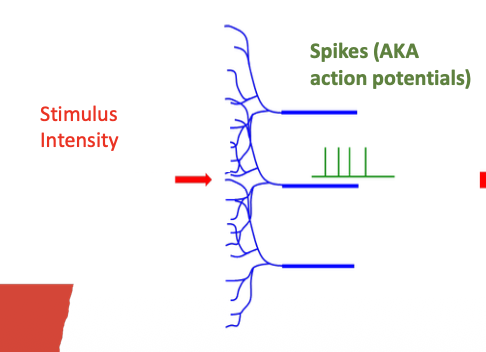
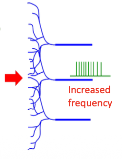
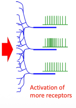
Stimulus intensity
Encoded by the frequency (rate) of the action potentials.( I I I or IIIIIIIII) & by the number of receptors activated (a stronger stimulus may activate a larger receptor area).
Stimulus intensity
Increased frequency
Activation of more receptors
Tonic (slowly adapting) receptors
They respond to the onset of the stimulus & keep sending signals as long as the stimulus exists.
Static property
Static property
They provide spatial & temporal information about the stimulus (size, shape, duration).
Phasic (rapidly adapting) receptors
Rapid discharge at stimulus onset & then silence even if the stimulus persists “adaptation”.
Dynamic property
Dynamic property
They inform about changes in stimulation, not steady states.
Topic receptor + phasix receptors
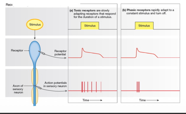
Tonic or slowly-adapting receptors:
Continuously transmit signals as long as the stimulus is present (where, how long).
Steady and sustained responses throughout the duration of the stimulus.
Allow continuous monitoring & adjustment of bodily functions.
Examples of Tonic or slowly-adapting receptors
Baroreceptors, in the walls of certain blood vessels (e.g., carotid), they sense blood pressure.
Nociceptors, pain.
Proprioceptors, body position.
Phasic or rapidly adapting receptors
They respond to changes in the stimulus intensity or quality.
Adapt to a constant stimulus, by decreasing their response over the time.
This allows these receptors to sense new or changes in the stimuli.
They filter out continuous stimulus & focus in detecting relevant changes in the environment.
Examples of Phasic or rapidly adapting receptors:
Olfactory receptors.
Pacinian receptors, involved in touch & vibration.
In order for the cerebral cortex to exercise its sensory functions
impulses must first be conducted to the sensory & association areas.
SOMATOSENSORY PATHWAYS
PNS (1st order neuron) → Medulla or spinal cord (2nd order neuron) → Thalamus (3rd neuron) → Sensory cortex
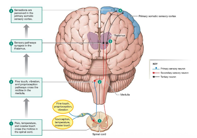
1st order neuron
Dendrites: Sensory receptors
Soma: Dorsal root ganglion
Synapse with the 2nd order neuron in the medulla oblongata
2nd order neuron
Soma in the medulla oblongata (gracile nucleus for lower body, cuneate nucleus for upper body)
Decussation (= opposite side crossing)
Synapse with 3rd orden neuron: thalamus
3rd order neuron
Soma: into the thalamus
Goes up to the 1st somatosensory cortical layer (also associative cortex).
POSTERIOR /DORSAL COLUMN TRACT
Fine touch, vibration & proprioception
SPINOTHALAMIC TRACT
Pain, temperature (lateral), coarse touch, & itching and pain (anterior)
TRIGEMINOTHALAMIC TRACT
Mechanosensitive information of the face