6. Diseases of the lens. Neoplasia of the eye.

0.0(0)
learnLearn
examPractice Test
spaced repetitionSpaced Repetition
heart puzzleMatch
flashcardsFlashcards
Card Sorting

1/45

encourage image

There's no tags or description

Looks like no tags are added yet.

Study Analytics
Name
Mastery
Learn
Test
Matching
Spaced

No study sessions yet.

46 Terms

1
New cards
What is the function of the crystalline lens?
Refracts incoming light rays to a point source on the retina
2
New cards

Describe the anatomy of the lens.

The crystallin lens is an avascular, transparent, & highly structured tissue.

It contains only a single layer of cuboidal cells, the anterior epithelium, inside the anterior lens capsule. Cortex, nucleus and posterior lens capsule.

The lens is attached to the ciliary body by fibres

3
New cards
What is the composition of the lens?
66% water and 33% proteins
4
New cards

Where in the eye has the most intensive refraction?

  1. Cornea

  2. Lens

5
New cards

What are examples of diseases of the lens?

  1. Congenital abnormalities

  2. Cataracts

  3. Lens luxation and subluxation

6
New cards
What are the congenital abnormalities of the lens?
  1. Aphakia

  2. Microphakia

  3. Lenticonus/lentiglobus

  4. Coloboma

  5. Lens luxation

  6. Vascular abnormalities (persistent pupillary membrane, PHTVL/PHPV)

7
New cards
What is aphakia?
Complete absence of the lens
8
New cards
What is microphakia?
Abnormally small lens
9
New cards
What is lenticonus/lentiglobus?
Deformity of the lens
10
New cards

What is coloboma of the lens?

Part of the lens is missing

11
New cards

What is lens luxation often in conjunction with?

Microphthalmia

12
New cards
What is PHTVL/PHPV?
Persistent hyperplastic tunica vasculosa lentis/Persistent hyperplastic primary vitreous, a vascular abnormality of the lens
13
New cards
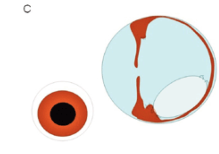
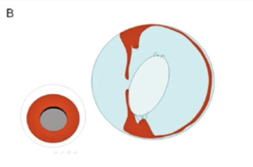
What are the grades of PHTVL/PHPV?
  • Grade 1: retro lens fibrovascular pigment/dots

  • Grade 2: dot + proliferation of retro lens tissue on the posterior lens capsule

  • Grade 3: plaque + persistent part of the hyaloid vessel

  • Grade 4: plaque + lenticonus

  • Grade 5: combination of Grades 3 and 4

  • Grade 6: combination of any grade with coloboma lentis, microphakia, retro lens pigment + bleeding
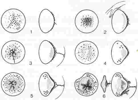
<ul><li><p>Grade 1: retro lens fibrovascular pigment/dots</p></li><li><p>Grade 2: dot + proliferation of retro lens tissue on the posterior lens capsule</p></li><li><p>Grade 3: plaque + persistent part of the hyaloid vessel</p></li><li><p>Grade 4: plaque + lenticonus</p></li><li><p>Grade 5: combination of Grades 3 and 4</p></li><li><p>Grade 6: combination of any grade with coloboma lentis, microphakia, retro lens pigment + bleeding</p></li></ul><p></p>
14
New cards
What are cataracts?
Increased opacity of the lens due to clumping and accumulation of proteins
15
New cards

How can cataracts be classified?

  1. Age

  2. Aetiology

  3. Consistency

  4. Localisation

  5. Degree of maturation

16
New cards
How are cataracts classified in relation to age?
  1. Congenital

  2. Developmental

  3. Juvenile

  4. Senile

  5. Acquired

17
New cards
How are cataracts classified by aetiology and pathogenesis?
  1. Primary (hereditary)

  2. Secondary (traumatic, due to intraocular diseases like uveitis, nutritional, radiation, diabetes, toxic, congenital abnormalities, senile)

18
New cards
How are cataracts classified by consistency?
  1. Soft

  2. Hard

  3. Watery

19
New cards
How are cataracts classified by localisation in the lens?
  1. Anterior capsular

  2. Anterior subcapsular

  3. Cortical (anterior or posterior)

  4. Equatorial

  5. Nuclear or perinuclear

  6. Posterior capsular

  7. osterior subcapsular

20
New cards
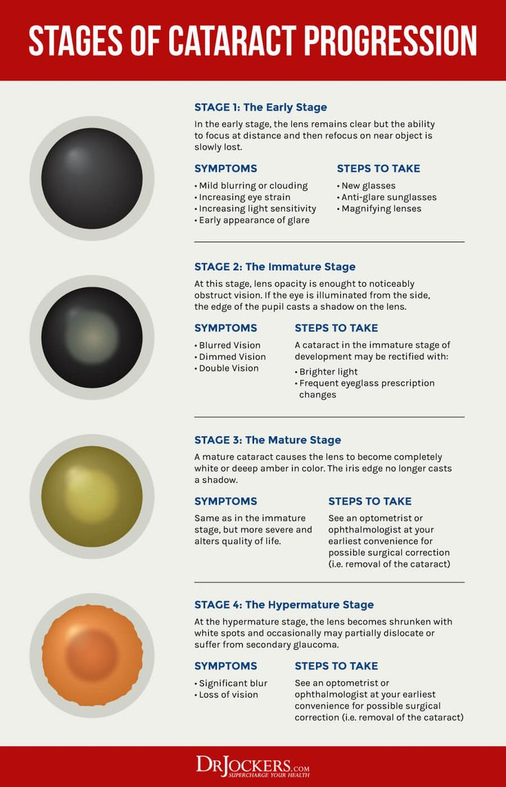
How are cataracts classified by degree of maturation?
  1. Incipient

  2. Immature

  3. Mature

  4. Hypermature

<ol><li><p>Incipient</p></li><li><p>Immature</p></li><li><p>Mature</p></li><li><p>Hypermature</p></li></ol><p></p>
21
New cards
What is an incipient cataract?
Focal opacity in the lens
22
New cards
What is an immature cataract?
Increased lens size and impaired vision
23
New cards
What is a mature cataract?
Totally opaque lens, absent fundic reflex, blindness
24
New cards
What is a hypermature cataract?

Wrinkling of the anterior lens capsule, crystal formation, liquefaction by proteolysis, and potential ventral settling of the lens nucleus

25
New cards
Which breeds are predisposed to congenital/hereditary cataracts?
Miniature Schnauzer, Boston Terrier, Old English Sheepdog, Welsh Springer Spaniel, West Highland White Terrier
26
New cards
What general diseases can contribute to cataract formation?
Diabetes, hypocalcaemia, and hypercupraemia
27
New cards
What nutritional deficiencies can contribute to cataract formation?
Arginine deficiency and use of inappropriate milk replacers
28
New cards
What external factors can contribute to cataract formation?
Various toxins and ionising radiation
29
New cards
What is the major clinical sign of cataracts?
Cloudiness of the lens. Other signs may include increased lens size and impaired/lost vision
30
New cards
What are the treatment options for cataracts?
  1. Spontaneous recovery (rare)

  2. Conservative management (topical mydriatics, aldose reductase inhibitors)

  3. Surgical removal of the lens

31
New cards
What are the surgical techniques for cataract removal?
  1. Extracapsular cataract extraction (leaving the capsule)

  2. Intracapsular extraction (removing the capsule)

  3. Phacoemulsification (using ultrasound to break up and aspirate the lens – the gold standard) with intraocular lens implantation
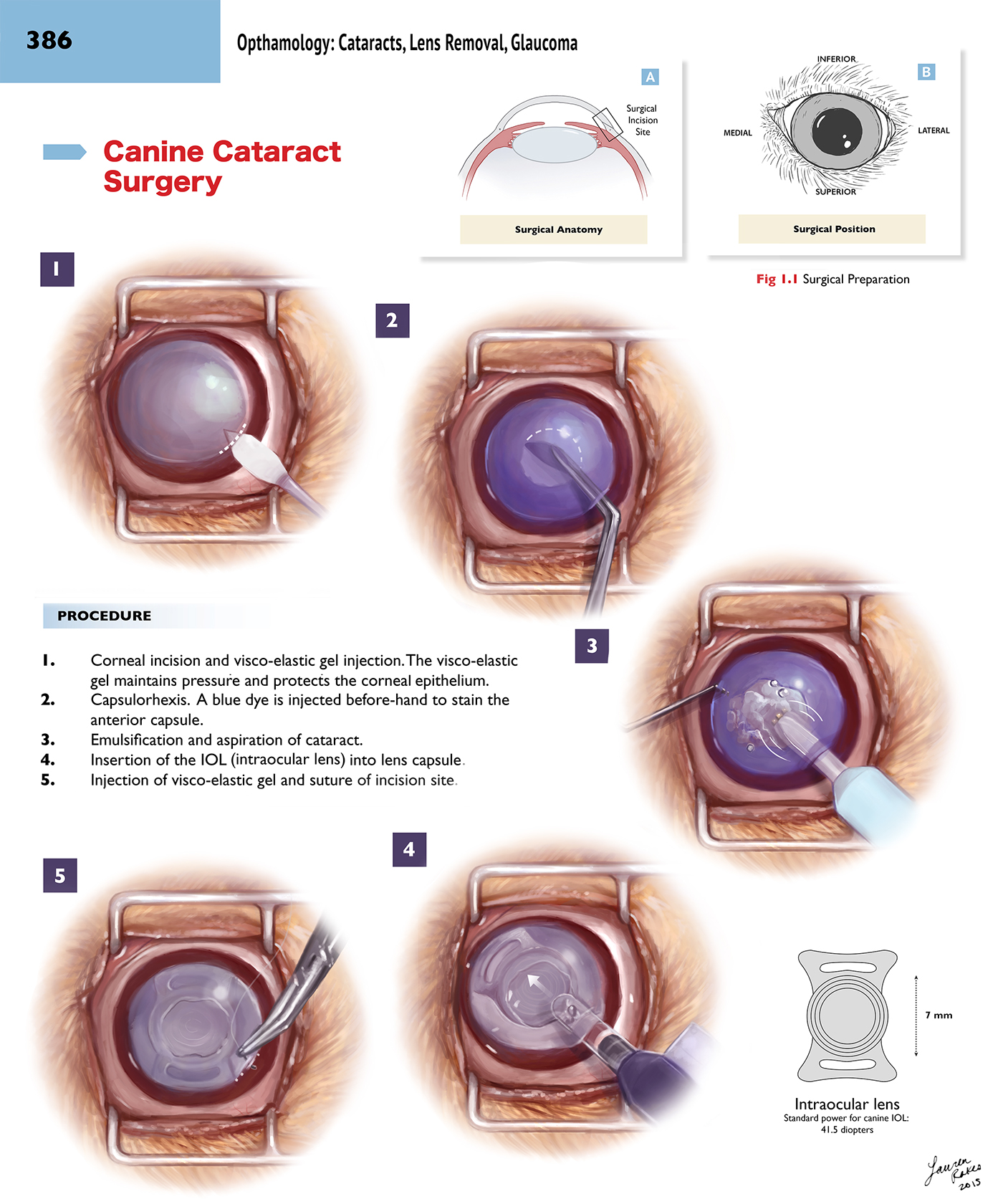
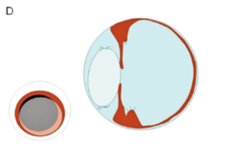
<ol><li><p>Extracapsular cataract extraction (leaving the capsule)</p></li><li><p>Intracapsular extraction (removing the capsule)</p></li><li><p>Phacoemulsification (using ultrasound to break up and aspirate the lens – the gold standard) with intraocular lens implantation</p></li></ol><p></p>
32
New cards
What is lens luxation/subluxation?
Dislocation (complete) or subluxation (partial) of the lens from its normal position due to pathological changes in the ciliary zonules
33
New cards
Which breeds are predisposed to lens subluxation?
Terrier breeds
34
New cards
What are the clinical signs of anterior lens luxation?
Acute signs including glaucoma and corneal oedema
35
New cards
What are the clinical signs of posterior lens luxation?
May be asymptomatic or cause ocular inflammation and glaucoma
36
New cards
What are the clinical signs of lens subluxation?
Instability of the iris and lens (half-moon-shaped edge)
37
New cards
How is lens luxation/subluxation treated?
Surgical removal of the lens by phacoemulsification or intracapsular lens extraction, depending on pain and vision status
38
New cards

What are examples of neoplasia of the eye?

  1. Iris melanoma

  2. Ciliary body adenoma/adenocarcinoma

  3. Uveal canine lymphoma

39
New cards
What are some treatment options for ocular neoplasia?
Surgical excision, radiation, cryotherapy, and enucleation
40
New cards
What is the most common eye tumour in dogs?
Iris melanoma
41
New cards
What are the clinical signs of iris melanoma?
Changes in iris pigmentation, potentially causing dyscoria and glaucoma. It can metastasise.
42
New cards
How is iris melanoma treated?
Laser coagulation
43
New cards
What are the clinical signs of ciliary body adenoma/adenocarcinoma?

Pink-red mass in pupil, swollen conjunctiva, prominent third eyelid, mydriasis, detached retina, and potential glaucoma
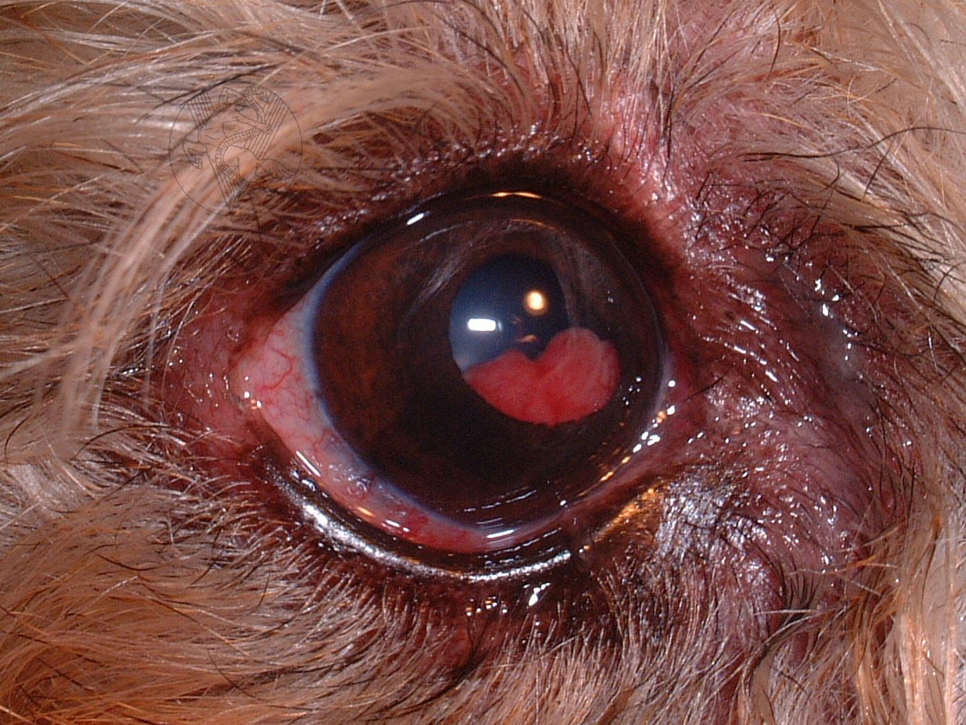
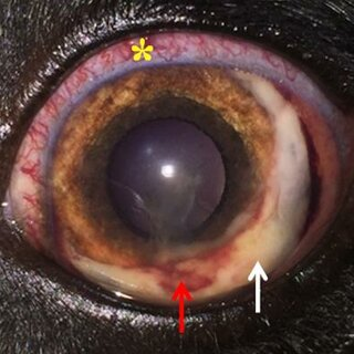
<p>Pink-red mass in pupil, swollen conjunctiva, prominent third eyelid, mydriasis, detached retina, and potential glaucoma</p>
44
New cards
How is ciliary body adenoma/adenocarcinoma treated?
Tumour removal
45
New cards
What is uveal canine lymphoma?
A secondary tumour (originating elsewhere) presenting as pink-white masses on the uvea
46
New cards
What are some other ocular tumours?
Dermoid, malignant melanoma on the cornea, uveal melanoma, and uveal lymphosarcoma