Biopsychology - Paper 2
1/112
Earn XP
Description and Tags
Name | Mastery | Learn | Test | Matching | Spaced | Call with Kai |
|---|
No analytics yet
Send a link to your students to track their progress
113 Terms
Motor neuron
Carry messages from CNS to muscles and glands (Short dendrites long axons)
Sensory neuron
Carry messages from PNS to CNS (Long dendrites short axons)
Relay neuron
Carry messages from sensory to motor neurons, or other relay neurons (Short dendrites short axons)
Dendrites
receive signals
Axon
carries impulses away from the cell body
Nodes of ranvier
Speed up transmission of impulse
Myelin sheath
protects axon
Terminal buttons
communicate with next neuron
Reflex arc
1. Stimulus presented
2. Sensory neurons send a message through peripheral nervous system
3. Message reaches the spinal cord, where it’s passed to a relay neuron
4. The message is either passed to a motor neuron or sent to the brain for further processing
5. Motor neuron carries the message to an effector
Synaptic transmission
1. The nerve impulse travels down the axon of the presynaptic neuron
2. When it reaches the end of the axon, chemical messengers called neurotransmitters are released from vesicles within the presynaptic neuron
3. These diffuse across the synapse
4. The neurotransmitters then bind to the receptors on the postsynaptic neuron
5. This stimulates the postsynaptic neuron to transmit a nerve impulse down its axon, to the next neuron
6. The neurotransmitters are deactivated by being reabsorbed back into the presynaptic neuron or by being broken down by enzymes in the synapse
Neurotransmitteres
Each neurotransmitter has its own specific molecular structure that fits perfectly into the postsynaptic site, like a lock and key. Neurotransmitters also have specialist functions.
Excitation
Some neurotransmitters have an excitatory effect on the neighbouring neuron. If a synapse is more likely to cause the postsynaptic neuron to fire, it is an excitatory synapse. This is like the accelerator
Inhibition
Some neurotransmitters have an inhibitory effect on the neighbouring neuron. If the message is likely to be stopped at the postsynaptic neuron it is an inhibitory synapse.
Summation
A neuron can receive both types of neurotransmitter at the same time. Likelihood of the cell firing is determined by adding up excitatory and inhibitory synaptic input which is summation. If the net effect on the postsynaptic neuron is inhibitory it will be less likely to fire. If the net effect is excitatory the neuron will be more likely to fire.
The nervous system
A complex system of nerve cells that carry messages to and from the brain and spinal cord to different parts of the body to help parts of the body communicate with each other. It is divided into the central nervous system and peripheral nervous system
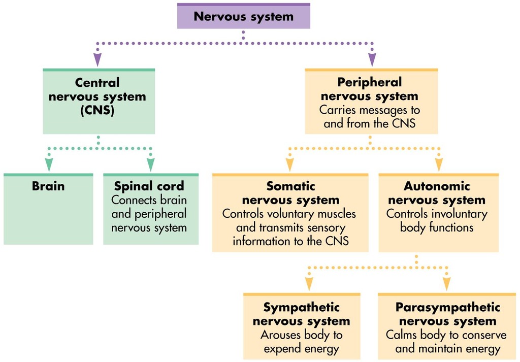
Central nervous system
Made of brain and spinal cord.
Brain - involved in psychological processes and is the centre of all conscious awareness.
Spinal cord - an extension of the brain responsible for the reflex action and relaying information between the brain and rest of the body
Peripheral nervous system
Transmits messages via neurons to and from the CNS. Function is to relay nerve impulses from the CNS to rest of the body back to CNS. It is subdivided into the Somatic and autonomic nervous system.
Somatic nervous system
Controls voluntary actions achieved by receiving information from the senses and carrying sensory and motor information to and from the CNS
Autonomic nervous system
Governs vital functions to the body such as breathing, heart rate etc. It controls involuntary actions and only carries motor information to and from the CNS. Subdivided into the parasympathetic and sympathetic.
Sympathetic nervous system
Involved in responses that help us deal with emergencies (fight or flight). Neurons travel to organs and glands to prepare body for rapid action. Causes the body to release stored energy and inhibits less important bodily processes e.g., digestion.
Parasympathetic nervous system
Involved in returning the body to a rest state once the emergency has passed. Bodily processes that are inhibited by the sympathetic branch are returned to normal
Endocrine system
Works with nervous system to regulate physiological processes. Endocrine communicates chemical messages which are hormones to organs. Regulate body’s growth, metabolism and function. Hormones released by glands
Glands
Organs secrete hormones to regulate bodily functions. Major gland is pituitary gland located in the brain. It controls the release of hormones from all other endocrine glands
Hormones
Chemicals that circulate in the bloodstream and influence target organs to regulate bodily activity. They are produced in large amounts but disappear quickly and have powerful effected. Incorrect timing of hormone release can result in dysfunction of bodily systems
Adrenal gland and Adrenaline
Triggers fight or flight by increasing heart rate, blood pressure etc
Testes and Testosterone
Causes the development of tests in the womb. A surge during puberty causes secondary sexual characteristics such as facial hair and deepening of the voice
Ovaries and Oestrogen and Progesterone
Help to regulate the menstrual cycle. Oestrogen is involved in repairing and thickening the uterus lining, progesterone maintains the lining
Pineal gland and Melatonin
Regulates the sleep-wake cycle, high levels cause drowsiness when daylight is low
Fight or flight response
When experiencing an acute stressor, the hypothalamus directs the sympathetic branch of the ANS to send neurotransmitters to the adrenal medulla. This results in the release of Adrenaline and Noradrenaline causing a fight or flight response by triggering physiological reactions.
These reactions can include activation of emergency functions like increased heart rate and blood pressure so O2 is pumped to the muscles for physical activity. Non emergency bodily processes are suppressed
Body in sympathetic state
- Increased heart rate
- Increased breathing rate
- Dilates pupils
- Inhibits digestion
- Inhibits saliva production
- Contracts rectum
Body in parasympathetic state
- Decreased heart rate
- Decreased breathing rate
- Constricts pupils
- Stimulates digestion
- Stimulates saliva production
- Relaxes rectum
Adrenaline and Noradrenaline
Increases: heart rate, blood pressure, glucose released, respiration, perspiration and blood coagulation. Decreases: digestion
Evaluation of fight or flight response
The fight or flight response may be different in females. Taylor et al found that females adopt a ‘tend and befriend’ response in stressful or dangerous situations. Women are more likely to protect their offspring and form alliances with other women rather than fighting or fleering. This suggests that research into ForF is gender bias as biological processes may only apply to males
Motor area
Frontal lobe and regulates movement
Somatosensory area
Parietal lobe and processes sensory info such as touch
Visual area
Occipital love and receives and processes visual info
Auditory area
Temporal lobe and analyses speech-based info
Language area
Broca’s and Wernicke’s
Localisation of function
Idea that specific functions have specific locations within the brain. If a certain area of the brain is damaged the associated function will also be affected. The brain is divided into two hemispheres. Outer layer is called the cerebral cortex
Auditory cortex
Two primary auditory cortices. Primary auditory cortex in both hemispheres receives information fro both ears. It is located in the temporal lobe. Damage to this area may result in partial hearing loss
Motor cortex
Movement is centred on the motor cortex which sends messages to the muscles via the brainstem and spinal cord. Responsible for generating voluntary motor movements. Each hemisphere controls movement in the opposite side of the body. Damage can result in loss of control over fine movements
Somatosensory cortex
Lies in motor cortex of the brain. Is where sensory information from the skin is represented. It perceives touch, so the number of neuronal connections needed dictates the amount of somatosensory cortex needed for that area of the body. located at the front of parietal lobe. Receives sensory information from the opposite side of the body
Visual cortex
Brain has two visual cortices. Is in occipital love which is at the back of the brain. Seen to be main visual centre. Each eye sendings info from the right visual field to the left visual cortex and vice versa.
Broca’s area
Responsible for speech production. Located in frontal lobe. Damage causes Broca’s aphasia which is characterised by speech that is slow, laborious, and lacking fluency.
Wernicke’s area
Damage to this area can lead to the inability to comprehend language and anomia. Area is important for understanding language and accessing words.
Localisation of function AO3: Case study evidence
Broca studied Tan’s brain in an autopsy. He found a lesion on his brain and concluded that this damage caused Tan’s aphasia and that this area of the brain controlled speech. Increases the validity of theory of localisation as it shows damage can show specific brain regions result in specific deficits.
Lacks external validity because it is difficult to conclude that speech production is localised to Broca’s area based on one individual case. Modern MRI of his brain showed damage to multiple brain regions
Localisation of function AO3: Brain scan research
Peterson et al used brain scans to demonstrate how Wernicke’s area was more active during listening tasks and Broca’s area during reading tasks.
Tulving et al have shown that episodic and semantic memories were recalled from different sides of the prefrontal cortex whilst procedural memory is associated with the cerebellum. This is positive as there is a wide range of evidence to support the idea that different areas of the brain have different functions.
Localisation of function AO3: Animal research
Lashley removed between 10-50% of the cortex un rats that were learning a maze and found that no area was more important in terms of their ability to learn the maze. This suggests that higher cognitive processes such as learning are not localised but distributed more holistically. Reduces the validity of theory of localisation. Difficult to generalise from animals to human behaviour. Learning may be localised in humans rather than animals.
Localisation of function AO3: Plasticity
When the brain is damaged and a particular function is lost, the rest of the brain is able to reorganise itself to recover the function. Turk et al discovered a patient who suffered damage to the left hemisphere but developed the capacity to speak in the right hemisphere. Suggests that functions aren’t localised and the brain can adapt.
Motor and somatosensory functions are highly localised but language functions appear not to be localised. Due to the high connectivity of the brain, no one area is independent.
Hemispheric lateralisation
Refers to the idea that two halves of the brain are functionally different and that certain mental processes and behaviours are mainly controlled by one hemisphere rather than the other. The two hemispheres are bridged by the corpus callosum. This is a bundle of fibres and is a communication pathway so that the hemispheres can exchange information.
Left hemisphere
Language processing. When someone has a stroke on the left side of their brain their speech is affected because this is were the Broca’s and Wernicke’s area are located
Right hemisphere
Dominant for facial recognition. A woman who had right hemisphere damage highlighted that the right hemisphere seems more adept for spatial relationships. The woman would often get lost suggesting that the right hemisphere deals with spatial info
Hemispheric lateralisation AO3: Supporting evidence
Fink used PET scans to identify which brain regions were active during a visual processing task. When participants were asked to focus on the forest as a whole, regions of the right hemisphere were more active. When required to focus on the finder detail (a specific tree) specific areas of the left hemisphere were more dominant suggesting visual processing is a feature of the connect brain
Hemispheric lateralisation AO3: Plasticity
When the brain is damaged the rest of the brain is able to reorganise itself to recover the function. Turk et al discovered a patient who suffered brain damage to the left side but developed an ability to speak in the right. This suggests that functions are not lateralised and the brain can adapt in certain areas.
Hemispheric lateralisation AO3: Animal research
Lashley removed 10-50% of the cortex in rats that were learning a maze and found that no area was more important in terms of learning the maze. This suggests that higher cognitive processes such as learning are not lateralised but distributed in a more holistic wave. This reduces the validity of the theory however it is difficult to generalise from animals for example learning may be more localised in humans than in animals
Hemispheric lateralisation AO3: Age
Lateralisation may be complicated by age. Research has shown that lateralisation of function may change over time. Szflarski found that language became more lateralised to the left hemisphere as children developed into adolescents but after 25 lateralisation decreased with each decade. This is a problem as it suggests hemispherical lateralisation is a much more complex process than many realise
Split brain research
Sperry studied 11 split-brain patients who had their corpus callosum cut to cure severe epilepsy. This was done to prevent electrical signals crossing between the hemispheres. Sperry’s work helped understand the function of the hemispheres and how they communicate
Sperry’s procedure - verbal recognition
A visual image e.g., a picture of a pencil was presented to the left visual field (which would be processed by the right) via a tachistoscope. The participants were asked to describe what they had seen. This would then be repeated using the right visual field.
Sperry’s procedure - touch recognition
Patient’s hands were screened so they could not see the objects in front of them. Participants would be asked to pick up an object using their right hand (processed by left hemisphere) and then asked to describe what they felt. This was repeated using the left hand
Sperry’s findings - verbal recognition
When a picture of an object was shown to the patient’s right visual field, the patient could easily describe what was seen. if the same object was shown to the left visual field, the patient could not describe what was seen and reported that there was nothing there. This is because language is processed in the left hemisphere and when a picture is presented in the left visual field this is processed by the right hemisphere which has a lack of language centres to be able to describe it. This allows us to infer that in the normal brain messages from the right hemisphere would have been relayed across hemispheres to the language centres in the left hemisphere to describe it.
Sperry’s findings - touch recognition
Patients couldn’t verbally describe objects projected in their left visual field, but were able to select a matching object from a grab-bag of different objects using their left hand (linked to RH). The objects were placed behind a screen so as not to be seen. The left hand was also able to select an object that was most closely associated with an object presented to the left visual field. In each case, the patient was not able to verbally identify what they had seen but could still understand what the object was, using the right hemisphere and select the object
Split brain research AO3: Controlled
Methodology praised for using highly standardised procedures conducted in a controlled environment to control possible extraneous variables. Images were flashed up for 1/10 of a second to ensure there was not time to spread info across both sides of the visual field. This is positive as it ensured the research measured what it intended to do which provides high internal validity.
Split brain research AO3: Sample
Findings came from a very unusual limited sample who were not well matched to a control group. Only 11 split brain Ps took part in all variations all of whom had a history of epileptic seizures and had received drug therapy for different amounts of time which may have caused unique changes in their brains. Some Ps may also have experienced more disconnection of the two hemispheres than others as part of their surgery. The control group consisted of Ps with no history of epilepsy making them poorly matched. This brings the conclusions of the research and support for lateralisation.
fMRI
Detect brain changes in blood oxygenation and flow in the brain. When brain is more active, consumes more o2 and to meet this increased demand blood flow is directed to the active area. It produces 3D images showing which part of the brain is involved in a particular process. The data can be used to identify which areas of the brain were being used when doing each task. They show 1-4s after an event and thought to be accurate within 1-2mm.
EEG
Measure electrical activity within the brain via electrodes that are fixed to an individual’s scalp. The scan recording represents the brainwave patterns that are generated from the action of neurons providing an overall account of brain activity. The electrodes measure the activity of the cells immediately under the electrode so using more gives a better picture. The scan recording represents the brainwave patterns that are generated from the action of millions of neurons. Real-time recording of brain activity within a millisecond.
ERPs
Stimulus presented to a participant. Using a statistical averaging technique, background brain activity from the original EEG recording is filtered out leaving only the event-related potentials. ERP is one of the most widely used methods in cognitive neuroscience.
Post mortem examniations
Brain is analysed following death. They are likely to have a rare disorder and have experienced unusual deficits in mental processes. Areas of damage within the brain are examined after death as a means of establishing the likely cause of the affliction. This may also involve comparison with a normal brain to see the difference.
fMRI AO3 - Less invasive
Less invasive than other techniques such as PET scans which use radiation or require instruments to be inserted into the brain. fMRI is usually risk free
fMRI AO3 - Spatial resolution
Good spatial resolution of 1-2mm. This is the smallest measurement that a scanner can detect. Greater spatial resolution allows psychologists to investigate the brain regions with greater accuracy. This helps researchers pinpoint specific responses and the exact source of brain activity.
fMRI AO3 - Expensive
Expensive to buy and maintain and they require trained operators. This makes research expensive and difficult to organise
fMRI AO3 - Temporal resolution
The accuracy of the scanner in relation to time. fMRI scans have a temporal resolution of 1-10 milliseconds. Psychologists are then unable to predict with a high degree of accuracy the onset of brain activity.
EEGERPs AO3 - General data EEG
only provides a measure of general brain activity which means that the brain response to a single stimulus or event of interest is not usually visible in the EEG recording.
EEG/ERPs AO3 - Spatial resolution
EEG/ERPs have poor spatial resolution. They only detect the activity in superficial regions of the brain. This means that they are unable to provide information on waht is happening in the depper regions of the brain, making this technique limited in comparison to the fMRI, which has a spatial resolution of 1-2mm.
EEG/ERPs AO3 - Cheaper
Cheaper in comparison to fMRI and therefore more widely available to researchers. This could help psychologists to gather further data on the functioning human brain, leading to greater understanding of sleeping and Alzheimer’s
EEG/ERPs AO3 - Temporal resolution
Good temporal resolution as it takes readings every millisecond, meaning it can record brain activity in real time. This leads to an accurate measurement of electrical activity when undertaking a specific task.
EEG/ERPs AO3 - Less invasive
Virtually risk free
Post mortem examination AO3 - Detailed structure
Provide a detailed examination of the structure of the brain that is not possible with other scanning techniques. Post-mortem examinations can access areas such as the hypothalamus and hippocampus, which other techniques would struggle to reach. This means that post mortem examinations provide researchers with an insight into these deeper brain regions which often provide a useful basis for further research.
Post mortem examination AO3 - Ethical issues
Issues of informed consent and whether a patient provides consent before his/her death, Many post mortem examinations are carried out on patients with severe psychological deficits who would be unable to provide fully informed consent and yet post mortem examinations have been conducted. This raises severe ethical issues surrounding the nature of investigations
Post mortem examination AO3 - Causation
The deficit a patient displays during their lifetime may not be linked to deficits in the brain. The deficits reported could have been the result of another illness and the psycholofyists would be unable to conlcude that it was caused by brain damage. Other confounding factors are medication a person may have been taking, the length of time between death and the examination and the age of person at death
Plasticity
The brain’s capacity to change or adapt because of new learning or brain trauma. New neural connections can be made at any time
Functional recovery
The brain learns to compensate for lost functions. The brain can be taught to learn how to use the working faculties and function to compensate for the ones that are lost forever. The brain can rewire itself by forming new synaptic connections. This is supported by axonal sprouting, recruitment of homologous areas and neuronal unmasking
Synaptic pruning
Rarely used connections are deleted and frequently used connections strengthened
Axonal sprouting
Growth of new nerve endings which connect with other undamaged nerve cells to form new neuronal pathways. When an axon is damaged its connection with a neighbouring neuron is lost. In some cases, other axons that already connect with that neuron will sprout extra connections to the neuron replacing the ones that have been destroyed. The brain can rewire itself by forming new synaptic connections close to the area of damage. This occurs for the most part two weeks after the damage happens. It helps replace function but only if the damaged axon and the compensatory axons do a similar job
Recruitment of homologous areas
Areas on the opposite side of the brain perform specific tasks. An example would be if the Broca’s area was damaged on the left side of the brain, a similar area on the right side of the brain would carry out its function.
Neuronal unmasking
Some of the brain’s neurons are ‘dormant’. These neurons are alive but are not doing their specific functions. When a brain becomes damaged, these dormant areas are unmasked. This opens connections in regions of the brain that are not normally activated which fives way to the development of new structures.
Factors affecting brain recovery - Age
Deterioration of the brain in old age, and this therefore affects the extent and speed of recovery. Marquez de la Plata et al found that following brain trauma, patients 40+ regained less function in treatment than younger patients and they were more likely to decline in terms of function for the 5 years following trauma
Factors affecting brain recovery - Gender
Research to show women recover better from brain injury as their function is not as lateralised. Ratcliffe et al examined 325 patients with brain trauma for their level of response for cognitive skills to rehabilitation. The patients were 16-45 at injury received rehabilitation at a care facility and completed a follow-up one year later. None of them had learning problems prior to the trauma. When assessed for cognitive skills, women performed significantly better than men on the tests of attention/working memory and language whereas men outperformed females in visual analytic skills.
Factors affecting brain recovery - Physical exhaustion, stress and alcohol consumption
When function is recovered in an individual it is important to remember that often the function is used with considerable effect and although the person can do a task, they are often fatigued by the effort. Other factors such as stress and alcohol consumption can affect the ability to use any function that has been regained.
Plasticity and functional recovery AO3 - Evidence
Maguire et al used an MRI scanner to scan the brains of London taxi drivers and found they had significantly more volume of grey matter in the posterior hippocampus than a matched control group. The volume of this area was also positively correlated with the amount of time they had been a taxi driver. This is positive as it supports the idea that the human brain can adapt as a result of learning and experience
Plasticity and functional recovery AO3 - Animal studies
Early evidence of neuroplasticity comes from an animal study conducted by Hubel and Wiesel. In this study, kittens had one of their eyes sewn up and the brain’s cortical responses were analysed. It was found that the area of the visual cortex associated with the shut-eye was not idle but continued to process information from the open eye. This is positive as it supports the idea that the brain can change or adapt as a result of experience
Plasticity and functional recovery AO3 - Practical applications
Understanding the processes involved in plasticity has contributed to the treatment and rehabilitation of brain injury patients. Following illness or injury to the brain, recovery tends to slow down after a few weeks so forms of physical therapy are usually performed to maintain improvements in functioning. This shows that although the brain may have the capacity to fix itself to a point, this process requires further intervention if it is to be successful
Plasticity and functional recover AO3 - Individual differences
Certain people have more of an ability to recover from trauma than others. Elbert et al showed that adults require far more intensive training than children after brain trauma. Schneider et al found that patients with a college education were 7X more likely than those who didn’t finish high school to be disability-free one year after brain injury. This suggests a number of factors contribute to brain plasticity and recovery from brain trauma, which makes it a complex area to study.
Plasticity and functional recovery AO3 - Negative consequences
Ramachandram and Herstein found that 60-80% of amputees have been known to develop phantom limb syndrome. These sensations are usually unpleasant, and painful and are thought to be due to cortical reorganisation in the somatosensory cortex because of limb loss. This negative plasticity is a problem as it shows the brain’s ability to adapt to changes doesn’t always have positive effects.
Biological rhythms
Biological rhythms are the natural cycle of change in our body’s chemicals or functions. These rhythms are governed by the body’s internal clocks (endogenous pacemakers) and external changes to the environment (exogenous zeitgebers)
Circadian rhythms
- Occurs once every 24 hours e.g., sleep-wake cycle
Hormone production - hormone release follows a circadian rhythm. The production and release of melatonin from the pineal gland follow a circadian rhythm with peak levels occurring during the hours of darkness. By activating chemical receptors in the brain, melatonin encourages feelings of sleep. When it is dark more melatonin is produced and when it is light again the production drops and the person wakes
Sleep-wake cycle - the circadian rhythm not only dictates when we should be sleeping but also when we should be awake. Light and darkness are the external signs that determine when we feel the need to sleep and when to wake up. The circadian rhythm also dips and rises at different times of the day so our strongest sleep drive usually occurs between 2-4AM and 1-3PM.
Circadian rhythms AO3 - Practical applications
Research into circadian rhythms provide an understanding of the consequences that occur when they are disrupted. Bolvin found that night workers experience reduced concentration around 6AM, leading to an increase in mistakes and accidents.
Circadian rhythms AO3 - Samples
Research on circadian rhythms usually involve small samples. For example, Siffre was the only person in his study. This is an issue because results cannot be generalised.
Infradian rhythms
- Last more than 24 hours e.g., menstrual cycle
The Menstrual cycle - it is governed by monthly changes in hormones that regulate ovulation. The typical cycle lasts 28 days. During each cycle, rising levels of oestrogen cause ovulation. After ovulation progesterone helps the womb lining to grow thicker readying the womb for pregnancy. If pregnancy doesn’t occur the egg is absorbed into the body and the womb lining comes away and leaves the body.
McClintock - 29 women with irregular periods were studied. Samples of pheromones from armpits were gathered from 9 of the women at different stages of the cycle. The pads were then rubbed on the upper lip of the other Ps. Stern and McClintock found that 68% of women experienced changes in their cycle which brought them close to the cycle of their odour donor
Infradian rhythms AO3 - Evolution
Research into infradian rhythms is supported by the theory of evolution. It may have been advantageous for women to menstruate together and become pregnant at the same time. In a social group, this would allow babies who had lost their mothers during child birth to have access to breast milk thereby improving their chances of survival. This suggests that the synchronisation of the menstrual cycle is an adaptive strategy.
Infradian rhythms AO3 - Limitations
There are many factors that may cause the menstrual cycle to change including stress, extreme dieting and exercise.