Kidneys 2
1/79
Earn XP
Description and Tags
UT 302 - Abdomen 1
Name | Mastery | Learn | Test | Matching | Spaced |
|---|
No study sessions yet.
80 Terms
___ hypertension is one of the most common causes of secondary hypertension
Renovascular
What causes renovascular hypertension?
It is mostly due to the narrowing of blood vessels in the kidney
___ affects 75 million adults in the United States and accounts for 8.6% of all primary care visits
Hypertension
Renovascular hypertension is one of the most common causes of ___ hypertension and often leads to resistant hypertension
secondary
What is renovascular hypertension?
Systemic hypertension that manifests secondary to the compromised blood supply to the kidneys, usually due to an occlusive lesion in the main renal artery
Renal artery disease
Correctable
Treatment will control or cure renovascular hypertension
Retention of renal mass
Stabilization of renal function
Why is duplex ultrasound is used to detect renal artery stenosis?
Provides anatomical as well as hemodynamic functional information
Low cost without risk of ionizing radiation or use of nephrotoxic contrast agents
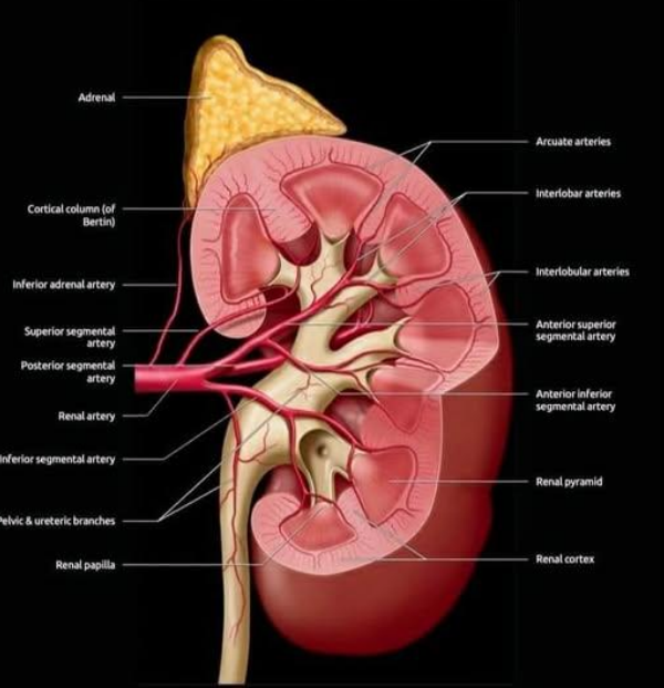
Kidney anatomy
Retroperitoneal organs
Located between 12th thoracic and 3rd lumbar vertebrae
Right kidney inferior to left
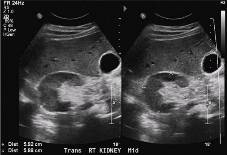
Kidney length is 9-13 cm and width is 5-7 cm
May decrease in size with age
T/F: Variation in size between a person's two kidneys is not common, and is therefore abnormal
False
The ___ kidney is typically slightly longer or larger than the ___ kidney
left; right
A size difference of up to ___ is considered within normal limits
1-1.5 cm
A variation of ___ or more or one kidney being significantly larger than the other may suggest an abnormality (which should be medically investigated)
2 cm
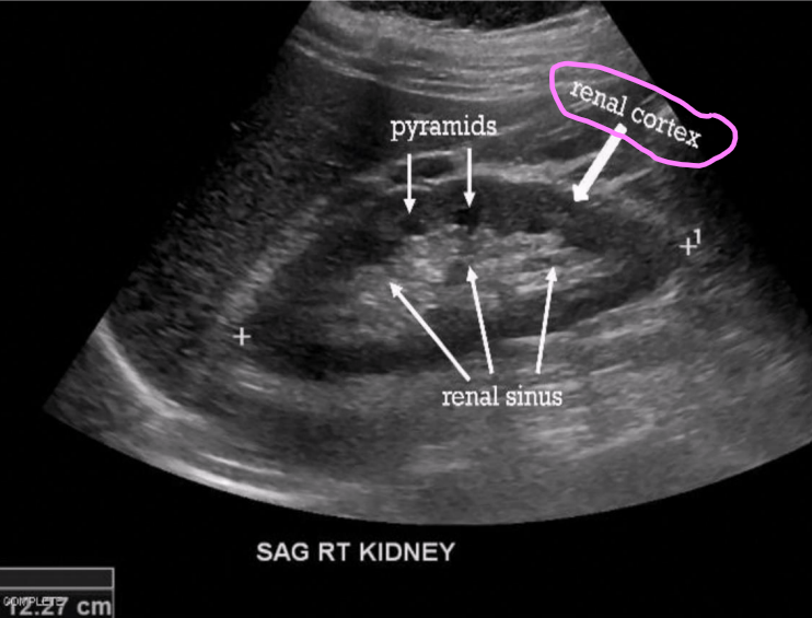
For sonographic examination, the kidney is divided into what four parts?
Renal hilum
Renal cortex
Renal medulla
Renal sinus
What is the renal hilum?
Area through which renal artery, vein, nerves, and ureter enter the kidney
What four structures pass through the renal hilum?
Renal artery
Renal vein
Nerves
Ureter
What is the renal sinus?
Cavity that contains the renal artery and veins and collecting and lymphatic system
Largely made up of fat and fibrous tissue
Appears brightly echogenic on sonographic imaging
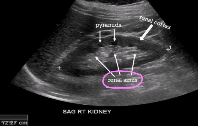
What two structures compose the renal parenchyma?
Medulla
Cortex
What is the renal medulla?
Contains 12–18 renal pyramids (triangular shaped)
Carry urine from cortex to renal pelvis
Lower echogenicity than cortex
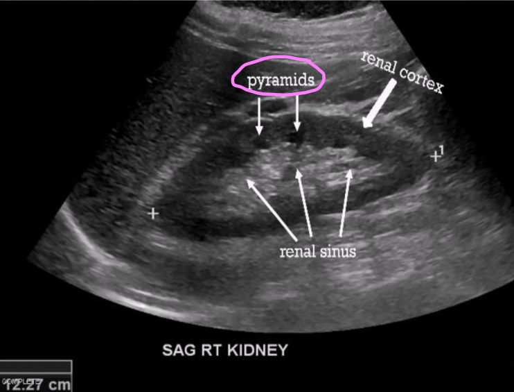
What is the renal cortex?
Outermost area of kidney
Lies just beneath renal capsule
Area in which urine is produced
Some cortical tissues extends between medullary pyramids (columns of Bertin)
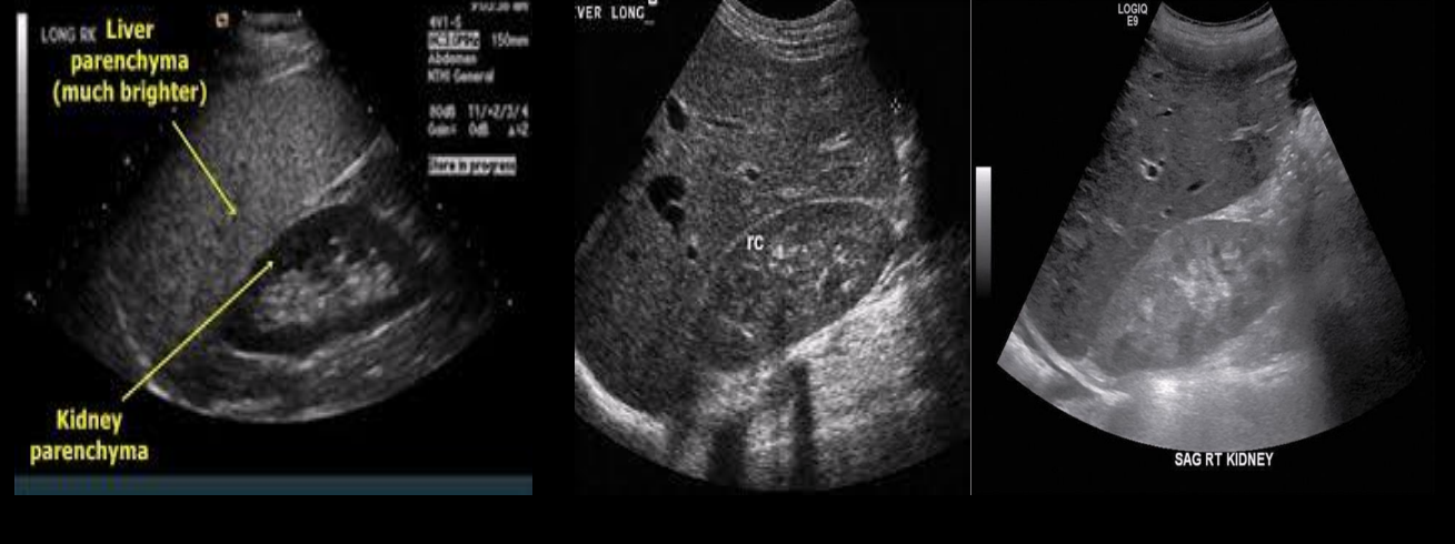
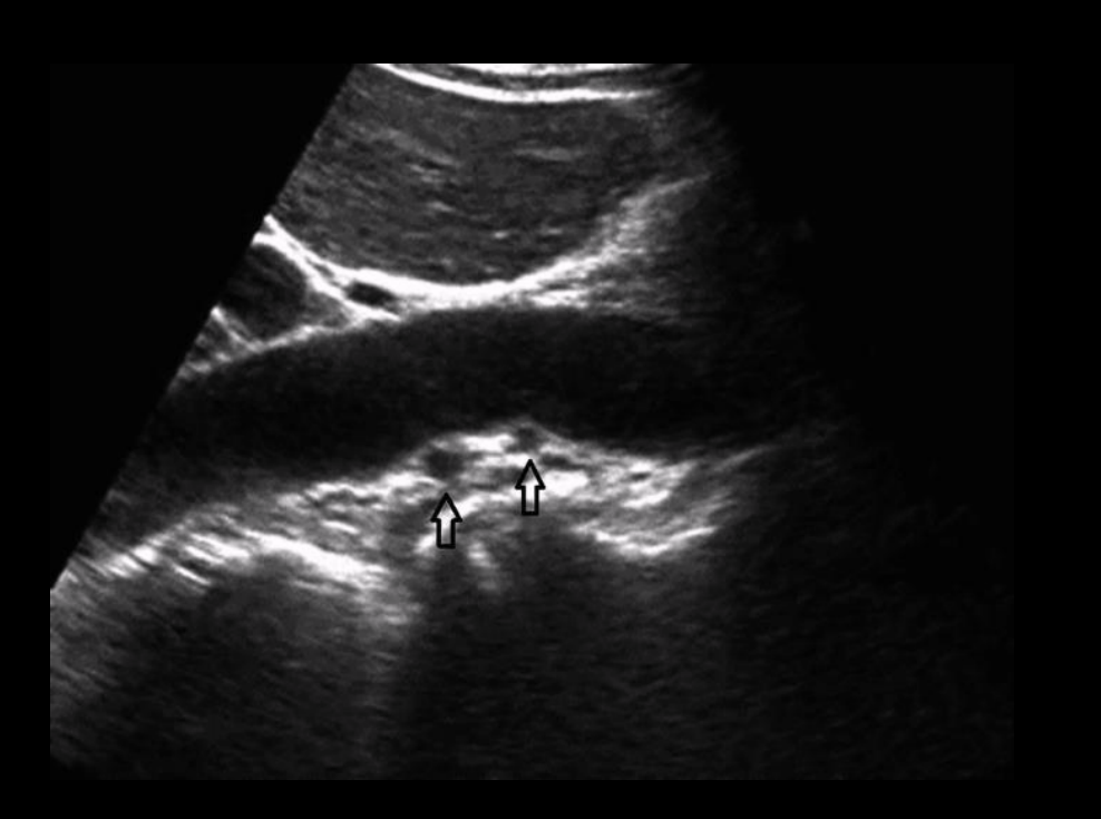
Which kidney has an abnormal echogenicity?
Right image (an adult kidney should not be hyperechoic to the liver)
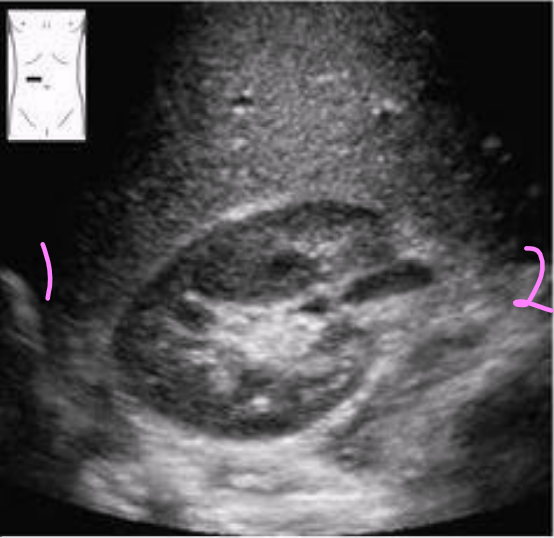
Label the image
Lateral
Medial
What is the transpyloric plane?
Important landmark for identification of kidneys
Located halfway between suprasternal notch and symphysis pubis
Cuts through lower border of first lumbar vertebrae, ninth costal cartilage, and pylorus
Renal arteries identified 2 cm below this plane

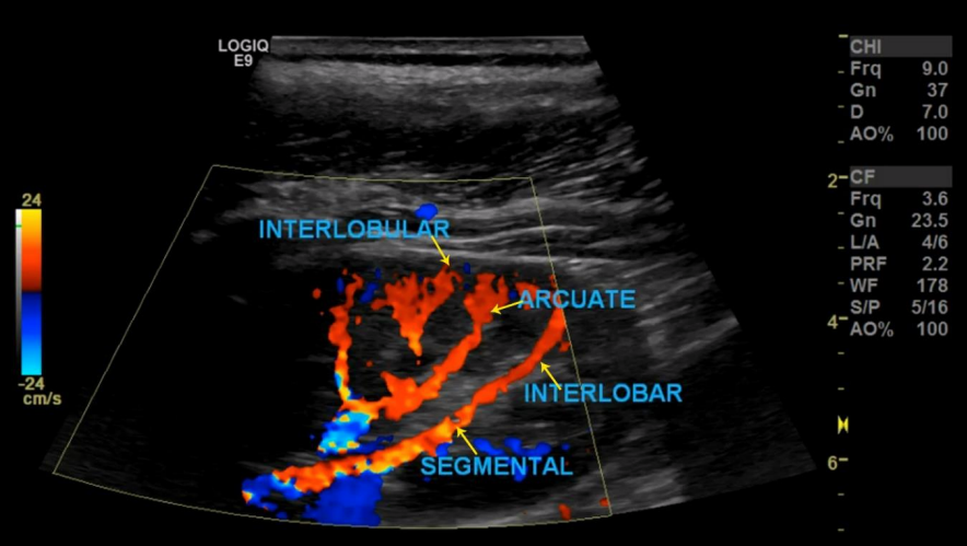
1
Renal artery

2
Interlobar arteries

3
Arcuate arteries

4
Interlobular arteries

5
Interlobular veins

6
Arcuate veins

7
Interlobar veins
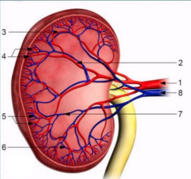
8
Renal vein
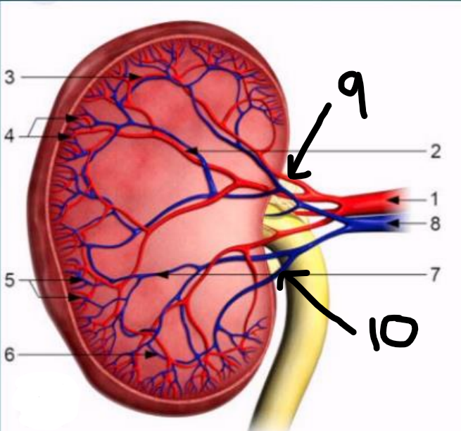
9
Segmental artery

10
Segmental vein
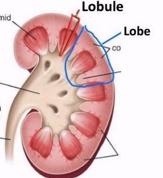
What is a lobe vs. lobule?
Lobe
Large, macroscopic anatomical division of an organ
Visible without a microscope
Lobule
Much smaller microscopic subdivision within a lobe
Only visible with a microscope
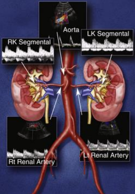
The renal arteries branch off …
anterior, lateral, or posterolateral from the abdominal aorta
Right renal artery
Longer than LRA
Travels posterior to the IVC and RRV
Left renal artery
Branches off aorta more superior (cephalad) than RRA
Shorter than RRA
What happens to the renal arteries?
Branch off aorta and travel to their respective kidneys
Enters renal hilum
Branches into segmental arteries (ant/post, sup/inf)
Branch into interlobar arteries
Branch into arcuate arteries
Branch into interlobular arteries

1
R renal artery
2
R renal vein
How is the renal artery arranged compared to other vessels when it enters the hilum?
Typically located posterior and inferior to the renal vein in the renal hilum
How is the renal vein arranged compared to other vessels when it enters the hilum
Typically located anterior and superior to the renal artery in the renal hilum
T/F: the renal vein exits the kidney in front of the renal artery, which enters behind the renal pelvis
True
The RRA is the only vessel that travels laterally ___ the IVC
under
Is it possible to have multiple renal arteries on each side?
There is usually one on either side, though multiples are quite common
Occur in 14-25% of the population
There can be duplicate, three or more
The main renal arteries usually enter the kidney at the ___, though accessory vessels may enter at the ___ or other surfaces of the renal parenchyma
hilum; poles
Renal arteries have ___ resistance waveforms
low
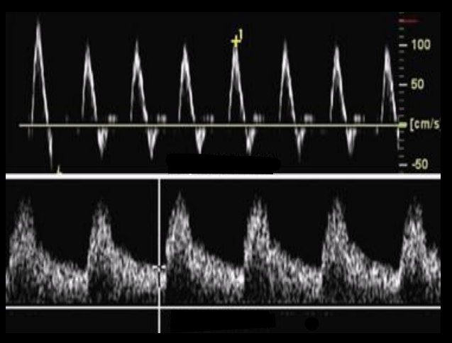
Which waveform is low-resistance and which is high-resistance?
Top: high-resistance
Bottom: low-resistance

1
Interlobular arteries
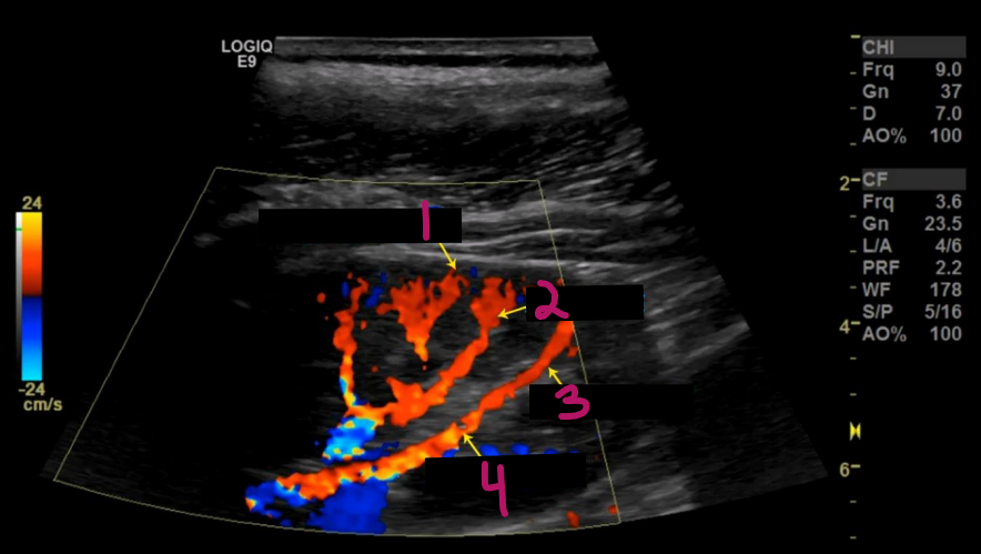
2
Arcuate arteries

3
Interlobar artery

4
Segmental artery
Intrarenal arterial anatomy consists of …
An arbor-like network of vessels coursing throughout the kidney
Once the renal artery enters the kidney it divides into
Segmental arteries (usually 5)
Interlobar arteries
Arcuate arteries
Interlobular arteries
Renal veins course __ in the renal hilum
anterior
The renal artery lies ___ the renal vein and the ureter
between
The right renal vein has a ___ course from the kidney to the IVC
short
The left renal vein courses ___ to the aorta just below the SMA and has a much ___ course than the RRV
anterior; longer
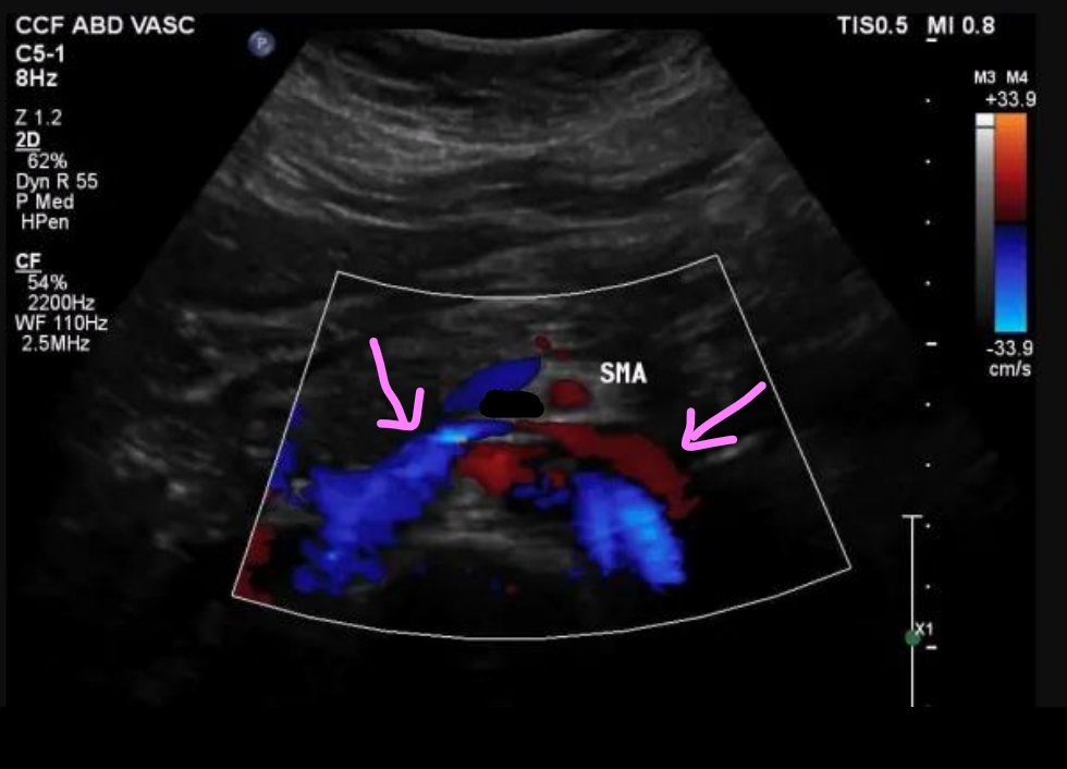
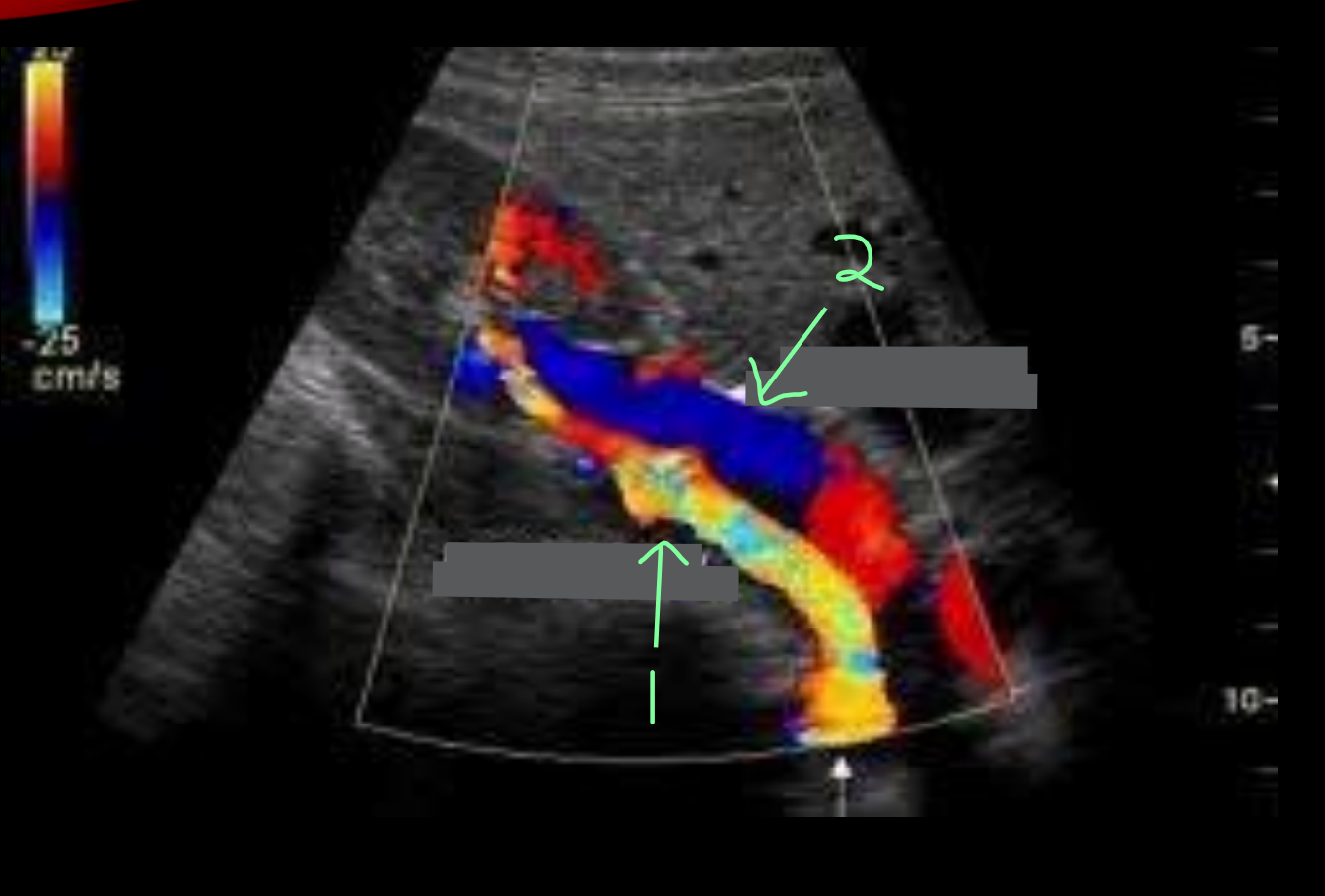
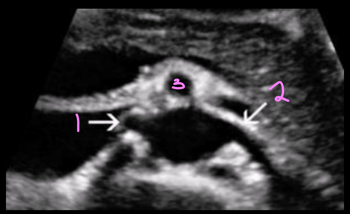
What vessel are the arrows pointing to?
LRV
The left renal vein is ___ and ___ to the renal artery
anterior; superior
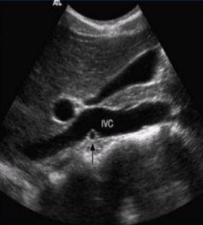
What is being shown in this image?
RRV draining into IVC
What are some renal anomalies?
Horseshoe kidney
Duplicate renal artery
RRA superior to IVC
Bifid LRV
Retroaortic LRV
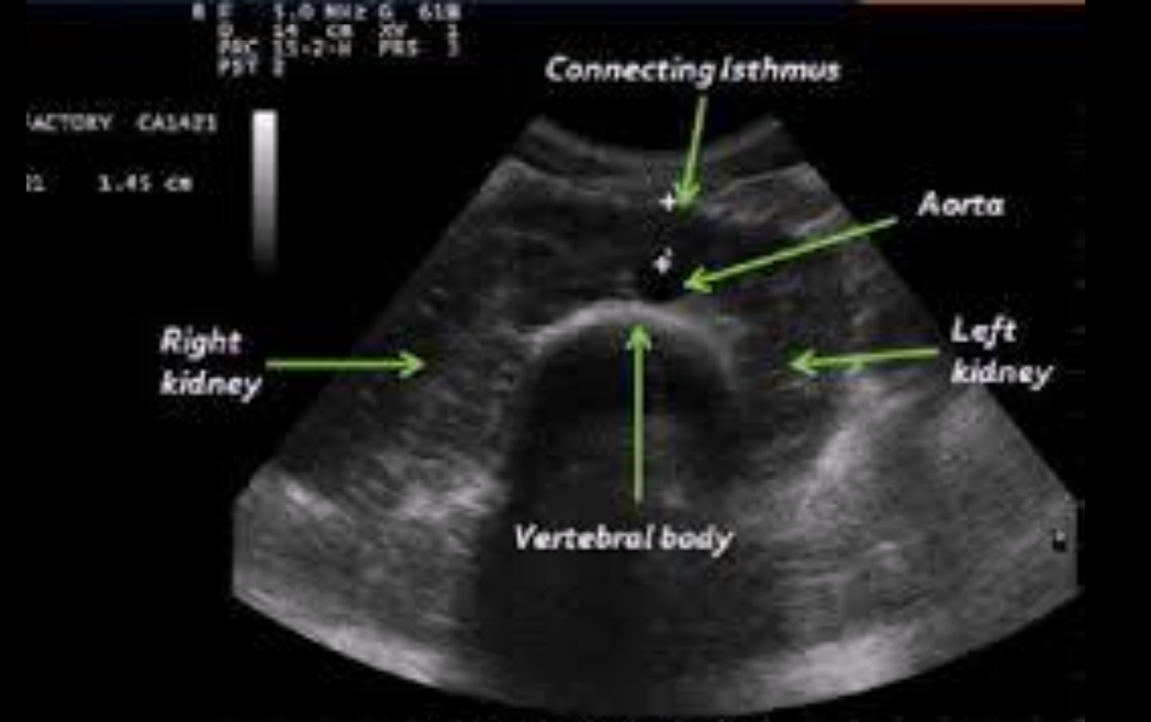
Horseshoe kidney
Kidneys are joined at the lower poles
Isthmus lies anterior to the aorta at the level of L4-L5
Duplicate renal artery
Often arises from aorta below main renal artery
Course to polar surfaces of kidney
What are the most common etiologies of renal arterial disease?
Atherosclerosis
Fibromuscular dysplasia
Atherosclerosis
Most common etiology of renal arterial disease
Lesions affect ostium and proximal 3rd of vessel
More common in men than women
Bilateral 30% of the time
Risk factors include age, hypertension, smoking, diabetes, hyperlipidemia, past history of CAD or PAD
Fibromuscular dysplasia
Second most common curable cause of renovascular disease
Nonatherosclerotic disease affects mid-to-distal segment of vessel
Occurs more commonly in women aged 25–50 years
Often bilateral
Produces “string of beads” appearance on ultrasound or angiogram
What are some other (less common) disease processes that can impact renal arteries?
Aortic dissection
Aneurysms of main or segmental renal arteries
Aortic coarctation proximal to renal arteries
Arteriovenous fistulae
Arteritis
Extrinsic compression by tumor or other mass
What do normal renal veins look like on US?
Anechoic lumens and respiratory phasicity on Doppler
What kind of flow occurs in the renal arteries in the presence of thrombosis?
Continuous, nonphasic low-velocity flow
What are some abnormalities of renal veins on US?
Acute thrombus
Partial venous obstruction
Recanalization
Collateralization
Extrinsic compression
What settings must be optimized in order to identify renal vein abnormalities?
B-mode
Doppler
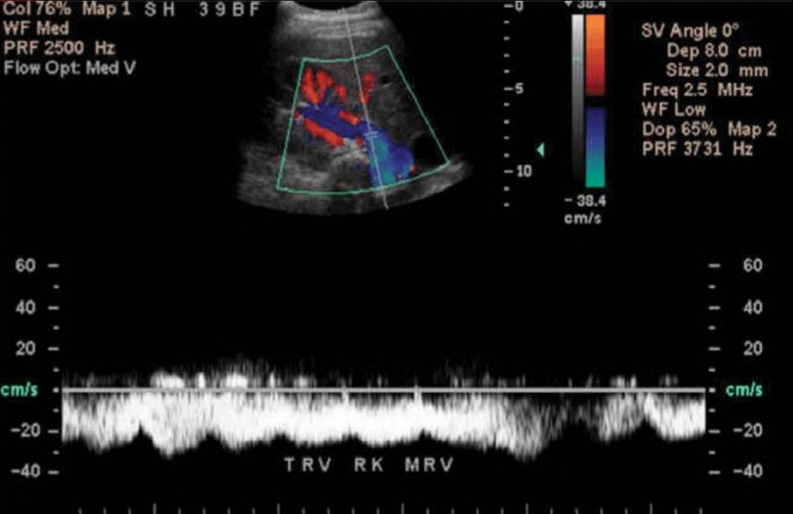
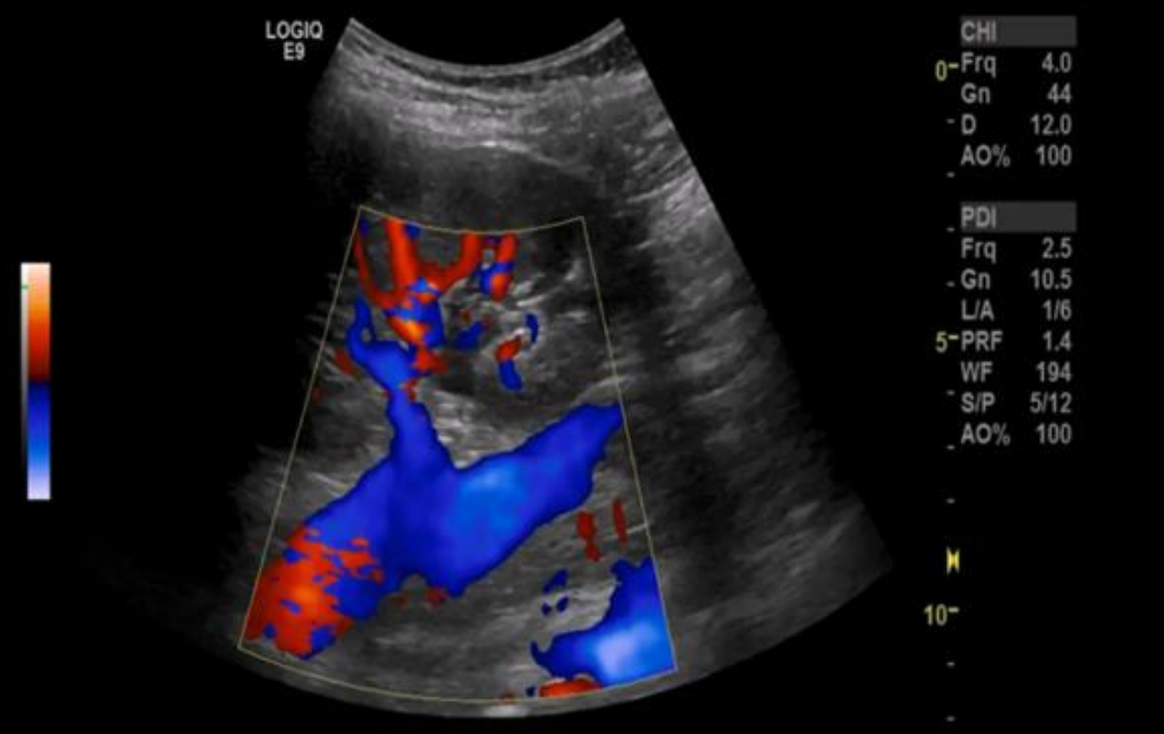
What does this image demonstrate?
Normal flow in mid renal vein of right kidney
How should patients be prepared for renal US?
Patients should fast 8–10 hours prior to exam
Reduces excessive abdominal gas
Exam is usually scheduled in the morning
Diabetic patients may be permitted to have dry toast and clear liquids if needed
Patients should refrain from smoking or chewing gum prior to exam to reduce amount of swallowed air
How should the patients be positioned for renal US?
Supine with head slightly elevated
Reverse Trendelenburg can be used to shift visceral contents into lower abdomen and pelvis
Right or left lateral decubitus positions used to evaluate kidneys and mid-to-distal renal artery segments
Prone position also works well to access kidneys
What should be examined during renal US?
Aorta
Celiac trunk
Proximal SMA
Renal ostia
Proximal-to-mid renal artery segments
How should the sonographer be positioned to perform a renal US?
Positioned to side of bed
Height of bed should be adjusted so that sonographer can scan without overextended arm
Patient should lie as close to sonographer’s side of bed as possible
Avoid overreaching
Sonographer should learn to scan with both hands
How should the kidneys be examined on US?
High-resolution ultrasound system
Phased or curved array 2–5 MHz transducer
Gray scale used to localize vessels and organs
Identify atherosclerotic plaque, aneurysmal dilation, and dissections
Color and/or power Doppler may help visualize arteries and veins (landmarks, regions of disordered flow, and vessel occlusion)
Spectral Doppler used for differentiation of normal and abnormal flow patterns
Optimization of color and spectral Doppler settings must be performed throughout exam
What are some helpful scanning techniques that are used to scan the renal arteries?
Access from midline, transverse plane
Lie inferior to left renal vein
Keep transducer perpendicular to abdominal wall
Angle slightly to right or left to create sagittal image of renal artery
Color flow used to help identify vessels
Vessels can usually be identified from ostium to mid segment
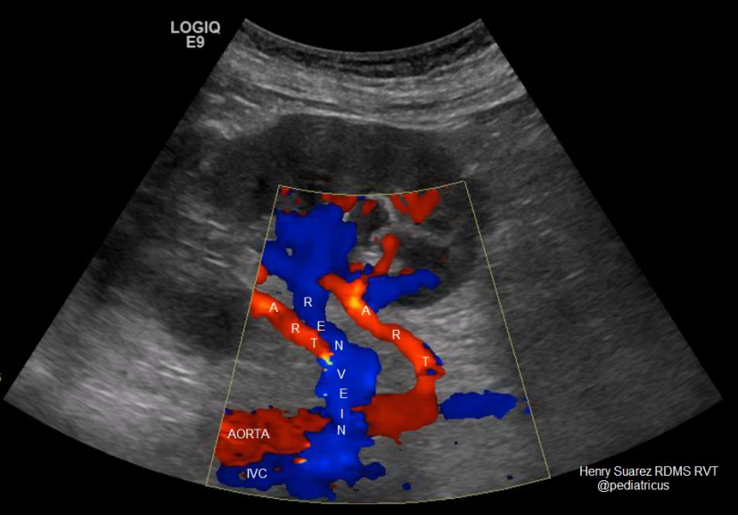
What are the landmarks in this image?
RRA
LRA
SMA
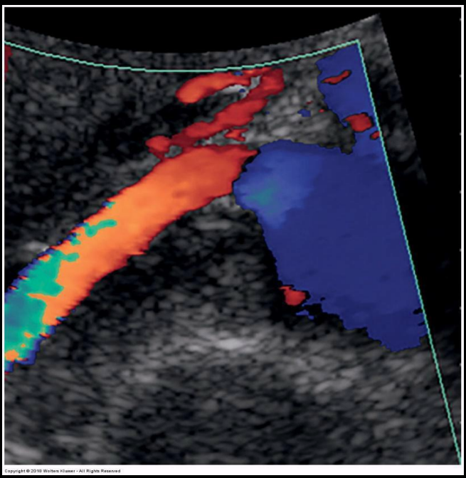
What does this image demonstrate?
Sagittal color-flow image of the RRA