Lecture 5/6 - Cells of the nervous system
1/215
There's no tags or description
Looks like no tags are added yet.
Name | Mastery | Learn | Test | Matching | Spaced |
|---|
No study sessions yet.
216 Terms
Like all organs in the body, the nervous system is composed of numerous WHAT
cells
We can infer their function by their WHAT, WHAT and their WHAT
We can infer their function by their LOCATION, SHAPE and their PARTS
How to visualize cells:
WHAT
Make WHAT
WHAT it to reveal features
How to visualize cells:
Fixed tissue
Make THIN SLICES
Stain it to reveal features
Fixation of tissue consists of two steps:
WHAT of normal life WHAT in the tissue (WHAT)
WHAT of the structure of the tissue (WHAT)
Fixation of tissue consists of two steps:
CESSATION of normal life FUNCTIONS in the tissue (KILLING)
STABILIZATION of the structure of the tissue (PRESERVATION)
Physiological cells:
WHAT
Physiological cells:
Unfixed tissue (live)
What basic elements are present in all cells
Golgi body
Lysosomes
Cell membrane
Mitochondria
Endoplasmic reticulum
Nucleus
Nuclear membrane
What basic elements are present in neurons
Axons
Dendrites
Dendritic spines
Cells are enclosed in a WHAT
Membrane
It forms a boundary between the WHAT and WHAT
It forms a boundary between the OTSIDE and INSIDE
Inside and outside of a cell is WHAT
Salt fluid
Fluid inside = WHAT
Cytoplasm
Fluid outside = WHAT
Extracellular fluid
Membrane is a WHAT barrier; only WHAT molecules and some WHAT can get through without aid
Membrane is a SMEI-PERMEABLE barrier; only FAT-SOLUBLE molecules and some GASES can get through without aid
The membrane contains WHAT that act as WHAT through which stuff (eg, ions) can (selectively) pass WHERE or WHERE of the cell
The membrane contains PROTEINS that act as CHANNELS through which stuff (eg, ions) can (selectively) pass INTO or OUT of the cell
Phospholipid:
Head = WHAT
Tail = WHAT
Phospholipid:
Head = HYDROPHILIC (loves water)
Tail = HYDROPHOBIC (fears water)
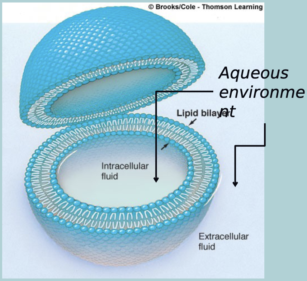
The phospholipid bilayer separates the two WHAT
Aqueous environments

Lipid character of bilayer limits WHAT across the cell membrane
Lipid character of bilayer limits DIFFUSION of WATER-SOLUBLE molecules across the cell membrane
The plasma-membranes hydrophobic (water fearing) interior prevents free (inward and outward) diffusion of WHAT (and other WHAT) substances, thus it constitutes as a WHAT membrane
The plasma-membranes hydrophobic (water fearing) interior prevents free (inward and outward) diffusion of CHARGED (and other HYDROPHILIC (water loving)) substances, thus it constitutes as a SEMI-PERMEABLE membrane
What are hydrophilic molecules
Small inorganic ions (Na+, Ca2+, Cl-)
Charged proteins
What are hydrophobic molecules
Lipid-soluble hormones (NO, O2, CO2)
Uncharged proteins
Golgi complex
Packaging and processing of proteins
Mitochondria
Converts sugar and oxygen into ENERGY, energy is in the form of adenosine triphosphate (ATP)
Endoplasmic reticulum and ribosomes
Site of protein synthesis
Nucleus:
WHAT structure inside the cell body
contain WHAT
Nucleus:
MEMBRANE-BOUND structure inside the cell body
contain DNA
DNA contains WHAT
Genetic information
DNA is the blueprint for all the WHAT a cell can make
protein
All neurons are interconnected to form WHAT
Coherent networks
How does information flow in a neuron
input zone
conduction zone
Output zone
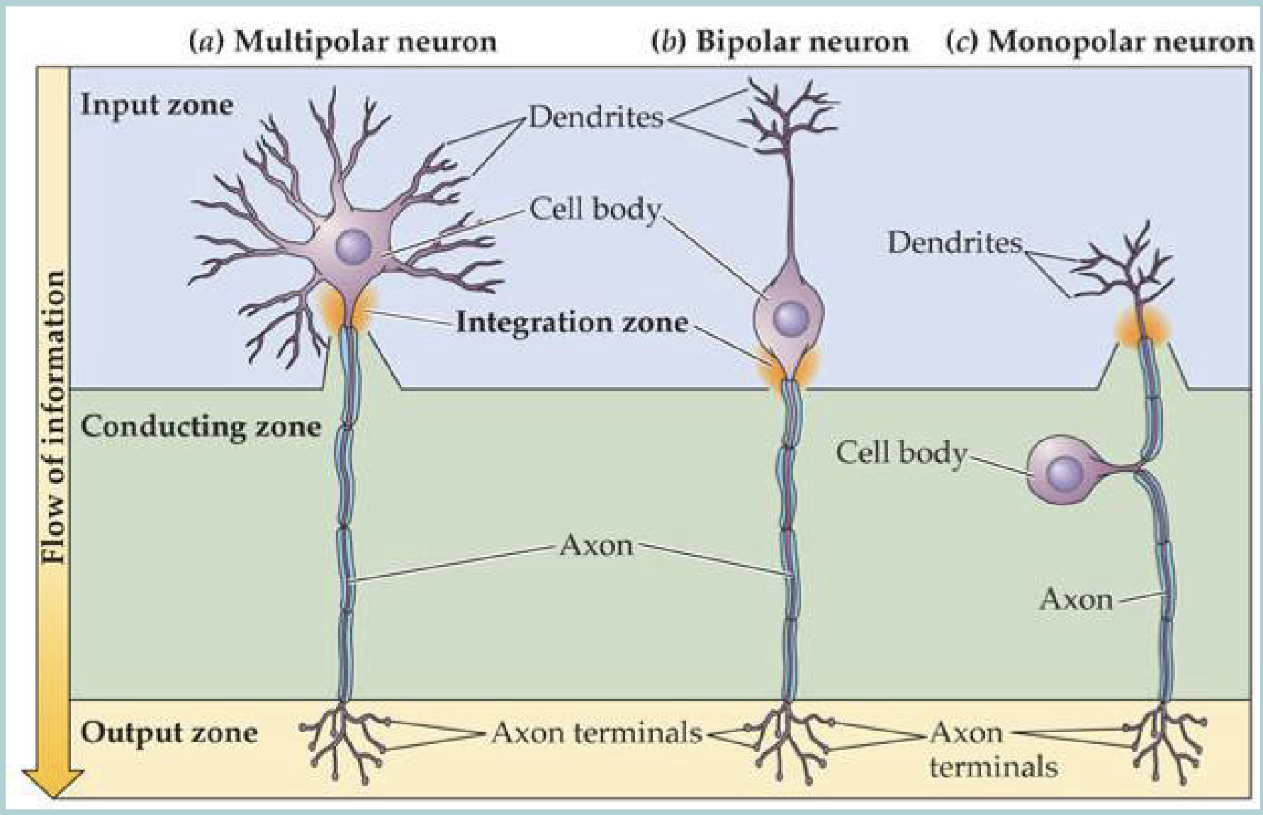
What are three neurons
Multipolar
Bipolar
Monopolar
Neurons are the WHAT units of the brain
Information processing
Neurons play many roles:
Receive information from WHAT receptors
Relay information to other WHAT and WHAT
Control WHAT → behaviour
Releases WHAT
Regulate local WHAT
Hold instructions for WHAT → memories
Regulate WHAT and WHAT
Regulates WHAT → homeostasis
Neurons play many roles:
Receive information from SENSORY receptors
Relay information to other NEURONS and CELLS
Control MUSCLES → behaviour
Releases HORMONES
Regulate local BLOOD FLOW
Hold instructions for BEHAVIOR → memories
Regulate THOUGHTS and EMOTIONS
Regulates UNCONSCIOUS PHYSIOLOGY → homeostasis
Core region of the neuron called the WHAT or WHAT
Core region of the neuron called the CELL BODY or SOMA
Branching extensions of the neuron called WHAT, collect information from WHAT
Branching extensions of the neuron called DENDRITES, collect information from OTHER CELLS
The neurons main root is the WHAT, which carries messaged to other WHAT
The neurons main root is the AXON, which carries messaged to other NEURONS
neurons only have HOW MANY axon that can branch, most neurons have many WHAT
neurons only have ONE axon that can branch, most neurons have many DENDRITES
Neurons are the functional units of the WHAT, they WHAT, WHAT, WHAT and WHAT information
Neurons are the functional units of the CNS, they RECEIVE, PROCESS, INTEGRATE and RELAY information
The axon starts at the WHAT
Axon hillock (right under the cell body)
The axon can branch into WHAT
Axon collaterals
Near the end of the axon are branches called WHAT
Teleodendria
Each teleodendria ends with a WHAT aka WHAT or WHAT
Each teleodendria ends with a PRE-SYNAPTIC TERMINAL aka BUTTON or BOUTTON
Button is close to WHAT of the next neuron
Dendritic spine
The teleodendria and the dendritic spine don’t WHAT there is a small gap called the WHAT
The teleodendria and the dendritic spine don’t TOUCH there is a small gap called the SYNAPSE
HOW MANY primary dendrites per cell
1-20
Each dendrites WHAT numerous times
Branches
This branching increases the WHAT of the cell
Surface area
Each dendrite is covered in WHAT taht also increases the WHAT
Each dendrite is covered in DENDRITIC SPINES taht also increases the SURFACE AREA
Dendrites collect information from other WHAT
cells
More surface area means more WHAT
Information exchange
What are the three types of neurons
Sensory neurons
Interneurons (majority)
Motor neurons
What do sensory neurons do
Input information to CNS
What are the two types of interneurons
Local - projects WITHIN the brain (receive and project info in the same brain region)
Projection - Projects BETWEEN the brain regions (receive and project info to other brain regions)
What do motor neurons do
Output signals from the CNS to the muscles
Morphology of a neuron can tell us about its WHAT
Function
The size of a neuron:
Neurons with long extension likely WHAT
Nuerons with short extensions likely WHAT
The size of a neuron:
Neurons with long extension likely RELAY INFORMATION
Nuerons with short extensions likely ENGAGE IN LOCAL PROCESSING
Shape of a neuron:
Complex dendrites integrate information from a WHAT
Simple dendrites integrate on a much smaller scale act as WHAT
Shape of a neuron:
Complex dendrites integrate information from a WIDE AREA
Simple dendrites integrate on a much smaller scale act as RELAYS
Sensory neurons:
WHAT relay cells
One simple WHAT
Dendrites can be:
WHAT
WHAT
Can be among the WHAT in the body
Sensory neurons:
EFFICIENT relay cells
One simple DENDRITE
Dendrites can be:
SMALL (eg. bipolar cells in retina)
LARGE (eg. sensory neurons for skin)
Can be among the LARGEST in the body
Technically every neuron between a sensory neuron and a motor neuron is a WHAT
Technically every neuron between a sensory neuron and a motor neuron is a INTERNEURON
What are the two types of interneurons
Long-distance projection
Local projection
What are the three most commonly studied interneurons:
WHAT
WHAT
WHAT
What are the three most commonly studied interneurons:
Stellate cell
Pyramidal cell
Purkinje cell
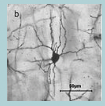
Stellate (star-shaped) cell:
WHAT
Many short WHAT extend around the WHAT
Short WHAT
Stellate (star-shaped) cell:
SMALL
Many short DENDRITES extend around the CELL BODY
Short AXON
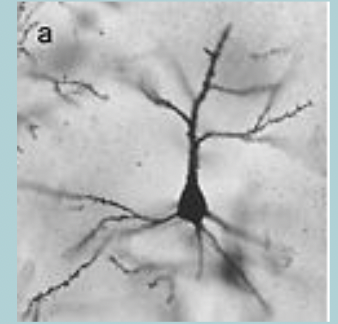
Pyramidal cell:
Has a long WHAT
A pyramid-shaped WHAT
two sets of WHAT, WHAT and WHAT
Carries information from the WHAT to the rest of the WHAT and WHAT
Pyramidal cell:
Has a long AXON
A pyramid-shaped CELL BODY
two sets of DENDRITES, APICAL and BASAL
Carries information from the CORTEX to the rest of the BRAIN and SPINAL CORD
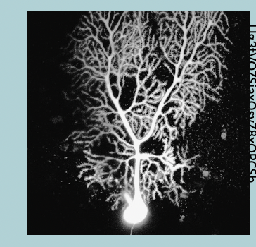
Purkinje cell:
Extremely branched WHAT that form a WHAT shape
Carried information from the WHAT to the rest of the WHAT and WHAT
Purkinje cell:
Extremely branched DENDRITES that form a FAN shape
Carried information from the CEREBELLUM to the rest of the BRAIN and SPINAL CORD
Motor neurons are located in the WHAT, WHAT and WHAT
Motor neurons are located in the CEREBRAL CORTEX, BRAINSTEM and SPINAL CORD
In motor neurons bushy dendrites collect WHAT
In motor neurons bushy dendrites collect INFORMATION
Motor neurons usually have large WHAT and WHAT that connects to WHAT
Motor neurons usually have large CELL BODY and AXON that connects to MUSCLES
All WHAT (outgoing) neural information must pass through WHAT to reach muscles
All EFFERENT (outgoing) neural information must pass through MORTOR NEURONS to reach muscles
Sensory neurons collect WHAT (WHAT) information from the body and connect to WHAT that process the information and pass it on to WHAT neurons
Sensory neurons collect AFFERENT (INCOMING) information from the body and connect to INTERNEURONS that process the information and pass it on to MOTOR neurons
The motor neurons efferent connections move WHAT and produce WHAT
The motor neurons efferent connections move MUCLES and produce BAHAVIOR
These three organizational aspects of neurons are this features of neuronal networks:
WHAT
WHAT
WHAT
These three organizational aspects of neurons are this features of neuronal networks:
INPUT
ASSOCIATION
OUTPUT
Information feeds into the WHAT
Dendrites
Dendrites feed information into the WHAT
Cell body
Information is relayed down the WHAT which branches at its ends
Axon
A neuron has thousands of WHAT (both WHAT and WHAT) but only a single WHAT
A neuron has thousands of INPUTS (both POSITIVE and NEGATIVE) but only a single AXON
The neuron processes and integrates the WHAT and (potentially) WHAT this information onwards depending on the average or summary of the WHAT
The neuron processes and integrates the INPUTS and (potentially) RELAYS this information onwards depending on the average or summary of the INPUTS
The axon terminals of the WHAT neuron come close to but do NOT contact the WHAT of the next WHAT cell forming a gap called a WHAT
The axon terminals of the PRE-SYNAPTIC neuron come close to but do NOT contact the DENDRITES of the next (POST-SYNAPTIC) cell forming a gap called a SYNAPSE
Terminal contains WHAT (WHAT and WHAT) that are released into the next cell to relay the signal
Terminal contains CHEMICALS (NEUROTRANSMITTERS and NEUROPEPTIDES) that are released into the next cell to relay the signal
In general the chemical synapse is WHAT: Only the WHAT cell influences the WHAT cell
In general the chemical synapse is UNIDIRECTIONAL: Only the PRE-SYNAPTIC cell influences the POST-SYNAPTIC cell
Most connections between neurons in an area are WHAT
local
Local interneurons
processing information
Axon collaterals can WHAT to regulate activity:
Typically regulate level of its own WHAT
turn off cell once it has WHAT its message
Axon collaterals can FEEDBACK to regulate activity:
Typically regulate level of its own ACTIVITY
turn off cell once it has SENT its message
Some cells send information to or receive information from other WHAT but most connections are WHAT
Some cells send information to or receive information from other BRAIN REGIONS but most connections are LOCAL
Signal from a pre-synaptic neuron either WHAT or WHAT the next post-synaptic neuron
Signal from a pre-synaptic neuron either EXCITES or INHIBITS the next post-synaptic neuron
The receiving neuron integrates all its various WHAT and determines the response:
If there is more excitation the neuron will WHAT the signal (yes) (++,---,+++)
If there is more inhibition the neuron will WHAT (no) (----, +++, ----)
The receiving neuron integrates all its various INPUTS and determines the response:
If there is more excitation the neuron will RELAY the signal (yes) (++,---,+++)
If there is more inhibition the neuron will REMAIN SILENT (no) (----, +++, ----)
What are the two views on how neurons code for behavior
Each neuron codes for a SPECIFIC aspect of behaviour
Neurons work together in COMPLEX NETWORKS to code for specific aspects of behavior (loss of one or more neurons would not effect behavior, network can still function)
Science favours which view
The network hypothesis
Ears on either side of a moth are connected to WHAT on opposite sides of the body
Side closest to a bat is WHAT the most
This affects the WHICH wing to steer the moth away form the from the bat
Ears on either side of a moth are connected to WING MUSCLES on opposite sides of the body
Side closest to a bat is ACTIVATED the most
This affects the OPPOSITE wing to steer the moth away form the from the bat
Neurons are not WHAT but change WHAT constantly:
rewire, make new WHAT
Neurons are not STATIC ENTITIES but change SHAPE constantly:
rewire, make new CONNECTIONS
Neurons are WHAT
Post-mitotic (can’t under go mitosis)
Most neurons are not WHAT this means most neurons live with you throughout your whole life
Most neurons are not REPLACED this means most neurons live with you throughout your whole life
Glial cells have between a WHAT to WHAT ratio with neurons
Glial cells have between a 1:1 to 10:1 ratio with neurons (way more glial cells)
Like neurons, glial cells have multiple WHAT
forms
Like neurons, glial cells play multiple WHAT
Roles
Unlike neurons, glial cells are constantly WHAT throughout life
Unlike neurons, glial cells are constantly RENEWED throughout life
They play the “WHAT” role in the nervous system
They play the “SUPPORTING ACTOR” role in the nervous system (neurons are still the main/ most important but need glial cells to help)
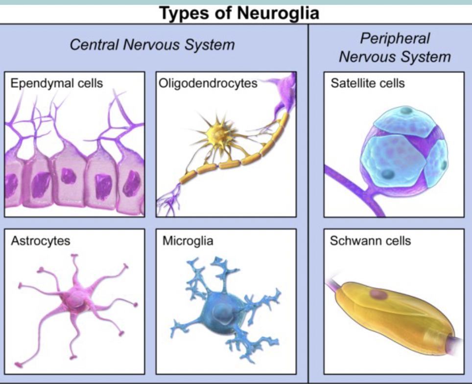
What are the six types of glia cells and what nervous system are they apart of
Ependymal cells (CNS)
Microglia (CNS)
Astrocytes (CNS)
Oligodendrocytes (CNS)
Satellite glial cells (PNS)
Schwann Cells (PNS)
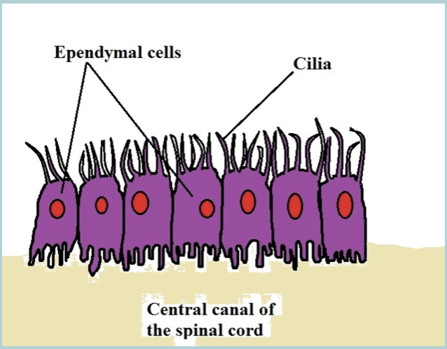
Ependymal cells
Line the VENTRICLES
Help generate CEREBROSPINAL FLUID (CSF) and create/assist the FLOW of the fluid
Microglia cells
IMMUNE SYSTEM and REPAIR in the CNS
Clear dead cells (including apoptotic neurons)
Activated by injury and toxins
Similar functions as WHITE blood cell (monocytes) and macrophages
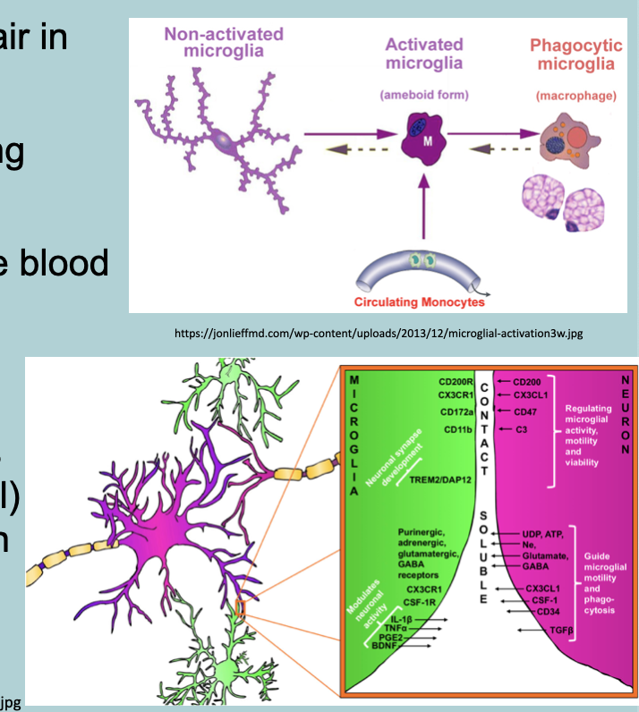
Extensive and essential WHAT between microglia and neurons, particularly in the WHAT
Extensive and essential COMMUNICATION between microglia and neurons, particularly in the SYNAPSE

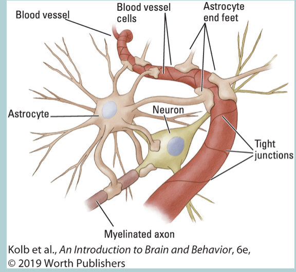
Astroglia (Astrocyte)
STAR shape
Provide STRUCTURAL support
Convey NUTRIENTS between BLOOD VESSELS and neurons
Regulate BLOOD flow
Part of the blood brain barrier
Can play a role in HEALING the brain after damage
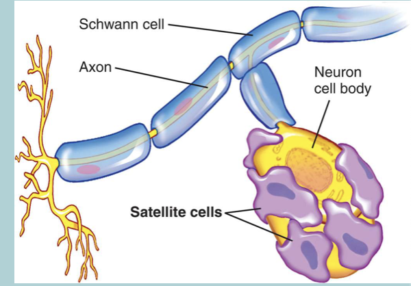
Satellite glial cells
Small cells that SURROUND neurons’ CELL BODIES located in the AUTONOMIC nervous system (ie. PNS)
They resemble the ASTROCYTES of the CNS and assist in regulating the EXTERNAL chemical environment
Very sensitive to INJURY and may exacerbate (increase) PATHOLOGICAL pain