Joints, Ligaments, Nerve Supply, and Blood Supply of Elbow Complex, Wrist, and Hand
1/28
Earn XP
Description and Tags
PT605 Anatomy, Oregon Tech
Name | Mastery | Learn | Test | Matching | Spaced |
|---|
No study sessions yet.
29 Terms
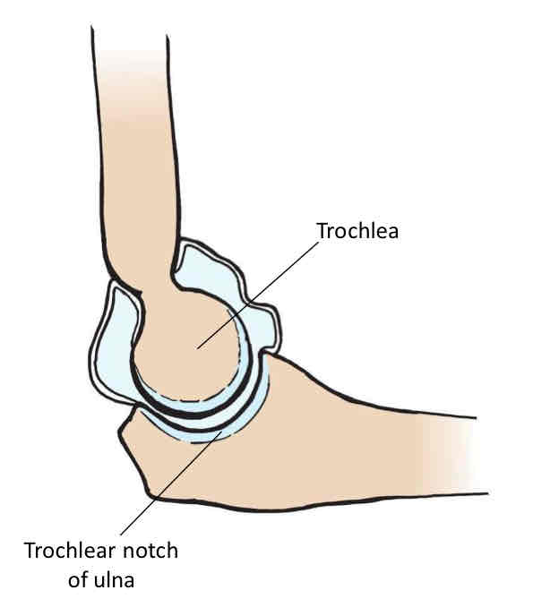
Ulnohumeral Joint
Diarthrodial synovial joint, hinge, uniaxial, flexion/extension.
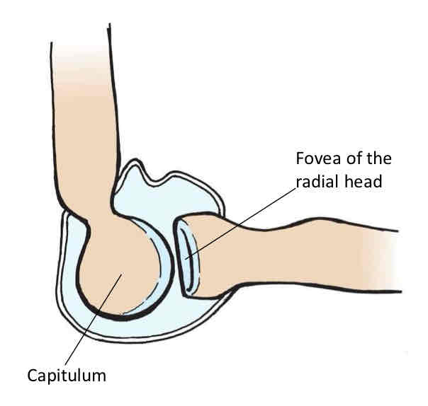
Radiohumeral Joint
Diarthrodial synovial joint, hinge, uniaxial, flexion/extension.
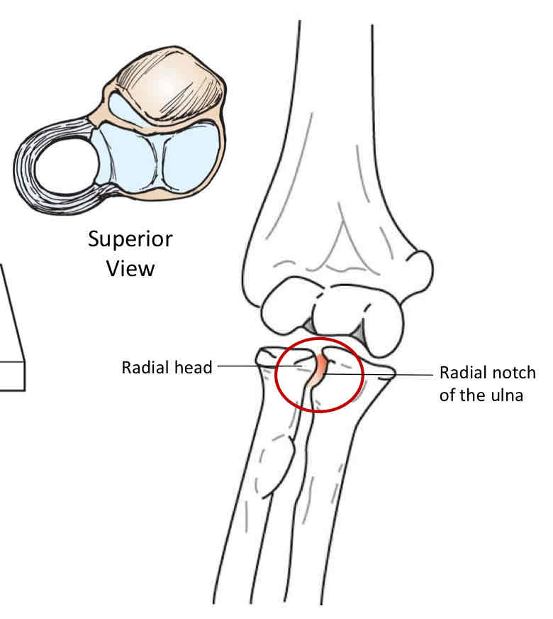
Proximal Radioulnar Joint
Diarthrodial synovial joint, pivot, uniaxial, forearm pronation/supination.
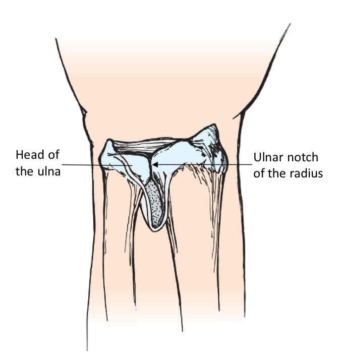
Distal Radioulnar Joint
Diarthrodial synovial joint, pivot, uniaxial, forearm pronation/supination.
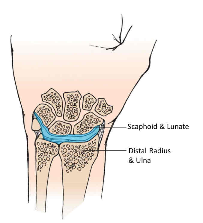
Radiocarpal Joint
Diarthrodial synovial joint, condyloid, biaxial, flexion/extension and radial/ulnar deviation.
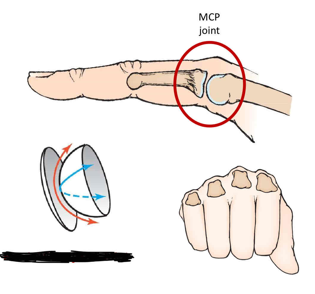
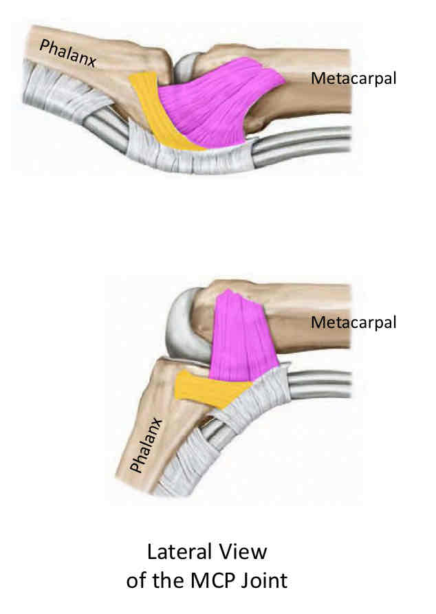
Metacarpophalangeal Joints
Diarthrodial synovial joint, condyloid, biaxial, MCP flexion/extension and MCP abduction/adduction.
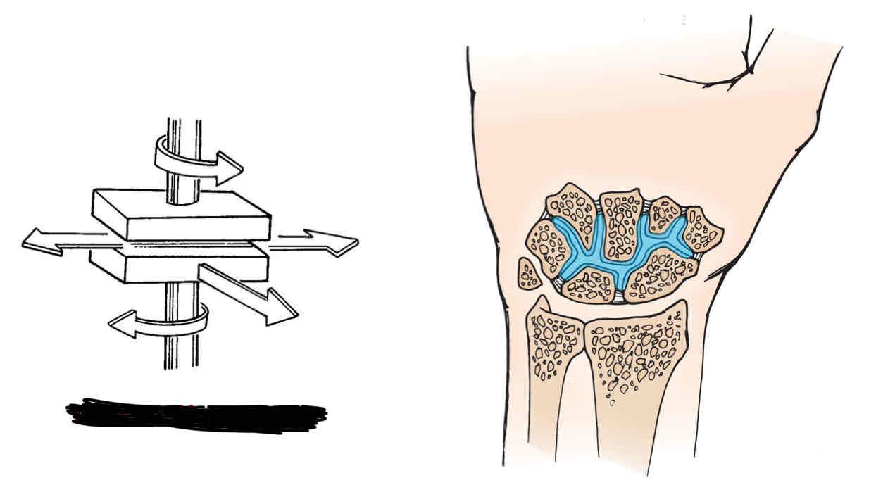
Intercarpal Joints
Diarthrodial synovial joint, plane, triaxial, glide in all directions during wrist motions.
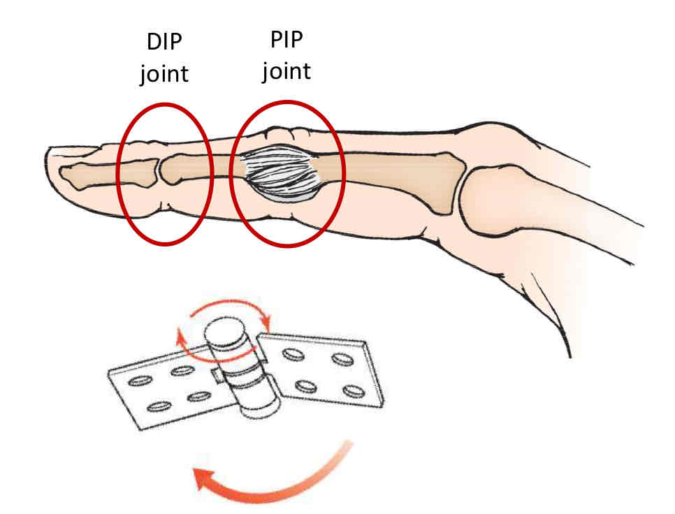
Interphalangeal Joints
Diarthrodial synovial joint, hinge, uniaxial, IP flexion/extension.
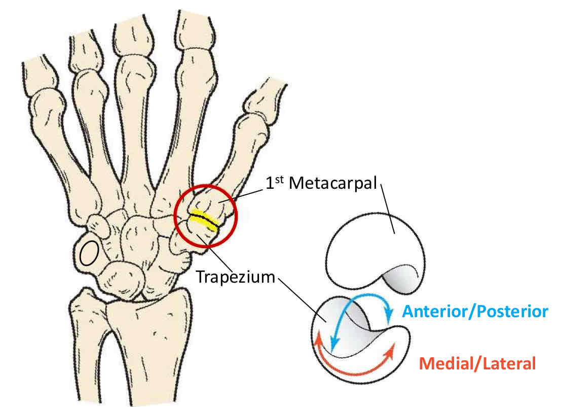
1st Carpometacarpal Joint
Diarthrodial synovial joint, sellar (saddle), biaxial, 1st CMC flexion/extension and 1st CMC abduction/adduction.
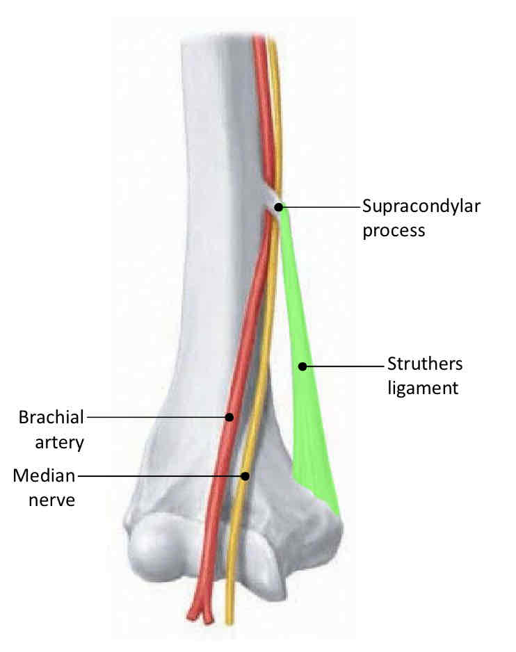
Arcade of Struthers
Formed by supracondylar process, medial supercondylar ridge, and Struther’s ligament (ligamentum Struther’s). Contents are brachial artery and median nerve.
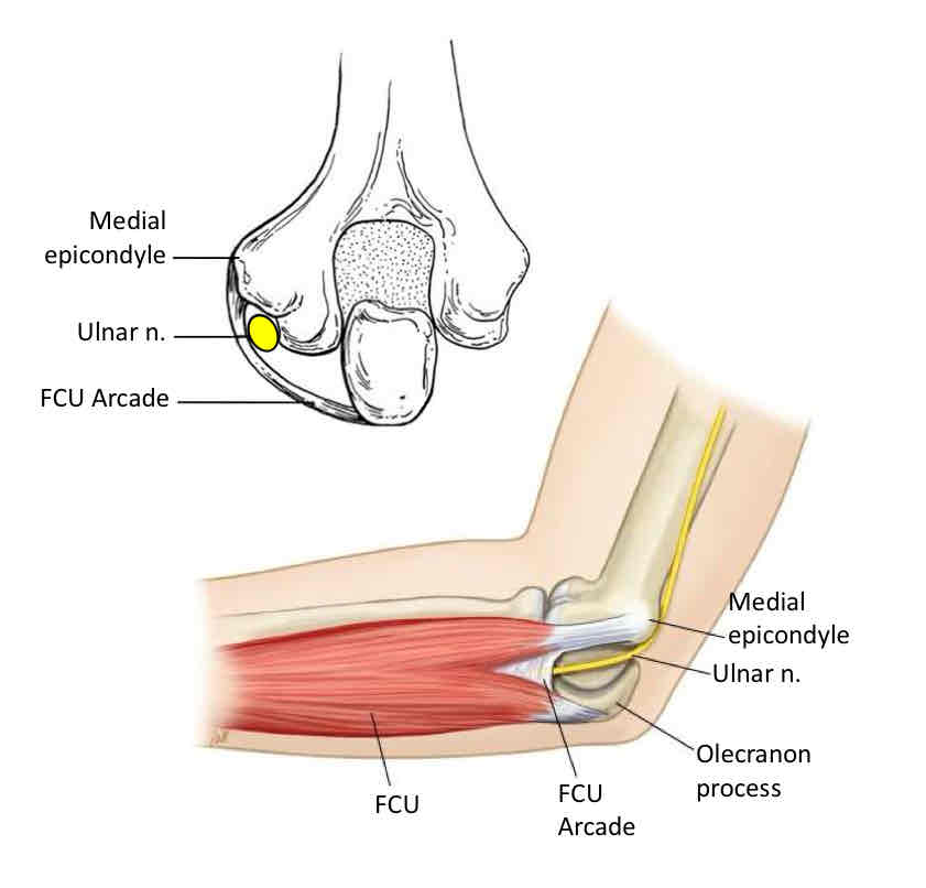
Cubital Tunnel
Formed by ulnar groove, two heads of FCU, and FCU arcade. Contents are ulnar nerve.
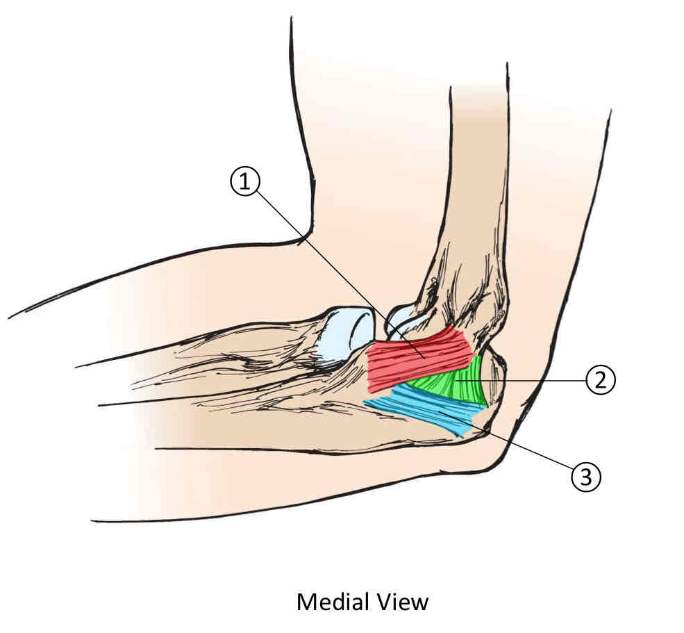
Medial Collateral Ligament
Formed by the anterior band, posterior band, and oblique band.
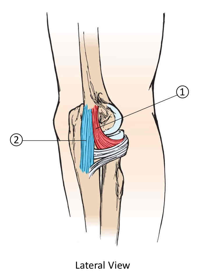
Lateral Collateral Ligament
Formed by the radial collateral ligament and lateral ulnar collateral ligament.
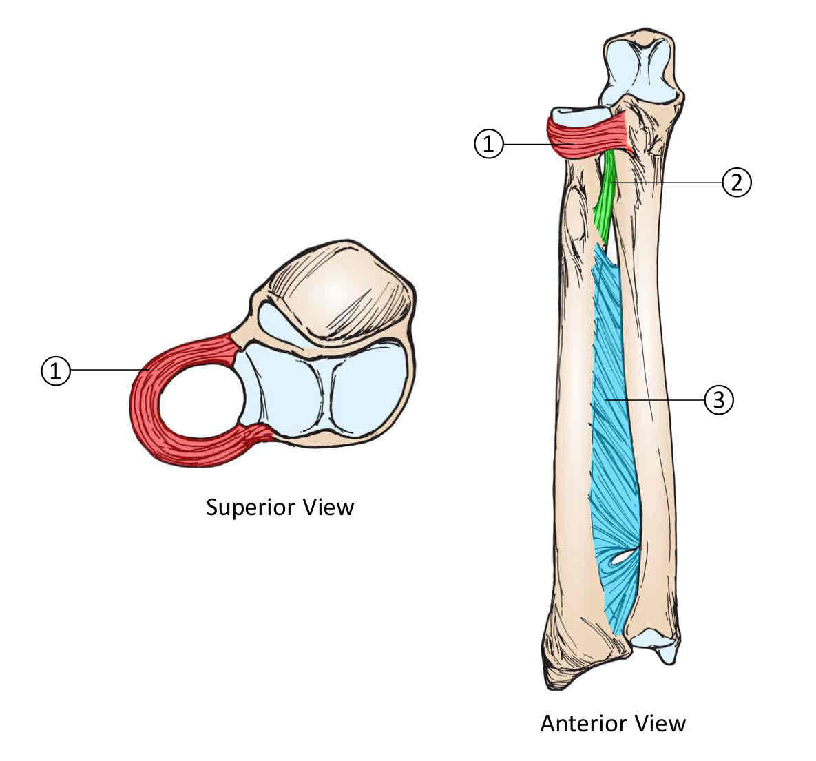
Forearm Ligaments
Formed by the annular ligament, oblique cord, and interosseous membrane.
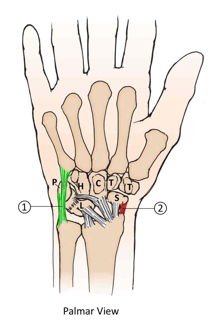
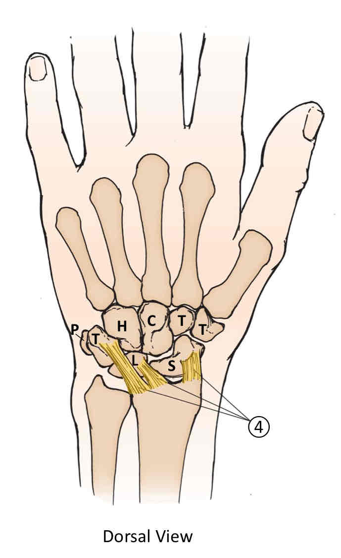
Collateral Ligaments of the Wrist
Formed by the ulnocarpal collateral ligament and radiocarpal collateral ligament.
Palmar Ligaments of the Wrist
Formed by the palmar radiocarpal ligament.
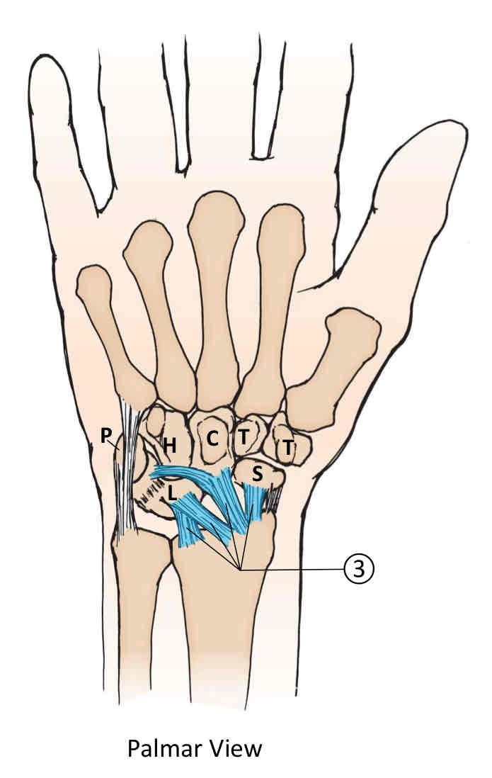
Dorsal Ligaments of the Wrist
Formed by the dorsal radiocarpal ligament.
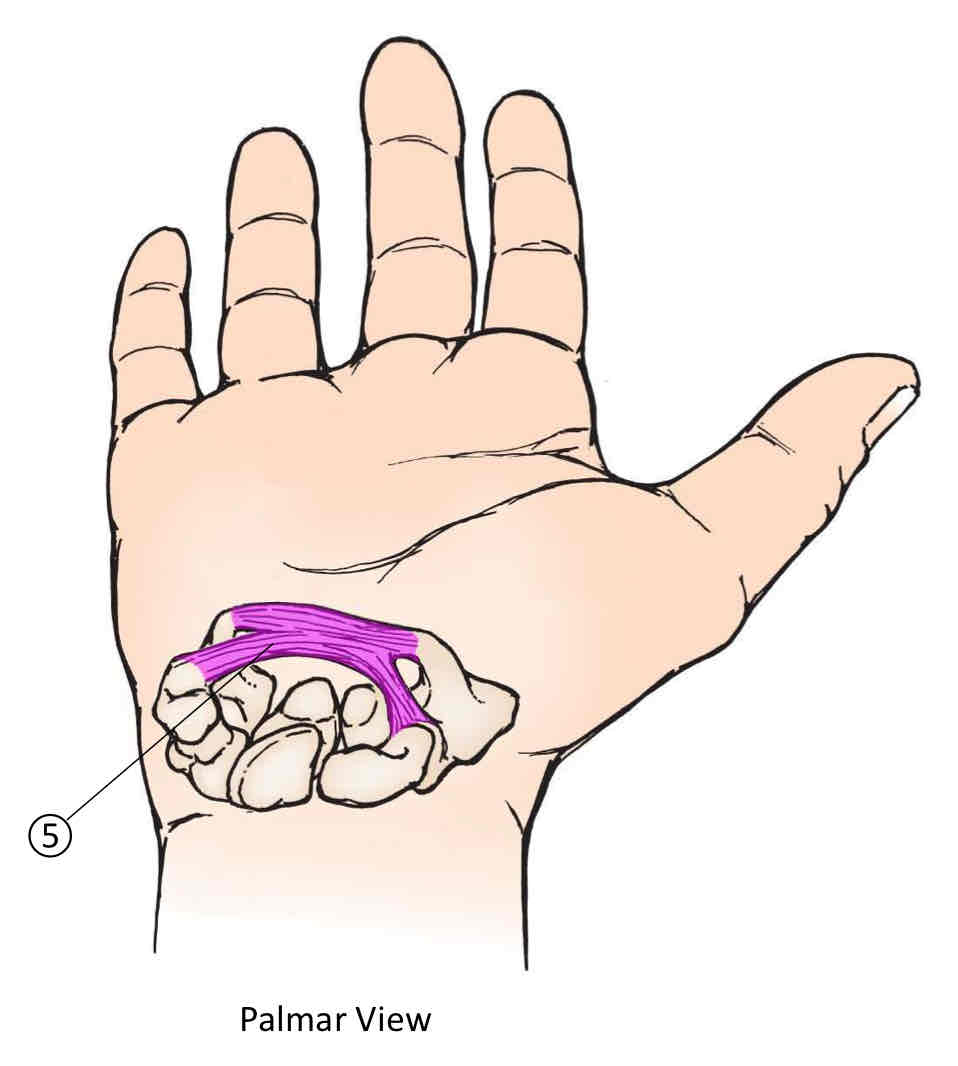
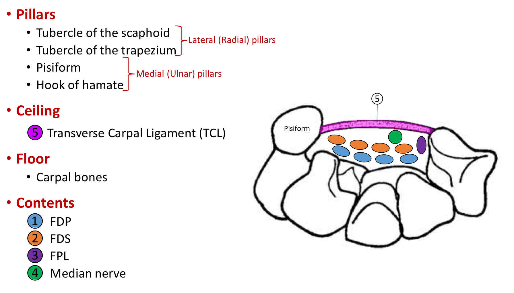
Carpal Tunnel
Formed by the transverse carpal ligament (ceiling), the tubercle of the scaphoid, the tubercle of the trapezium, pisiform, and the hook of hamate (pillars), and carpal bones (floor). Contents are FDP, FDS, FPL, and median nerve.
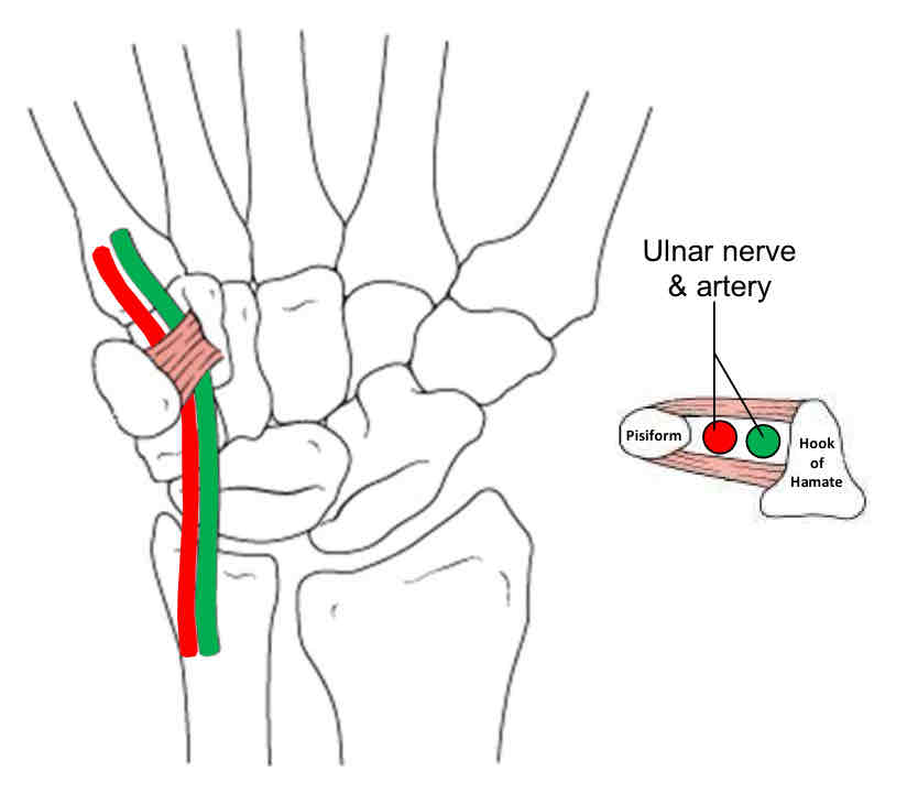
Guyan’s Canal
Formed by the pisohamate ligament (ceiling), and the hamate and triquetrum (floor). Contents are the ulnar nerve and ulnar artery.
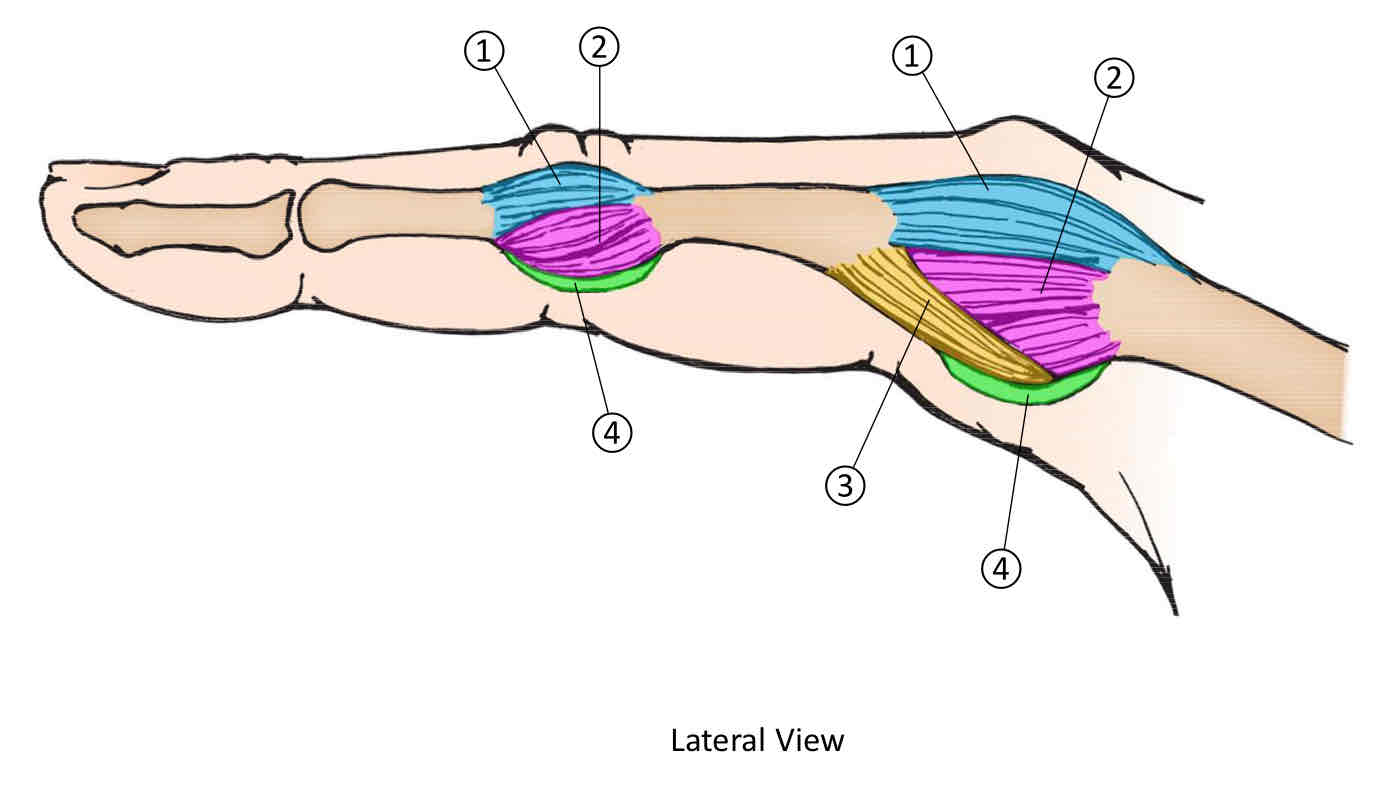
Supporting Finger Structures
Formed by the joint capsule, collateral ligament, glenoid ligament, and volar plate.
Finger Ligaments
Formed by the collateral ligament and glenoid ligament.
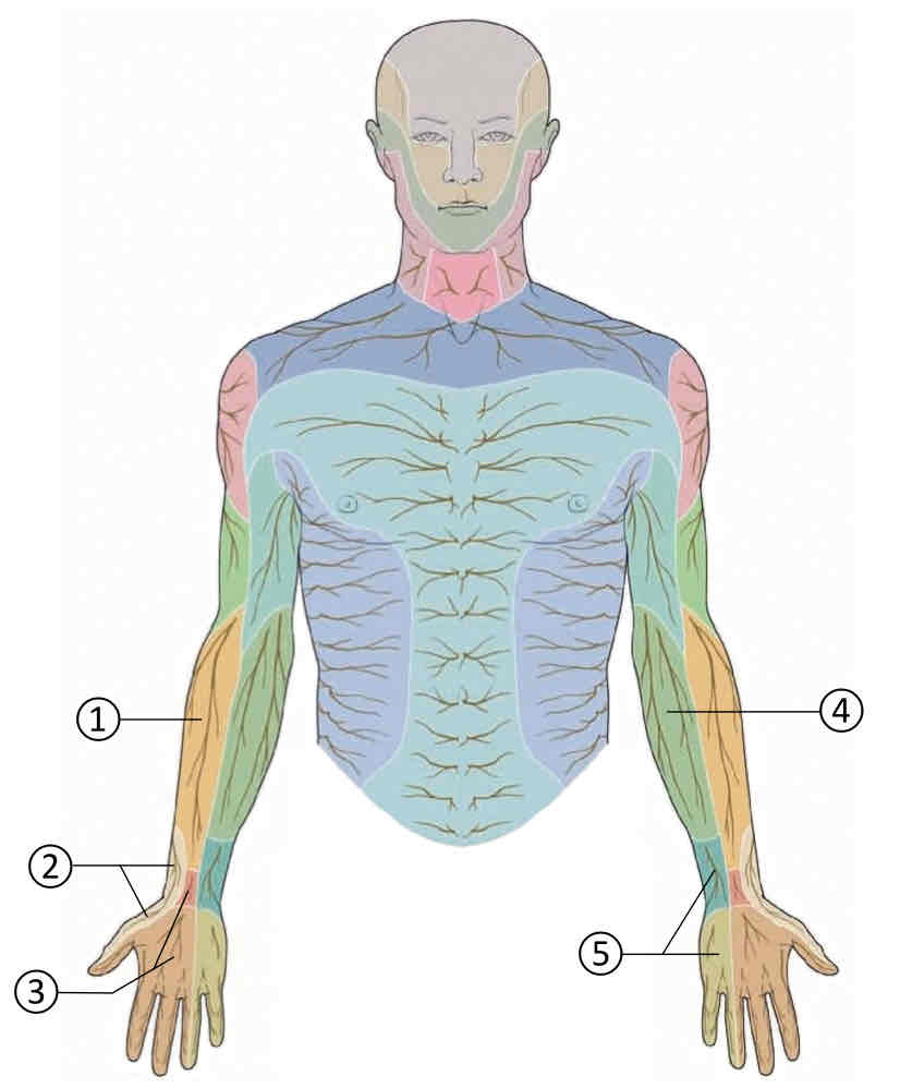
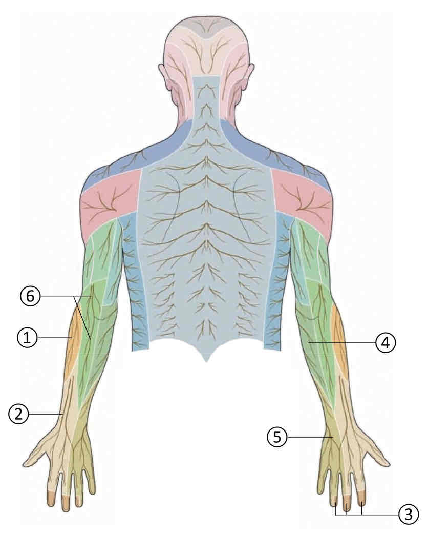
1 Lateral Antebrachial Cutaneous Nerve
Musculocutaneous nerve


2 Superficial Radial Nerve
Radial nerve


3 Sensory Branch of the Median Nerve
Median nerve

4 Medial Antebrachial Cutaneous Nerve
Medial cord of brachial plexus


5 Sensory Branch of the Ulnar Nerve
Ulnar nerve


6 Posterior Antebrachial Cutaneous Nerve
Radial nerve

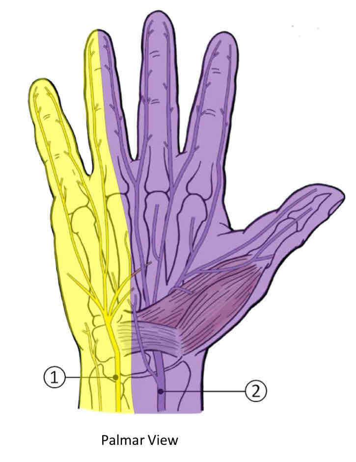
Sensory Nerves of Palmar View of the Hand
Ulnar nerve and median nerve.
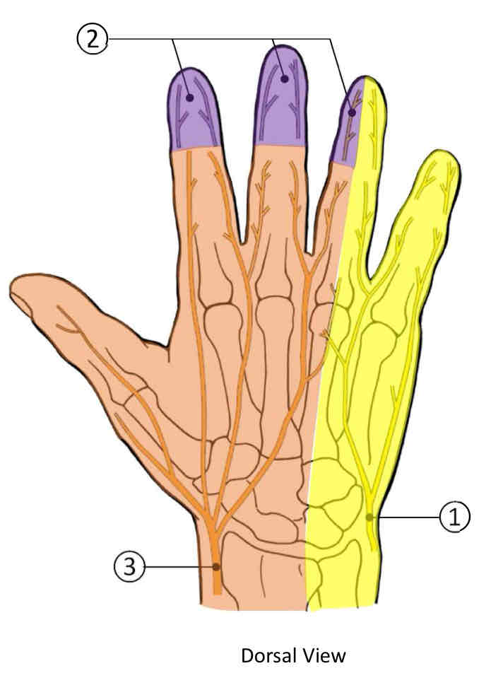
Sensory Nerves of Dorsal View of the Hand
Ulnar nerve, median nerve, and superficial radial nerve.