Anatamy and Physiology
1/95
There's no tags or description
Looks like no tags are added yet.
Name | Mastery | Learn | Test | Matching | Spaced | Call with Kai |
|---|
No analytics yet
Send a link to your students to track their progress
96 Terms
5 cellular functions
growth
reproduction
absorbtion
excretion/secretion
metabolism
tissue
aggregation of cells and intercellular substances specialized to perform a particular function
4 types of tissue
epithelial tissue
conective tissue
muscle
nervouse tissue
surface epithelial
sheets of agregated cells
covers external surfaces of the body
Glandular epithelium
forms secretory cells of glands
sits inside connective tissue
epithelial tissues (4 things)
cells are diverse
capable of mitosis
contacts basement membrane
avascular
Basal
in contact with underlying connective tissue
apical
in contact with surface
lateral
in contact with other epithelial cells
squamous epithelial cell
lining of internal surfaces
dehydrates quickly
cuboidal epithelial cells
lines ducts of kidneys and thyroid glands
secretion and absorbtion
columnar epithelial cells
lines absorbtive surfaces (respiratory and digestive tract)
excretes hormones out of body
Stratified Squamous
only superficial cells have squamous shape
if keratanized, no nuclei present
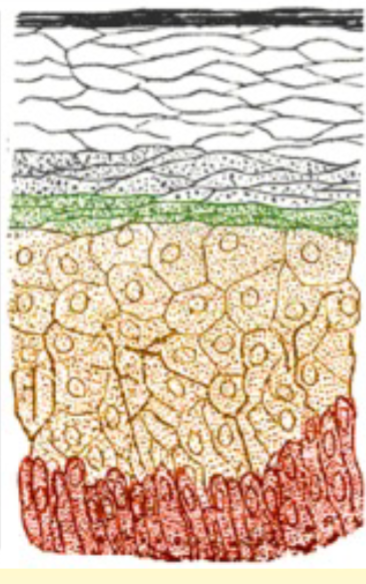
stratified squamous layers
Stratum corneum
stratum granulosum
stratum spinosum
stratum basale
endocrine
secretes products without ducts
exocrine
utilizes ducts to transport materials
glandular epithelial shapes
tubular
alveloar
tubulo-alveolar
merocrine gland
secretory product from cytoplasm to cell membrane
no loss of cell
apocrine gland
secretory product from cytoplasm to cell membrane
partial cell loss
Holocrine gland
secretory product from cytoplasm to cell membrane
whole cell is secreted
serous secretion
clear and watery fluid
may contain enzymes
lacrimal glands
mucoid secetion
mucous in nature
thicker substance
salivary glands'
vaginal secretion
4 types of connective tissue
proper
support
adipose
blood
properties of CT
Diverse
heterogeneous population of cells
unique etracellular materials
3D
Functions of CT
connection
insulation
repair
defense
regeneration
support
locomotion
mesenchymal stem cell
mother stem cell to all types of CT
-blast
immature cell
-cyte
mature cell
collagen fibers
fibro, osteo, and chondro
high tensile steength
hyaline cartilege, dermis, tendons, and ligaments
reticular fibers
fibro
delicate network
arranged around budles of collagenous fiber, smooth muscle
elastic fibers
fibroblast
stretch and rebound
arteries, veins, nuchal ligament
ground substance
secretory sproduct of the cell
defines type of CT
holds cells and fibers together
transports nutrients
stores water
glycosaminoglycan
ground substance
“amino-sugar” combined with carbohydrates
polysaccharide, mucopolysaccaride
glycosaminoglycan distribution
hyaluronic acid (synovial fluid, loose CT)
Chondriotin-4-sulfate(cartilage, bone, cornea)
keratin sulfate (cartilage, bone, crnea, nucleus pulposus)
proteoglycan
ground substance
combines glycoaminoglycan with proteins
absorbs water
supports
proteoglycan distribution
loose CT
Dense irregular CT
synovial fluid
division of proper CT
loose (areolar, adipose)
Dense ( irregular, regular)
elastic
Special (reticular, pigmented)
Areolar /loose CT
mesenchymal, fibroblast, fibrocyte
collagenous, reticular, elastic
Dense CT
mesench. fibroblast, fibrocyte
collagenous
regular and irregular
elastic CT
mesench. fibro
elastic
surounds blood vessels
reticular Special CT
reticular fibers + elastic + collagenous
found in spleen and lymphnode
pigmented special CT
Areolar CT with added pigmented cells
Adipose CT
supported by Areolar CT
energy storage
insulation
absorbtion of cuncussive forces
mechanical protection
white Adipose tissue
large fat droplet
lipids mobilized into blood as energy
brown adipose tissue
smaller cell
numerous fat droplets
metabolism releases heat to elevate temperature of tissues and warm blood
3 types of cartilage
fibro-cartilage
hyaline cartilage
elastic cartilage
properties of cartilage
mesenchym. chondro
collagen fibers
ground substance: amorphous proteoglycans
avascular, support, covers end of bone, template for bone growth
cartilage growth
appositional
interstitial
hyaline cartilage
covers end of bone
serves as bone forming site
supportive structure (nose, trachea, bronchii)
lacunae
space
fibrocartilage
transition from dense CT to haylaine cartilage or bone
ligamnets and tendons
shock absorber
intervertebral disc
elastic cartilage
elastic fibers
supprt with elastiity
ear pinna, laryngeal cartilage
epiglottis
3 parts of a long bone
diaphysis
epiphysis
metaphysis
diaphysis
houses bone marrow
provides support compact/corticle bone
epiphysis
site of articulation
covered in hylaine or articular cartilage
metaphysis
corticle bone with cancellous bone
epiphysial plate
bone physiology
mesenchymal, osteo
collageneous fibers
osteoblast
bone forming cells
secrete collagen fibers and ground substnce
osteoclast
derived macrophage in blood
break down and remodel bone
compact/corticlw bone
dense
resists compression’shaft of long bones
plate paters
growth in diameter
cancellous/spongy bone
arranged in spicules or trabeculae
porus
resists compression
end of long bones
between layers of compact bone in skull
3 envelopes of bone
periosteum
endosteum
osteonal endostum
periosteum
outer covering/capsule of bone
endosteum
inner shaft
defines marrow cavity
osteonal endosteum
connects periosteum to endosteum
ossification methods
inntramembranous
endochrondral
heteroplastic
ossification
deposition of calcium salt on osteoid (laying down ne bone)
calcification
deposition of calcium salts in tissue (disease)
intramemberanous ossification
osteoprogenitor cells to osteoblasts
osteoid to bone
cancellou sbone and flat bones of skull
endochondral ossification
replacement of hyaline cartiladge with bone
heteroplastic ossification
bone is formed outside of skeletal system
achondrodysplasia
short limbs
types of fractures
greenstick
complete
comminutes
epihyseal
fracture repair
blood clot- connective tissueew
osteoblast proliferation
osteid formation
mineralization
remodeling
fibrous joint
suture
syndesmosis
gomphosis
cartilaginous joint
synchondrosis
symphyses
gomphosis
periodontal ligament
synchondrosis
joined by hyaline cartalige
symphysis joint
pelvis and mandible
synovial joint parts
articular surface
aticular cartilage
joint capsule
condylar synnovial joint
hinge joint
ellipsoid synovial joint
wrist and ankle
right angle
spheroid synnovial joint
shoulder and hip
saddle synovial joint
fingers
layers of the skin
epidermis
dermis
subcutaneous tissue
melanocytes
produce melanin which cause pigmentation
melanocytes
located in stratum basale of epidermis
papillary layer
dermal papillae project into epidermis
dense in vascular and nerve suppy
collagenous and elastic CT
reticular layer
reticular CT
skin thickness
subcuticle layer
superficial fasia, subcutis, hypeosermis
areolar CT
fat tissue
sebaceous glands
oily secretion that waterproofs
hair shaft parts
medulla
cortex
cuticle