L4- cell cycle regulation and evasion of anti growth signalling
1/44
There's no tags or description
Looks like no tags are added yet.
Name | Mastery | Learn | Test | Matching | Spaced | Call with Kai |
|---|
No analytics yet
Send a link to your students to track their progress
45 Terms
what 3 interrelated processes control cell growth
cell proliferation
cell differentiation
cell apoptosis
cancer- elevated cell growth
cells in the mass have diminished differentiation, reduced apoptosis and/or increased proliferation
describe cell growth in healthy tissue
number of proliferating cells remains constant

How does blocking apoptosis and differentiation lead to aberrant growth?
Proliferation rate remains unchanged
Apoptosis is blocked
Differentiation is blocked
Cells continue cycling instead of exiting the cell cycle
Result: abundance of proliferating cells increases, causing aberrant growth
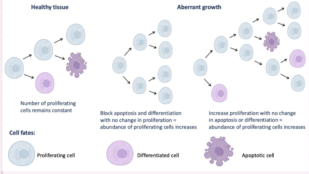
How does increased proliferation lead to aberrant growth without changes in apoptosis or differentiation?
Proliferation rate increases
Apoptosis remains unchanged
Differentiation remains unchanged
More cells enter and remain in the cell cycle
Result: abundance of proliferating cells increases, causing aberrant growth

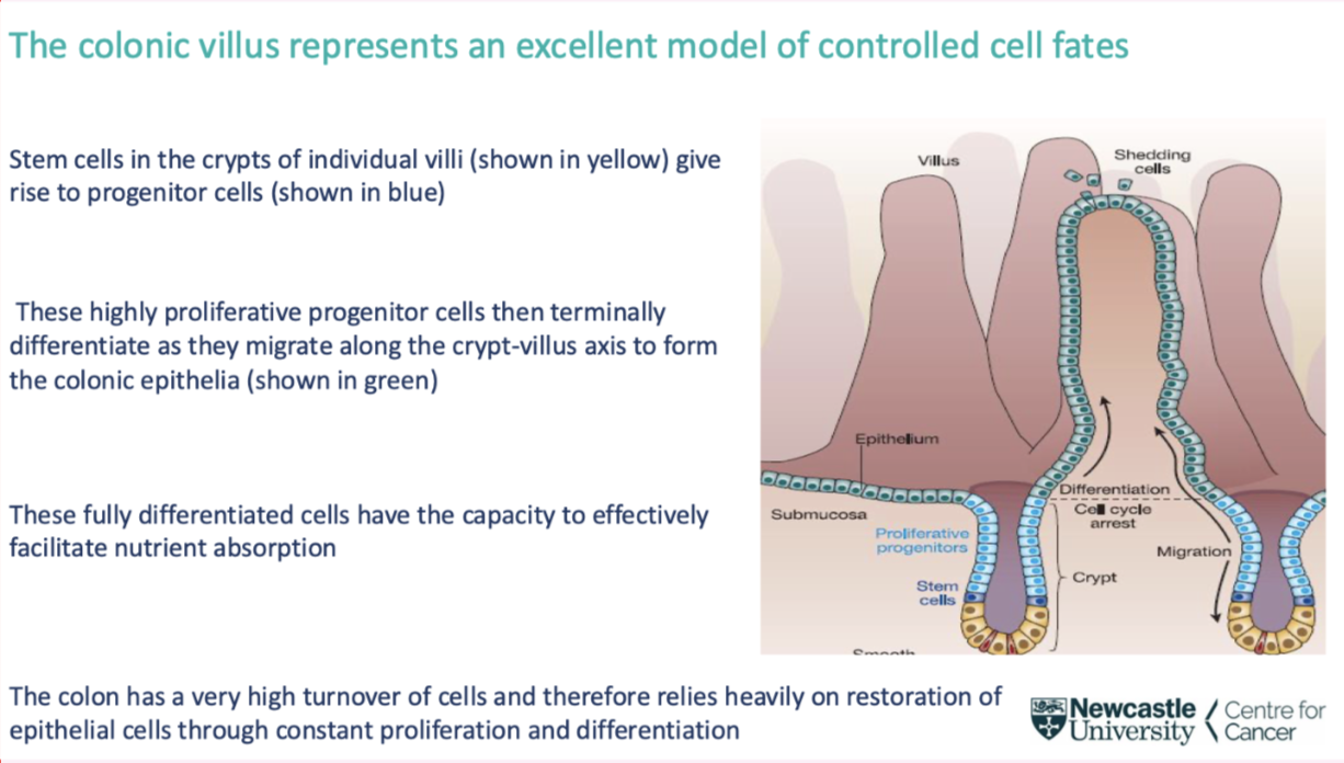
which tissue is a good representation of interplay between stem and progenitor cells
Colon- good example of interplay between stem cells progenitor cells and fully differentiated cells
Undifferentiated cells in stem cell crypts- can differentiate and partially differentiate into progenitor cells (mostly useless)
They move up and acquire more characteristics of colon cell- fully differentiated when they reach the villus- able to absorb food at this point
This process is under tight control
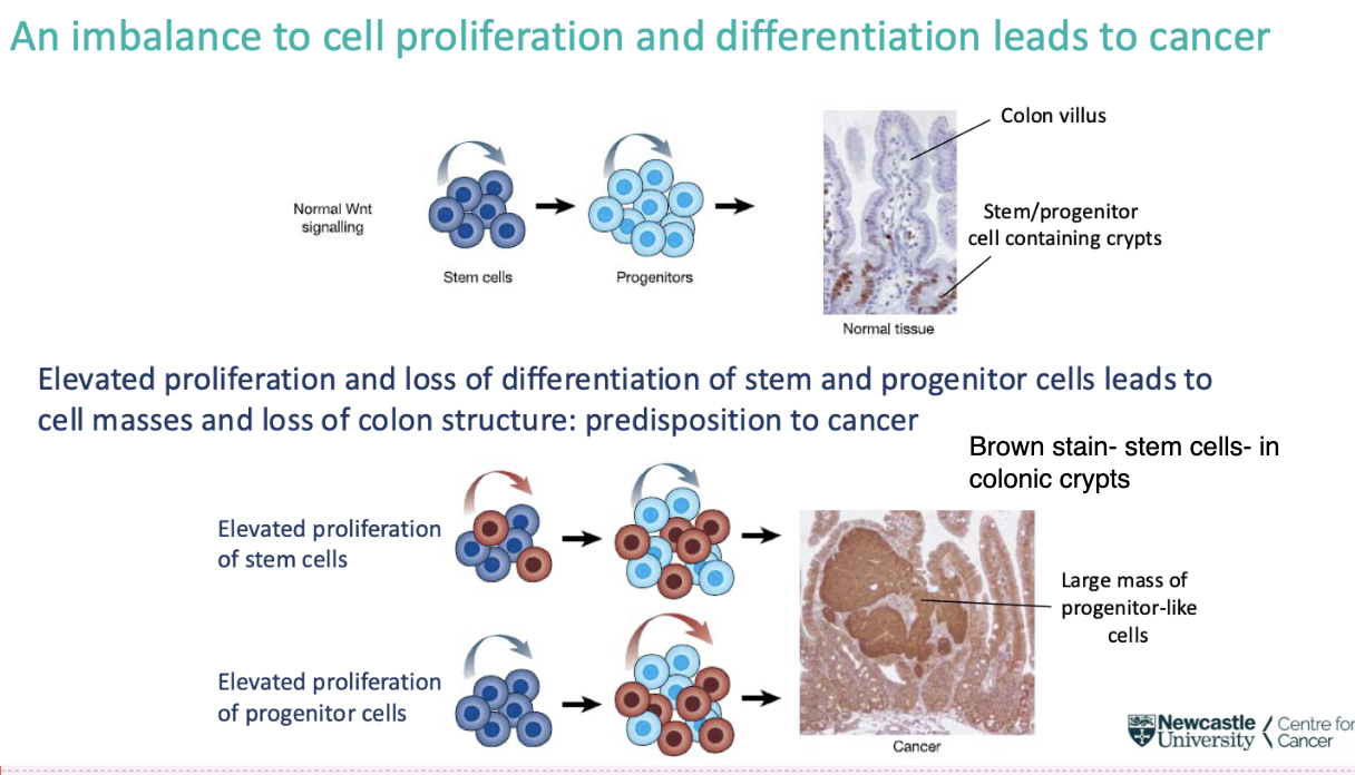
what does an imbalance between cell proliferation and differentiation lead to
cancer
In colon cancer- deregulated stem cell renewal capacity cell masses and loss of colon structure- predisposition to cancer
Outgrowth of poorly differentiated cells, crypts erupt into masses, lose functionality of colon
Example of imbalance of homeostasis of stem and progenitor cell differentiation
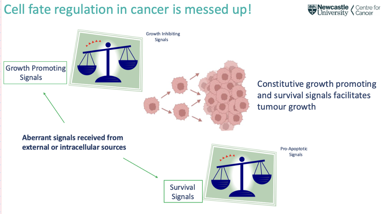
what controls cell fate
Controlled by signals (positive and negative)
Growth promoting and inhibitory
Cell enters G1 if promoting outweighs inhibitory
Very strict signals that regulate this
Inhibitory- p53
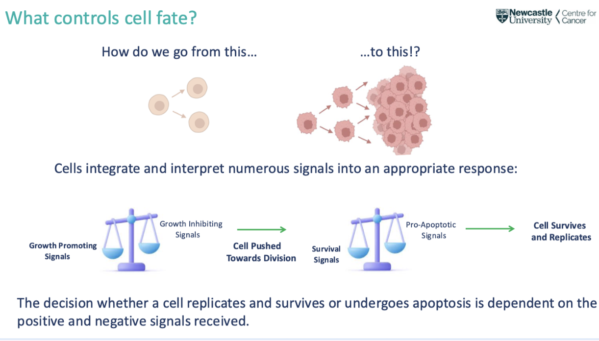
what happens to cell signalling in cancer
In cancer- survival and growth signals outweigh the inhibitory- tumour masses
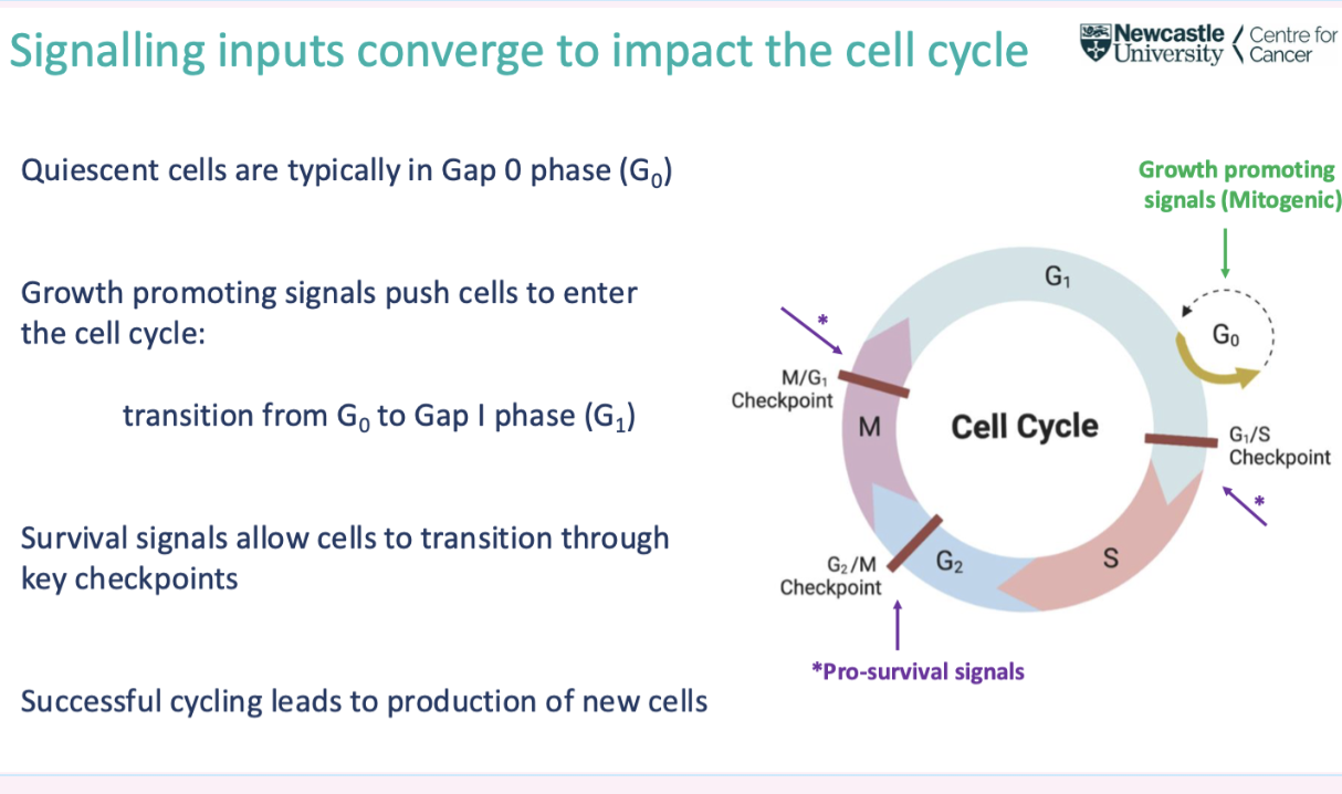
what are mitogenic signals
Mitogenic signal- drives cell through mitosis
Lots of cells in G0 phase- need mitogenic signal to enter G1
G1 prepare for division- generate organelles to prepare for S phase
Checkpoints need to be reached to enter next phase
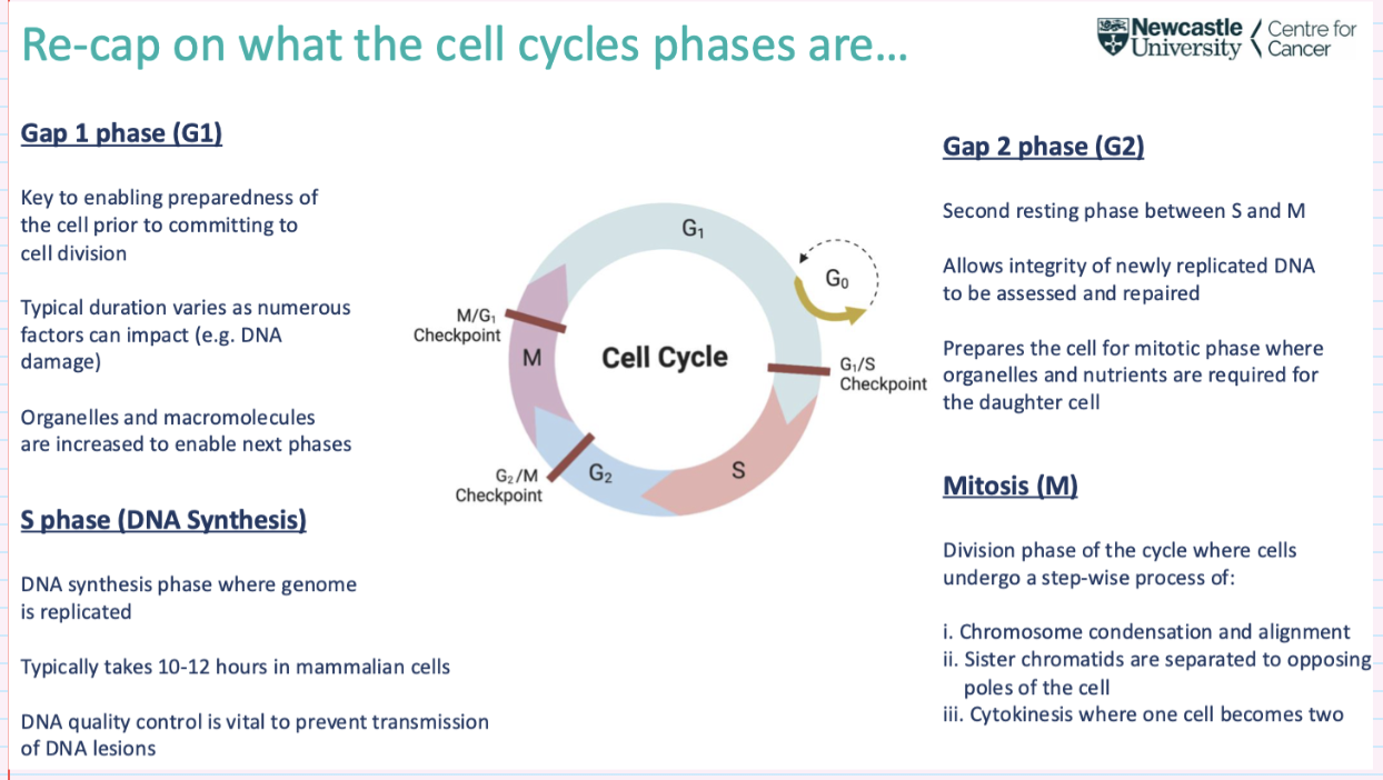
give an overview of the cell cycle phases
gap 1 phase- G1
S phase (DNA synthesis)
gap 2 phase- G2
mitosis- M
what are the main regulators of the cell cycle
CDKs
cyclin dependent kinases- a family of serine/threonine kinases which phosphorylate key substrates to enable cell cycle progression

what structure do CDKs form
They form a holoenzyme complex with their respective cyclin protein which are required to enable activation of CDK catalytic activity

what are CDKs controlled by
Each cognate CDK-cyclin complex is itself controlled by specific inhibitors which help govern progression through the cell cycle: these are called CDK inhibitors (CKIs)
prevent hyperactivity
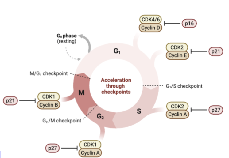
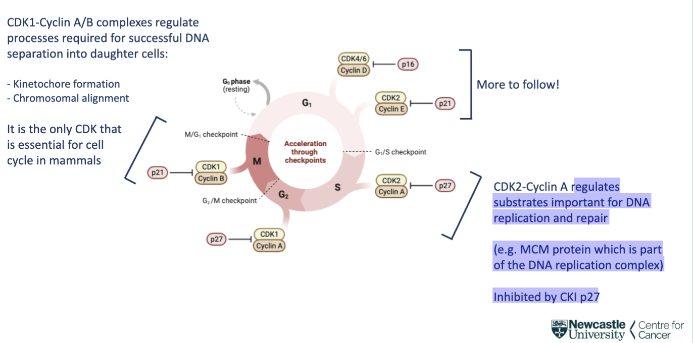
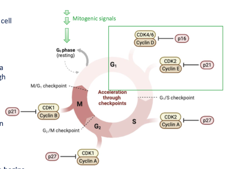
what process does CDK1-Cyclin A/B complex regulate
regulates processes required for successful DNA separation into daughter cells:
- Kinetochore formation
- Chromosomal alignment
It is the only CDK that is essential for cell cycle in mammals
inhibited by CKI p21

what process does CDK2-Cyclin A complex regulate
regulates substrates important for DNA replication and repair (e.g. MCM protein which is part of the DNA replication complex)
Inhibited by CKI p27
how were CDKs discovered
by pioneering studies in yeast and frog oocytes
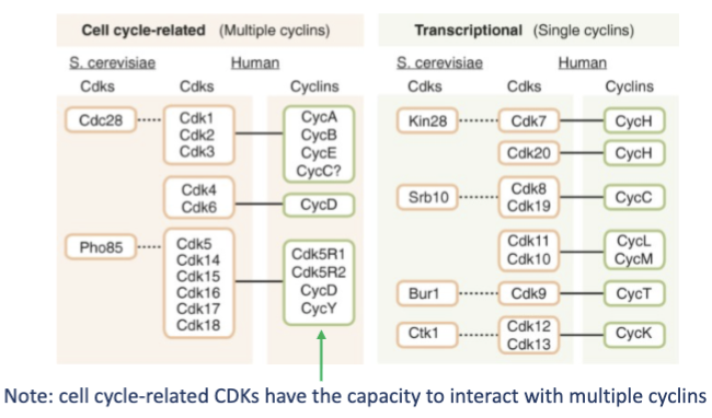
how many CDKS are encoded in the human genome, what types are there
There are 20 CDKs encoded in the human genome and are classified into two separate groups:
1. Cell cycle-related
2. Transcriptional
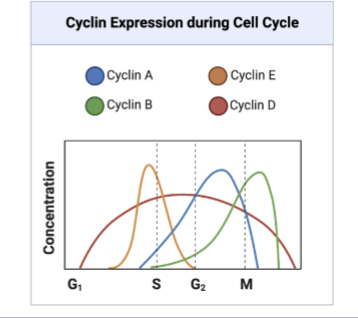
how do levels of CDKs and cyclins change throughout the cell cycle
Cellular CDK levels do not fluctuate throughout the cell cycle but cyclin levels do to control CDK activity at distinct phases
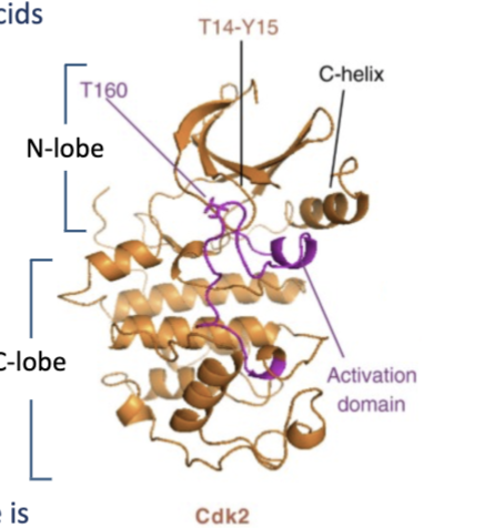
describe the structure of CDKs
250-1500 amino acids
catalytic site called activation domain which phosphorylates substrates
In the cyclin unbound (apo) form, the activation domain is inaccessible
when bound to appropriate cyclin, there is an allosteric shift in structure which pulls apart the C-helix to expose the activation domain
where are substrates of the CDK-cyclin holoenzymes phosphorylated
Substrates of CDK-cyclin holoenzymes are typically phosphorylated on S/T-P-X-K/R sequences
Ser/Thr – Pro – anything – Lys/Arg
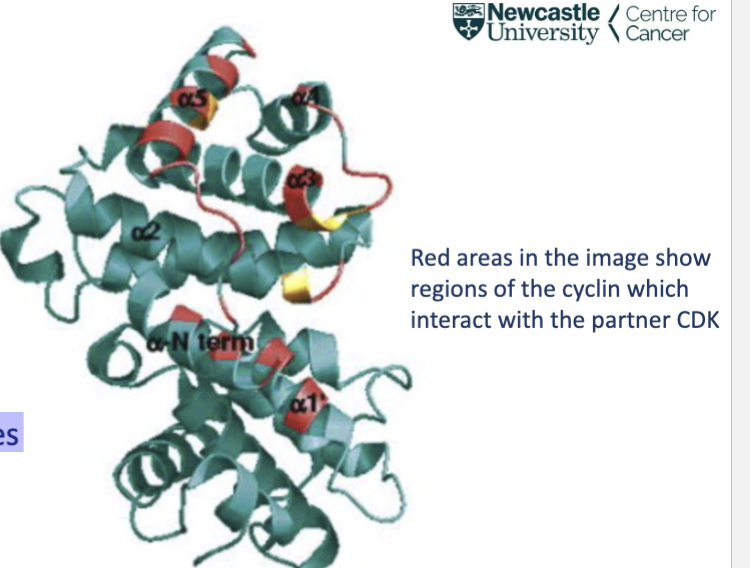
describe the structure of cyclins
30-95 kDa- 29 human cyclins
defined by the cyclin box
A 100 amino acid sequence that assembles as a 5 ∂-helix stack
The cyclin box is important for making contacts with the partner CDK
what are the 2 key mechanisms of CDK-cyclin complex regulation
they are involved in preventing prolonged (and potentially cancer driving) activation of CDK complexes:
i. Phosphorylation-dependent regulation
ii. CDK inhibitors (CKIs)
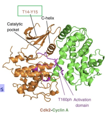
give an example of Phosphorylation-dependent regulation of CDK-cyclin complex regulation
Threonine 14 and Tyrosine 15 reside within the G loop of CDKs
They are phosphorylated by the kinase enzyme Wee1
Phosphorylation of these residues reduces interaction with cyclins and kinase activity
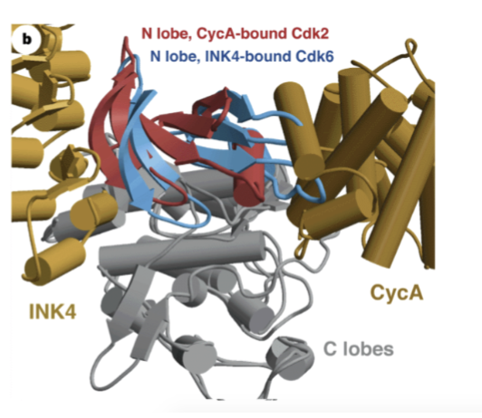
give an example of CDK inhibitor (CKIs) regulation of CDK-cyclin complex regulation
p16 (INK4), p21 and p27 are well characterised CDKis
They selectively interact with specific CDK-cyclin complexes and block binding of CDK enzymes to their cognate cyclin protein
For p16 (INK4) interaction with CDK4/6-cyclin D complexes, p16 distorts the structure of the cyclin binding region of CDK4/6 and the ATP-binding site to prevent catalytic activity
what happens to CDK-cyclin complex regulation in cancer
they are commonly dysregulated
Genes encoding Cyclin D1 and CDK4/6 are commonly amplified in numerous cancer types:
More CDK4/6-cyclin D complexes uncouple the requirement for mitogenic signalling
The CKI p16 gene is commonly lost or mutated which allows cells to pass through G1 unhindered
More rapid cell cycling; higher risk of cancer
which molecules control G1 cycling
CDK4/6-cyclin D and CDK2-cyclin E complexes
For cells to come out of G0 and enter G1, the cell must receive mitogenic signals
These signals are typically received by plasma membrane receptors and transmitted through the cell via kinase cascades (e.g. MAPK)
activation of the MAPK pathway leads to an increase in cyclin D and CDK4 levels in the cell
Cyclin D interacts with CDK4/6 and the cycle begins
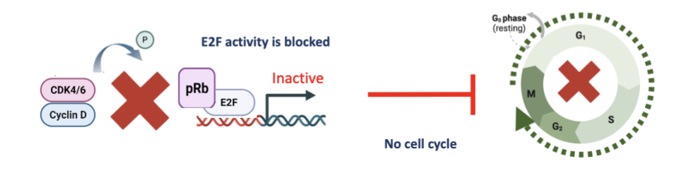
How does CDK4/6 and CDK2 activity enable cycling through G1 to S phase?
Once CDK4/6-cyclin D complex is active, it phosphorylates pRb which is a tumour suppressor
This phosphorylation blocks interaction between pRb and the transcription factor E2F.
E2F is then able to activate transcription of genes important for transition to S-phase such as:
Cyclin E: This interacts with CDK2 which further phosphorylates pRB to block it’s activity and support elevated E2F transcriptional function
Numerous genes involved in DNA synthesis which prepare the cell to allow replication of DNA in S-phase
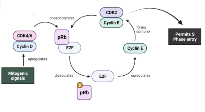
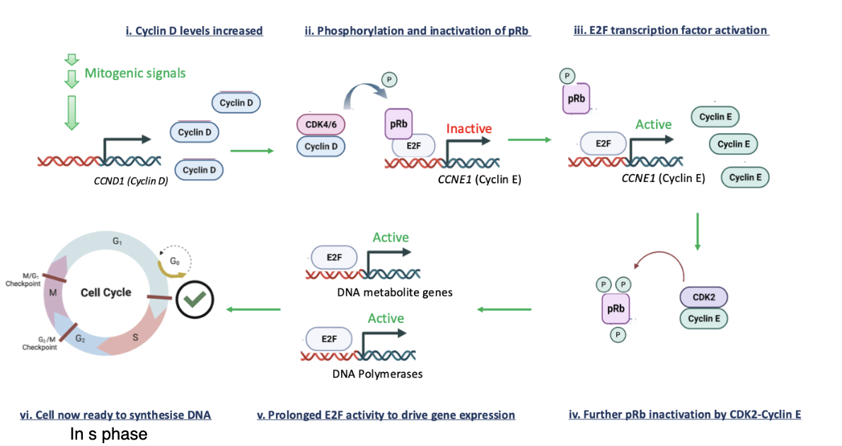
what are the 6 steps of control of G1 entry and exit to S phase
Cyclin D levels increase
phosphorylation and inactivation of pRB
E2F transcription factor activation
cell now ready to synthesise DNA in S phase
prolonged E2F activity to drive gene expression
further inactivation by CDK2-cyclin E
Mitogenic signal drives all this so can enter S phase
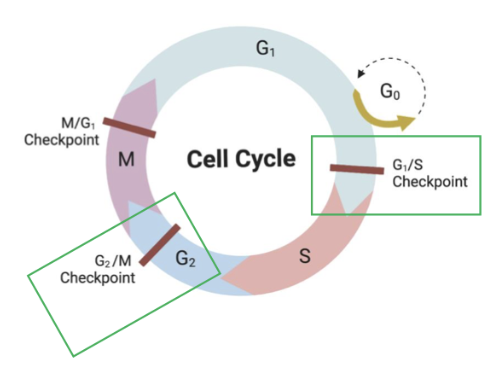
why is a G1/S checkpoint required
If damage is unrepaired prior to S-phase, this will lead to transmission of genetic lesions into cell progeny which could contribute to cancer development
Likewise, errors during DNA replication (e.g. replicative stress) need to be resolved before the cell can undergo mitosis: G2/M checkpoint
Need to also see how good the new copy of DNA is before its given to daughter cell
why is p53 deemed the guardian of the genome
as it controls key restriction points that permits transitions between G1/S and G2/M only if the DNA is error free and replicated without issue
how does p53 levels change when DNA is damaged
p53 levels are increased so that it can regulate an appropriate cell fate
Steady-state levels of p53 are low in cells to prevent unwanted interference to the cell cycle
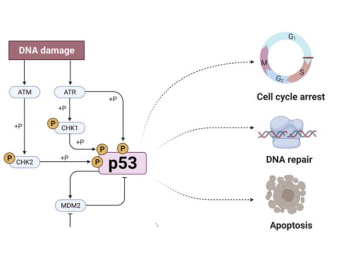
how does p53 control cell fates upon sensing DNA damage
DNA damage is ‘sensed’ by kinases ATM and ATR
These directly phosphorylate p53 to increase p53 cellular levels
P53 phosphorylation stops mdm2 from being able to degrade p53
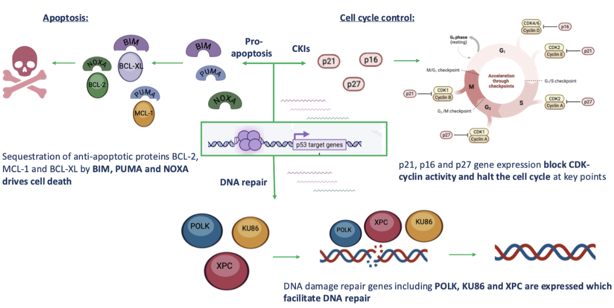
p53 can then act as a transcriptional regulator to control expression of genes involved in:
i. Cell cycle arrest
ii. DNA repair
iii. Apoptosis
Depending on the extent of DNA damage, p53 will decide whether repair is feasible or if the cell should die
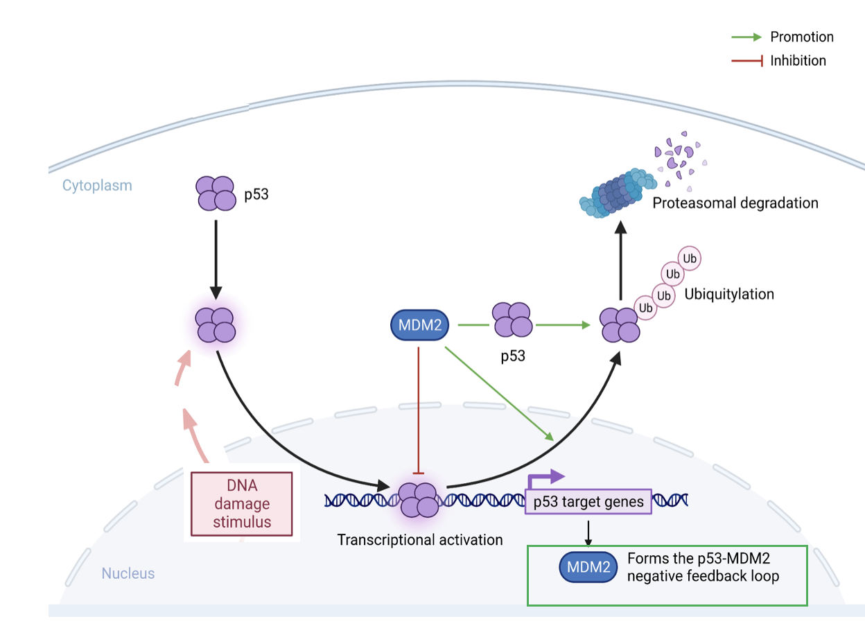
what happens to p53 in the absence and the presence of DNA damage
p53 levels are kept low by MDM2
MDM2 is an E3 ubiquitin ligase enzyme which poly ubiquitinates p53 to cause it destruction by the proteasome
Upon DNA damage, p53 phosphorylation by ATM/ATR blocks MDM2 interaction and allows p53 to tetramerise and move to the nucleus
p53 then binds and activates target genes to regulate cell fate
what genes does p53 control
Directly increases expression of Cdks
Want to elongate G1 to give enough time for DNA to be repaired
Before transitioning into S phase
why is pRb and p53 relevent to cancer
pRb was the first tumour suppressor identified and as the name suggests is strongly associated with retinoblastoma:
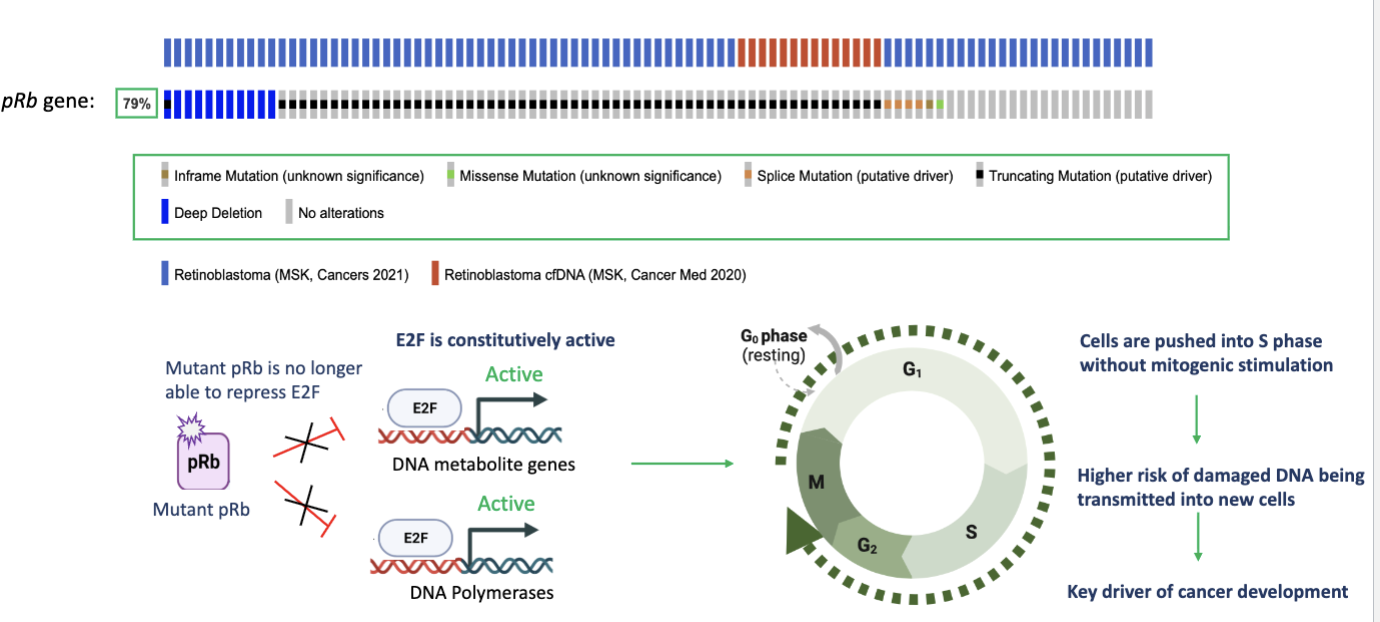
where does pRb get its name from
name from retinoblastoma
High number of patients with pRB mutations
Cell pushed into proliferation constantly
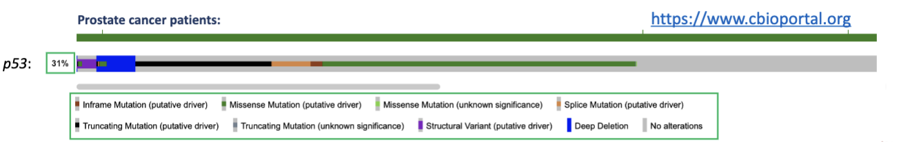
in what type of cancer is p53 the most commonly mutated gene
prostate cancer
These mutations are deleterious to p53 function; hence in cells that are p53 mutant:
i. They are unable to pause G1-S transition if DNA damage is present
ii. They have reduced capacity to repair DNA
iii. Their apoptotic potential is compromised
Interestingly, when p53 is wild-type, MDM2 gene amplification is often found in cancer which blocks p53 activity
ultimately, loss of p53 allows transmission of potentially cancerous genetic lesions to be transmitted to progeny
what are 2 key strategies for anti cancer treatments regarding cycle cycle regulators
1. Inhibition of CDKs using CDK4/6 as an example
2. Reactivation of wild-type p53 (in cells which have high MDM2 levels)
what is the rational for inhibiting CDK 4/6 in anti cancer treatments
Aberrant CDK4/6 activity either though cyclin D or CDK4/6 gene amplification drives cell cycle without appropriate mitogenic signals
The kinase activity of CDK4/6 is therefore a tractable therapeutic target…this is helped by knowing its structure
By blocking the kinase activity of CDK4/6, pRb phosphorylation will be diminished and cells will not go through
High levels of Cdk protein in cells so might not need mitogenic signal to enter cell cycle
describe CDK 4/6 inhibition with Ribociclib
Ribociclib is an ATP analogue which engages with the activation domain of CDK 4/6
it is a competitive ATP inhibitor which stops CDK 4/6 kinase activity
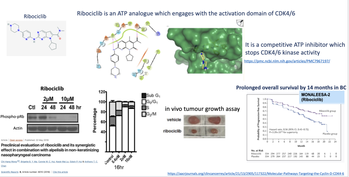
how doe most kinase inhibitors work
Make reduction in the phosphorylation in pRB so E2F can no longer bind and transcribe factors for G1- S transition
describe the reactivation of wild type p53
In many tumours, p53 is not mutated but components of the negative regulatory pathway are amplified:
this stops p53 function
Block interaction between p53 and mdm2 no more ubiquitination of p53 so more active in cells and more control
In these tumours there is an opportunity to reactivate wild-type p53 which would enable cell cycle blockade
Key to these strategies is the interaction between p53 and the negative regulator MDM2:
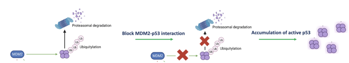
how does Nutlin-3a/RG7112 stop MDM2 binding to p53
Stops mdm2 from ubiquitinating p53 so p53 levels increase
Accumulation of p53 as no longer degraded
As negative feedback loop- mdm2 transcription also goes up since -53 increases
Mutant p53 cant do anything cand bind DNA or drive anti proliferative effects
Only efficacy In patients who are wild type p53
So in clinical trial- do sample of only wild type patients as won’t make a difference In those with mutated