Looks like no one added any tags here yet for you.
What is the hierarchy of muscle structure?
Muscle
Fascicles
Muscle Fibers (Cells)
Myofibrils
Thick and Thin Filaments
What is the hierarchy of the connective tissue that surrounds muscle?
Epimysium - surrounds the entire muscle
Perimysium - surrounds muscle fascicles (groups of fibers)
Endomysium - surrounds individual muscle fibers
Of the three connective tissue layers, which layer contributes the most to the resistance to stretch?
Perimysium
All can contribute to tightness, but the perimysium contributes the most
What is part of the epimysium’s physical make-up and how does it impact its function?
Tough outer layer that contains an abundance of collagen
This makes it resistant to stretch
Which of the three layers of connective tissue surrounding a muscle contains blood vessels and nerves?
Perimysium
The endomysium is just outside of the ___________.
The endomysium is just outside the sarcolemma
The sarcolemma is the area of…
Metabolic exchange between the capillaries and muscle fibers
T/F: The endomysium transmits some of the force to the tendon
True
What is the basal lamina?
The scaffolding that surrounds the muscle cell
If the basal lamina is damaged, what is not able to occur?
The muscle is not able to heal properly —> will lose its contractile abilities!!
It will not be replaced with muscle
It will instead heal/be replaced with CT
Scarring / Type I Collagen
Muscle is ____-nucleated
Muscle is multi-nucleated
Where are muscle cells located in mature muscle? How do they adapt?
On the outer edges of the muscle
The nuclei adapt at differing rates along the length of the muscle
Where are satellite cells located? How do these cells replicate?
On the outside of the basal lamina
The cells divide and one daughter cell enters the muscle while the other one remains on the outside
What is the POWERHOUSE OF THE CELL?
MITOCHONDRIA
What type of respiration/metabolism does the mitochondria use?
Aerobic metabolism/respiration
What does the mitochondria generate?
ATP
How does mitochondria density vary?
Varies depending on the cellular processes of the muscle
Cytoplasm also allows for ATP generation through…
Anaerobic glycolysis
What is a byproduct of anaerobic glycolysis?
Lactic acid
How quickly can we get rid of lactic acid?
~1 hour (quickly!)
T/F: Lactic acid causes delayed onset muscle soreness
False! Our bodies get rid of lactic acid within ~1 hour (and lactic acid can also be used as an energy source)
When we say that someone “hits the wall”, what are we referring to?
An individual who ran out of ATP (really only occurs in ultra-marathoners)
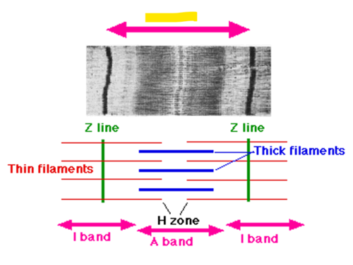
What is a sarcomere?
The contractile unit of a muscle fiber
Where are the Z-lines and what is their function?
Located at the end of sarcomere with supportive proteins to hold actin and myosin in place
Where is the A-band?
The space of the sarcomere where both myosin and actin are
Where is the I-band?
The space of the sarcomere where actin is
During contraction the A-band…
Stays the same
During contraction the I-band…
Shortens
Sarcomeres have more proteins besides actin and myosin. What is their function?
To stabilize the cell
With actin and myosin, which is the thin filament and which is the thick filament?
Actin — Thin Filament
Myosin — Thick Filament
Actin binds with ______ to generate force and _______ the sarcomere.
Actin binds with myosin to generate force and shorten the sarcomere
What is the function of tropomyosin?
Stabilizes the actin filament
What is the function of troponin?
Influences the position of tropomyosin with a bond to Ca2+
What is the position of the Troponin / Tropomyosin complex at rest?
At rest (without Ca2+) the Troponin/Tropomyosin complex is covering up the binding site of myosin
Therefore it controls whether or not contraction occurs (also plays a role in controlling the speed of contraction)
What are the two components of myosin?
Heavy chains
Light chains
Where are the heavy chains and light chains located?
Heavy chains are located on the ends of myosin
Light chains are located in the middle of myosin
What is the function of the heavy chains of myosin?
Molecular motor for muscle contraction
This is where the ATP is attaching and where the actin binding site is
What is the function of the light chains of myosin?
Influences the contraction velocity of the sarcomere
Modulates the kinetics of cross-bridge cycling
Myosin has multiple heavy and light chains, so as actin comes across myosin, its moving ________ and _______.
Myosin has multiple heavy and light chains, so as actin comes across myosin, its moving laterally and twisting
What is the function of nebulin?
Holds actin in place
What is titan? What is part of its function?
Titan is a non-contractile protein that provides passive tension within the muscle fiber (helps with stiffness of the muscle)
It also helps hold myosin in place
What is the function of dystrophin?
Gives the muscle and musculotendinous junction support
Maintains alignment and arrangement of the Z-line
What is desmin and what is its function?
Desmin is a non-contractile protein that stabilizes the alignment of adjacent sarcomeres
Generally, non-contractile proteins will…
Generate passive tension when stretched
Provide internal and external support and alignment of muscle fibers
Help transfer the active force of the muscle
Titan bears most of the _______ load in muscle
Titan bears most of the passive load in muscle
T/F: Titan is the largest protein ever discovered
True
Titan plays a critical role in…
Organizing the developing sarcomere (due to it’s tremendous length)
Titan is ideally situated to serve as a _______ for altered muscle mechanical conditions such as….
Titan is ideally situated to serve as a “sensor” for altered muscle mechanical conditions such as chronic length change or chronic force change
Titin is a(n) __________ filament, but unlike actin, myosin, or desmin, which are filaments composed of ___________ molecules, titin is a _________ molecule filament.
Titin is an intramuscular filament, but unlike actin, myosin, or desmin, which are filaments composed of polymerized molecules, titin is a single molecule filament.
What is PVEK?
Elastic component of titan that is important for maintain stiffness in eccentric contractions
ATP has to be present for the myosin head to be ________ and for Ca2+ to ________.
ATP has to be present for the myosin head to be cocked and for Ca2+ to leave
Why/how does rigor mortis occur?
Occurs due to Ca2+ being stuck in the cell (since there is no ATP present to transport it out)
Muscle is a(n) _________ tissue!
Muscle is an excitable tissue!
What is the function of the cell membrane?
Prevents or allows ions in or out of the cell by a system of membrane receptors and protein channels
When would it be appropriate to directly stimulate a muscle contraction (bypassing an alpha motor neuron)?
With a patient who has a spinal cord injury and cannot perceive pain
What are the three types of protein channels that the membranes of muscle and nerve cells have?
Chemical Dependent (Ligand) Channel
Voltage Dependent Channel
Potassium Channel
When does a chemical dependent (ligand) channel open?
Opens only when a particular chemical neurotransmitter fits into a very specific receptor (ex. sodium channel)
When does a voltage dependent channel open?
Opens only when the charge difference across the membrane becomes -50mV
When does a potassium channel open?
Opens when a specific chemical neurotransmitter fits into a receptor on the outside of the membrane or when the inside voltage reaches +30mV
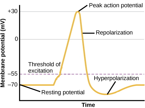
What is the resting potential of a cell membrane?
-70 mV (the difference in electrical charge between the inside and outside of cell membrane)
What is occurring during depolarization?
Occurs during the process of an action potential when sodium is rushing into the cell causing the interior to become more positive (about -55mV)
What is the charge of the action potential?
+30 mV (neural impulse; brief electrical charge that travels down the axon)
What is occurring during repolarization?
Return of the cell to resting state, caused by re-entry of potassium into the cell (while sodium exits the cell)
What is occurring during hyperpolarization?
Movement of membrane potential of a cell away from resting potential in a more negative direction (the refractory period of contraction)
When Ca2+ is released from the sarcoplasmic reticulum, what follows?
This exposes the the binding sites on the thin filament (actin)
Ca2+ binds to troponin complex
Tropomyosin is pulled aside to expose binding site
What is the contraction cycle?
Exposed binding sites on actin allow the contraction cycle to occur
Cross-bridge binds actin to myosin
Cross bridge pulls actin filament (power stroke)
ADP and Phosphate released from myosin
New ATP binds to myosin, causing linkage to release
ATP splits, which provides power to “cock” the myosin cross-bridge
What occurs during muscle relaxation (at the level of the sarcomere)?
Active transport of Ca2+ back into sarcoplasmic reticulum
This requires ATP —> makes myosin binding sites unavailable
What are the steps for muscle contraction? (11 steps)
Motor neuron releases neurotransmitter (Ach)
Neurotransmitter interacts with muscle membrane receptor
Receptors opens sodium channel (allowing Na+ to slowly move into cell)
Movement of Na+ into cell causes the electrical potential to change from -70mV (resting potential) toward an excitatory post-synaptic potential of -50mV (threshold to open voltage channel)
Voltage channels open allowing Na+ to flood the cell, resulting in electrical potential change of +30mV
The electrical potential change is depolarization
The +30mV is the action potential
This wave of depolarization spreads across the muscle membrane, also depolarizing the T-tubular system
When T-tubules depolarize, Ca+ is released from sarcoplasmic reticuli
Ca+ diffuses throughout muscle cell cytoplasm to interact with troponin causing the tropomyosin on the actin to uncover the myosin-binding sites
Allows myosin cross-bridges on the myosin to interact with myosin-binding sites on the actin molecule pulling the actin and myosin molecules closer to one another
Results in shortening of the myofibril as the stacks of actin and myosin move closer to one anothe
As all the myofibrils in a particular muscle cell shorten, the entire muscle cell contracts
What is rate coding?
Frequency of an action potential; the cell must meet a certain threshold to produce a smooth contraction
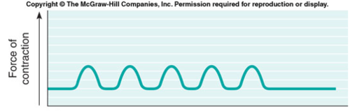
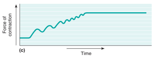
The following image represents….
A muscle contraction after a discrete stimuli
The following image represents…
A muscle where the stimuli are delivered more frequently (muscle does not have time to completely relax). Contraction force increases as the individual twitches are more frequent.
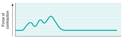
The following image represents…
A more complete fusion of twitches because the stimulus are delivered as a faster rate (Tetanus). This leads to a smooth continuous contraction of maximal force.
What is the typical minimum frequency to achieve tetanus?
35 - 50 Hz
(35 Hz for a smaller muscle to 50 Hz for a larger muscle)
What is a motor unit?
A motor neuron and all of the muscle fibers it innervates
What are the different muscle fiber types?
Type I
Type IIa
Type IIx
When considering the different muscle fiber types, most muscle is split…
50/50 (composed evenly)
What muscle fiber type would postural muscles (like the soleus and erector spinae) have?
Type I (slow-twitch)
T/F: You can change muscle fiber type through training
False; you cannot change muscle fiber type through training, you can only make tissue more aerobic
What are the characteristics of a Type I muscle fiber?
Slow contraction time
Small motor neuron size
High resistance to fatigue
Used for aerobic activity
Low force production
High mitochondrial, capillary, and oxidative density
Low glycolytic capacity
Fat is major storage fuel
What are the characteristics of a Type IIa muscle fiber?
Fast contraction time
Large motor neuron size
Intermediate resistance to fatigue
Used for long-term anaerobic activities
High force production
High mitochondrial, capillary, and oxidative density
High glycolytic capacity
Phosphocreatine and glycogen are the major storage fuel
What are the characteristics of a Type IIx muscle fiber?
Very fast contraction time
Very large motor neuron size
Low resistance to fatigue
Used for short-term anaerobic activities
Very high force production
Low mitochondrial, capillary, and oxidative capacity
High glycolytic capacity
Phosphocreatine and glycogen are major storage fuel
What is Henneman’s Size Principle?
Increased force demands leads to increased number of motor units to be activated through either:
Increased amount/number of motor units
Increased frequency of motor unit firing
In what order are the different muscle fiber types typically recruited (according to Henneman’s Size Principle)?
Type I —> Type IIa —> Type IIx
(however, does not always work this way, think about if were are going to do a sprint, all would be recruited simultaneously)
When does Henneman’s Size Principle not really apply?
When we have to perform an activity with power and intensity
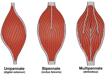
What is muscle architecture?
Arrangement of fibers relative to the axis of force generation
Fiber diameter is similar (regardless of the muscle)
Design of muscle
How does the muscle produce force?
How is the muscle able to generate torque?
T/F: Muscle mass is always directly related to the functional aspect of the muscle
False
Muscle mass may or may not be directly related to any functional aspect of the muscle (large muscle does not always = advantage)
What is the best way to estimate the amount of force a muscle can produce?
Arrangement of fibers is most critical part of understanding force production of a muscle
What is muscle length?
The distance measured from the proximal tendon to the distal tendon
What is fiber length? In comparison to the muscle, how long is it typically?
Measurement of a single fiber length
~1/3 length of entire muscle
What is pennation angle?
The angle between the tendon and the fiber orientation
What is the typical pennation angle?
Usually between 0 - 30 degrees
How does pennation angle affect the amount of force of a muscle?
Force generated will be less along the tendon
BUT, design allows us to pack more muscle into cross sectional area → so overall force production is greater (than a fusiform muscle of equal size)
What is the physiological cross-sectional area?
The amount of active proteins available to produce a contraction
How is the physiological cross-sectional area measured?
Measured by perpendicularly cutting through the muscle fibers
Physiological cross sectional area is proportional to…
Maximal force production
What are the three types of pennation forms?
Unipennate
Bipennate
Multipennate
T/F: PSCA will almost always be greater than CSA
True
What is fusiform muscle built for?
Speed/Velocity
What is pennate muscle built for?
Force production