3a exchange and transport systems
1/21
Earn XP
Description and Tags
RP5
Name | Mastery | Learn | Test | Matching | Spaced | Call with Kai |
|---|
No analytics yet
Send a link to your students to track their progress
22 Terms
surface area to volume ratio
an organism’s surface area to volume ratio affects how quickly substances are exchanged. small organisms have a larger surface area to volume ratio than large organisms
substance exchange in single celled organisms
in single celled organisms, substances diffuse directly into the cell. the diffusion pathway is small so diffusion is quick
substance exchange in multicellular organisms
diffusion across the outer membrane is slow due to some cells being deep within the body and a low surface area to volume ratio
therefore these organisms have specialised exchange organs. the mass transport system carries substances to these organs
how different bodies affect heat exchange
small organisms lose lots of heat because of their large surface area to volume ratio. they have a high metabolic rate to generate lots of heat. this also applies to animals with a compact shape
behavioural and physiological adaptations to aid exchange
animals with high surface area to volume ratio lose more water by evaporation. some animals have adapted their kidney strucutre to lose less urine
small animals living in cold area eat high energy foods for their high metabolic rates
small mammals can have fur and hibernate
larger organisms in hot areas find it hard to keep cool. they often develop features to maximise their surface areas or stay in the water
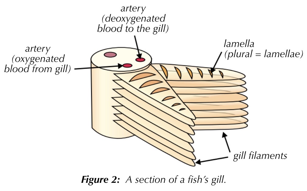
gill structure
water enters the fish through its mouth and passes out through the gills. each gill is made out of lots and lots of thin plates called gill filaments which give it a large surface area
the gill filaments are covered in tiny lamellae, which further increase surface area. this speeds up diffusion
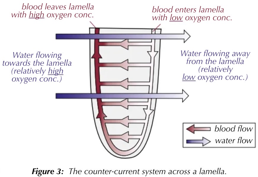
the counter-current system
in fish gills, blood flows through the lamellae in one direction and water flows over them in the other direction
water with a higher oxygen concentration always flows next to blood with a lower oxygen concentration. therefore a steep concentration gradient is always maintained
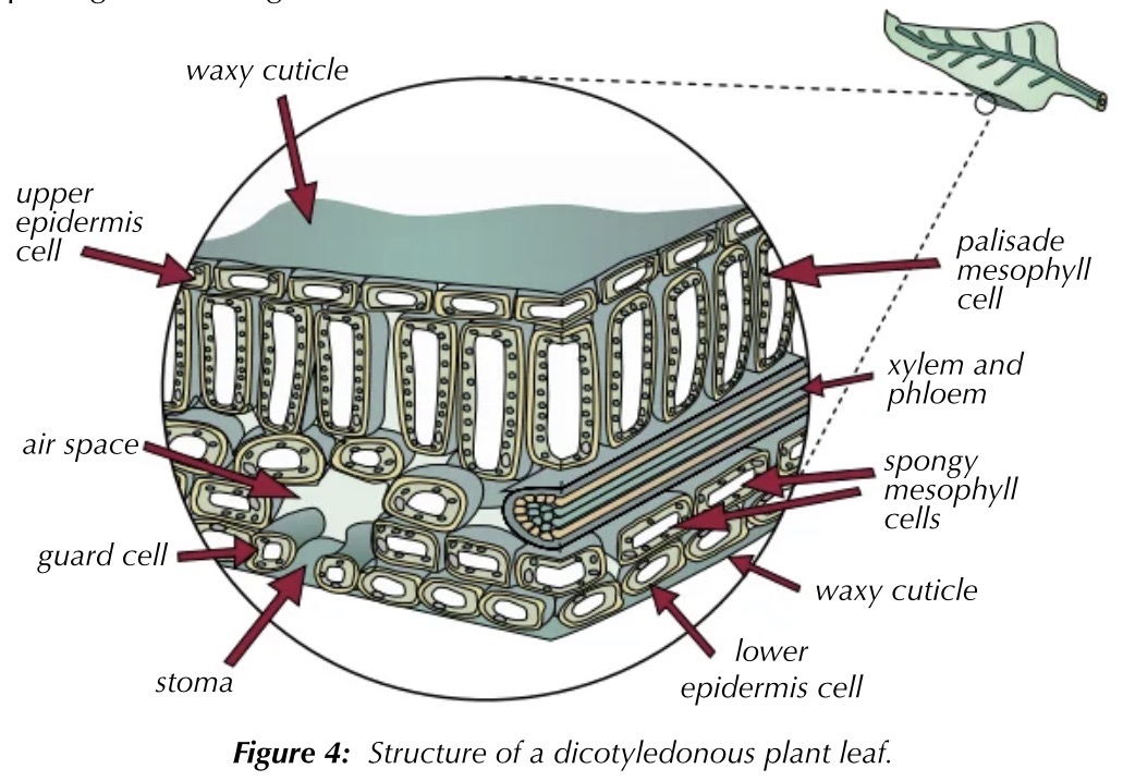
gas exchange in dicotyledonous plants
plants need CO2 for photosynthesis and O2 for respiration
the main gas exchange surface in plants is the mesophyll cells, which have a large surface area. mesophyll cells are inside the leave and are reached through pores in the epidermis (stomata). guard cells allow stomata to open and close
gas exchange in insects
terrestrial insects (land insects) have small air filled pipes called tracheae. air moves into the tracheae through small pores called spiracles, that have thin permeable walls that go to individual cells, so oxygen diffuses directly
CO2 from the cells moves down its own concentration gradient toward the spiracles to be released
controlling water loss
exchanging gases can often lead to a loss of water
if insects are losing too much water, they close their spiracles. they also have a waxy waterproof cuticle
plants’ stomata are kept open during the day for gas exchange. water causes guard cells to be turgid, opening the pores. dehydrated flaccid guard cells close the pores
xerophyte adaptations
stomata sunk in pits trap water vapour, reducing the concentration gradient of water between the leaf and the air, reducing evaporation
hairs on the epidermis also trap water vapour
curled leaves with the stomata inside protect them from wind
less stomata means less water loss
thicker waxy waterproof cuticles reduce evaporation
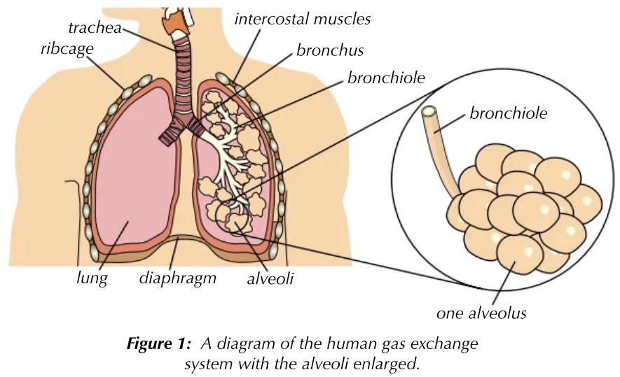
lung structure
as you breathe in, air enters the trachea. this spilts into two bronchi, one leading to each lung. these branch off into bronchioles. the end of the bronchioles add small air sacs called alveoli. this is where gases are exchanged
the intercostal muscles are between the ribs (internal and external)a
inspiration and expiration
during inspiration the external intercostal and diaphragm muscles contract, causing the ribcage to move up and out, increasing the volume of the thoracic cavity. lung pressure then decreases to below atmospheric pressure, forcing air into the lungs
during expiration the external intercostal abd diaphragm muscles relax, causing the ribcage to move down and in, and the diaphragm curves in, decreasing the volume of the thoracic cavity. lung pressure then increases to above atmospheric pressure, forcing air out of the lungs
normal expiration is passive
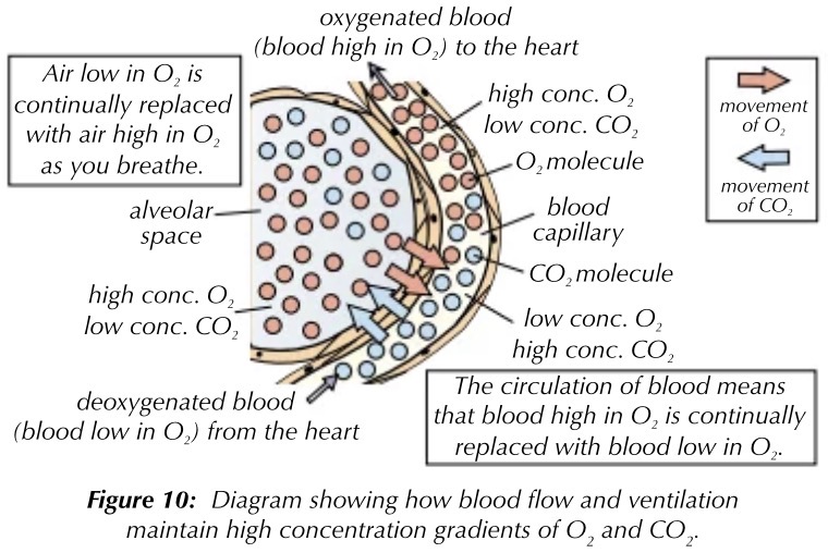
gas exchange in the alveoli and alveoli structure
the wall of each alveolus is made from a single layer of thin, flat cells called alveolar epithelium. this contains elastin, allowing the alveoli to recoil
O2 diffuses out of the alveoli, across the alveolar epithelium and the capillary endothelium, into haemoglobin. CO2 diffuses into the alveoli from the blood. there is a steep concentration gradient
tidal volume, ventilation rate, FEV1, FVC definitions
tidal volume: the volume of air in each breath - usually around 0.4/5 dm³ in adults
ventilation rate: the number of breaths per minute - about 15 breaths usually
forced expiratiory volume 1: the maximum volume of air that can be breathed out in 1 second
forced vital capacity: the maximum volume of air it is possible to breathe forcefully out after a really deep breath in
pulmonary tuberculosis
a lung disease caused by bacteria. immune system cells build a wall around the bacteria in the lungs after being infected, forming small, hard lumps known as tubercules. infected tissues within the tubercules die and the gas exchange surface is damaged, so tidal volume is decreased. this may lead to fibrosis
reduced tidal volume means that less air is inhaled with each breath. this increases ventilation rate
symptoms include a persistent cough, coughing up blood/mucus, chest pain, shortness of breath, fatigue
fibrosis
fibrosis is the formation of scar tissue in the lungs, usually as a result of infection of exposure to asbestos/dust. scar tissue is thicker and less elastic, so lungs are less able to expand
FVC and tidal volume are reduced and there is a reduction in the rate of gas exchange. sufferers have a quicker ventilation rate
sympoms include shortness of breath, a dry cough, chest pain, weakness
asthma
asthma is a respiratory condition where the airways become inflamed and irritated, usually due to an allergic reaction. during an asthma attack, the bronchiole lining contracts and mucus is produced, restricting the airways. less oxygen enters the alveoli and blood.
reduced air flow reduces FEV1
symptoms include wheezing, a tight chest, shortness of breath. symptoms come suddenly during an attack and can be reduced using drugs that relax the bronchioles (inhalers)
emphysema
disease caused by smoking or long term exposure to air pollution, since foreign particles become trapped in the alveoli. this causes inflamation, attracting phagocytes, which produce an enzyme that breaks down elastin in the alveoli. loss of elastin means the alveoli can’t recoil to expel air well, leaving it trapped. the surface area of the alveoli is also reduced, decreaing rate of gas exchange
people with emphysema have an increased ventilation rate to increase the amount of oxygen reaching their lungs
symptoms include shortness of breath, wheezing
required practical 5
dissection of a mammalian lung
wearing a lab coat, lay the lungs on a cutting board
put the lungs in a clear plastic bag, to stop bacteria being released into the room. attach a piece of rubber tubing to the trachea and pump air into the lungs
the trachea is supported by rings of cartilage. cut the trachea lengthways down the gap in the rings, using dissecting scissors or a scalpel
cut down one of the bronchi and cut off a piece of the lung
wash your hands after
required practical 5
dissection of a bony fish
wearing a lab coat, place the fish on a cutting board
gills are located on each side of the fish’s head. theyre supported and protected by gill arches and an operculum
push back the operculum and use scissors to carefully remove the gills. cut each gill arch through the bone at the top and bottom
you can see the gill filaments
required practical 5
dissection of a large insect
fix the insect to a dissecting tray using dissecting pins
cut and remove a piece of the exoskeleton to view the tracheae
fill the abdomen with saline solution, and you will see a network of thin, silvery-grey tubes - the tracheae
you can view the tracheae under an optical microscope, where you will see chitin also