Lecture 7: Sound
1/37
There's no tags or description
Looks like no tags are added yet.
Name | Mastery | Learn | Test | Matching | Spaced | Call with Kai |
|---|
No analytics yet
Send a link to your students to track their progress
38 Terms
What are sound waves
longitudinal oscillation
sinusoidal wave
compression of air
travels through a medium
period and wavelength
period → time taken for one up-down cycle
must move a distance of one 1 length
wavelength → distance between two identical points on a repeating wave
what is the relationship between frequency, wavelength, and velocity
distance/time = speed = wavelength/period
the frequency (in cycles per second or Hz) is 1/period
speed = wavelength x frequency
Trends within frequencies of sound
shorter length of wavelength → higher freq of sound
has to be same medium to compare
longer length of wavelength:
bend more
more time to oscillate compared to shorter
speed
depends on the medium the waves are travelling through
adult human voice and its frequencies and wavelength
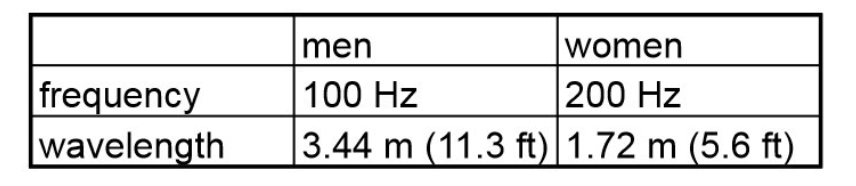
Humans can hear sounds from 20Hz - 20,000 Hz
Explain the ascending auditory pathway, and key features, especially in comparison to the somatosensory pathway
Comparison:
each hemisphere receives input from both ears
each ear projects info to both hemispheres
more bilateral system
information is relayed through the ear
from the spiral ganglion, it goes through the CN VII (cochlear nerve)
synapses at the rostral medulla
central cochlear nucleus synapses at the mid-pons and forms the superior olivary complex
the middle and dorsal cochlear nucleus synapses next at the caudal midbrain and decussates at the mid-pons
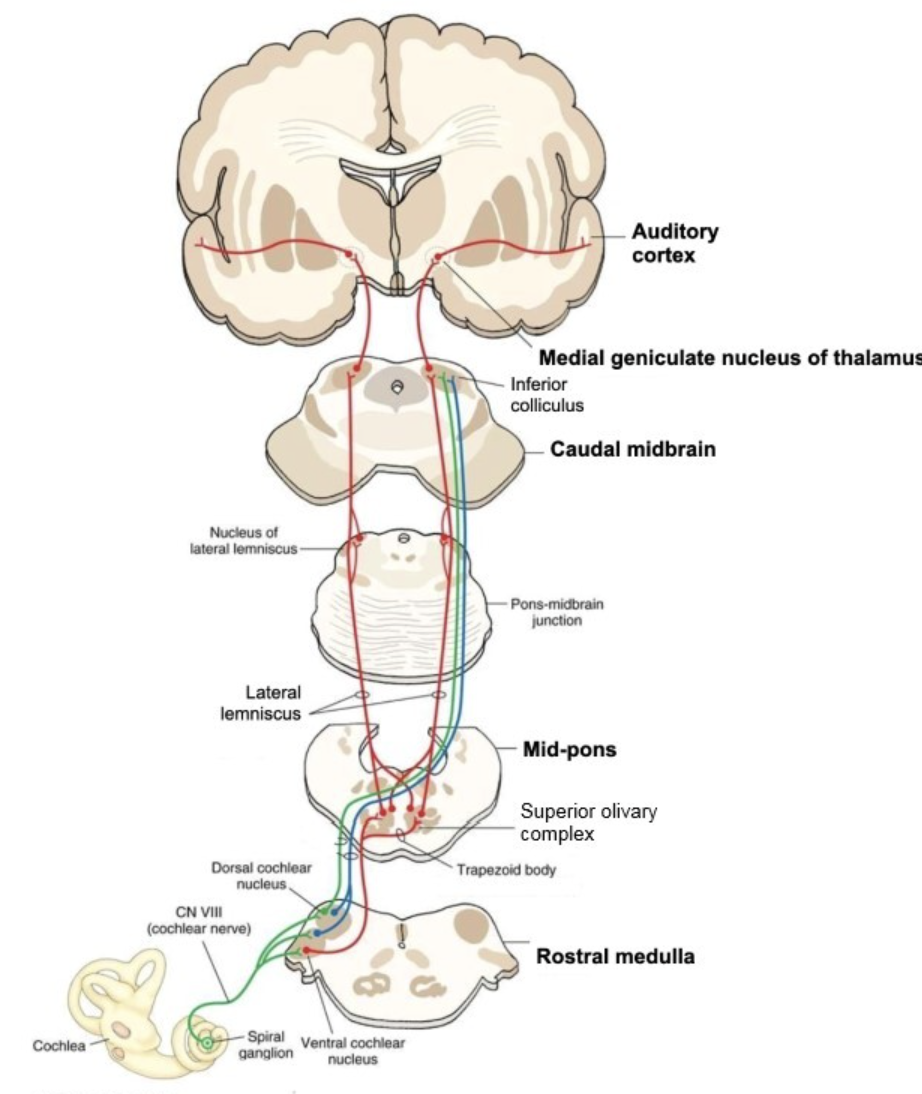
Auditory cortex
two parts:
primary
secondary (belt areas)
In primary:
spilt up to regions where different ranges of frequencies are heard
500-16,000Hz
apex →base of cochlea
The superior olivary complex contains how many and which nuclei for what specific role?
2 nuclei for sound localization:
lateral superior olive (LSO)
medial superior olive (MSO)
Explain the early auditory pathway
sound starts in cochlea
converts sound vibrations into neural signals
auditory nerve enters the brainstem, it synapses in the cochlear nuclei which has 3 divisons:
Dorsal cochlear nucleus (DCN)
Posteroventral cochlear nucleus (PVCN)
Anteroventral cochlear nucleus (AVCN)
information goes bilaterally to the superior olive
pathways from the cochlear nuclei project to both sides of the brainstem
bilaterally helps the brain to compare:
time differences
intensity differences
mid pons: first place where input from both ears meet
two major structures
MSO
LSO
overall:
Cochlea → auditory nerve → cochlear nuclei → superior olive
Cochlear nuclei send signals to both sides
Superior olive compares the two ears → sound localization
Superior Olivary Nuclei: LSO
monitors interaural intensity difference for high-freq sounds
the head blocks sounds that have wavelengths smaller than the diameter of the head
freq=speed/wavelength = 344m/s (speed of sound) / <20cm (diameter of human head) » 2kHz
high freq sounds of greater than 2000 Hz are blocked by the head = sound shadow
ear closest to sound hears it well, the other does not
How does the LSO determine where a sound is coming from → what is this called and how does it work
Interaural intensity differences (IIDs)
sound reaches one ear louder than the other
creates:
strong input to left cochlear nucleus
weaker input to right cochlear nucleus
The LSO receives signals by each cochlear nucleus sending two projections:
excitatory projection to the ipsilateral LSO
left cochlear nucleus → left LSO (glutamatergic - excitatory glutamate)
excitatory projection to the contralateral MNTB
left cochlear nucleus → right MNTB (glutamatergic)
MNTB is an inhibitory interneuron (receives glutamate)
MNTB neurons send GABA/glycine inhibition to the ipsilateral LSO
Left cochlear nucleus → Right MNTB → Inhibits Right LSO

the LSOs
left:
strong excitation from left cochlear nucleus
receives weak inhibition from right MNTB
left LSO fires a lot
right:
receives weak excitation
receives strong inhibition from left MNTB
right LSO firing suppressed
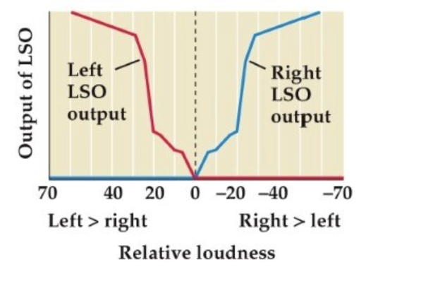
Overall:
LSO decides sound direction based on loudness differences
it uses:
ipsilateral excitation
contralateral inhibition (via the MNTB)
Superior Olivary Nuclei: MSO
Medial superior Olive
monitors interaural time difference for low-frequency sounds
the head does not block sounds that have wavelengths greater than the diameter of the head

low frequency sounds (less than 2000Hz) are not blocked
wave length has to be greater than 20cm
since there is no sound shadow, time difference is detected
How does the MSO compute sound location - what is this called and how does it work
Interaural time differences
→ Jeffress Model, 1948
sound reaches left ear first
action potential begins travelling toward MSO
axons from each ear have different lengths → different conduction delays
hence signals from the left ear can be delayed enough so that they arrive simultaneously with signals from the right ear at certain MSO neurons
each MSO neuron fires strongly only when both ears’ inputs arrive at the same time
due to: strong, fast EPSPs, precise timing, voltage-gated channels that prefer synchronized input
different MSO neurons will get simultaneous input at different ITDs
ex:
A neuron on the far left MSO fires when left ear early
A neuron on the far right MSO fires when right ear early
A middle neuron fires when both ears equal timing
brain reads out which MSO neuron fired → sound location
called a place code
which MSO neuron fires tells you:
how big the ITD is
which ear the sound was closer to
where the sound source is
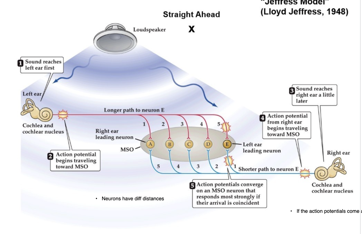
Overall:
if sound reaches right ear later than the left ear, the AP travelling towards the MSO will take the route that will allow for both of them to converge at the MSO neuron at the same time
this allows for this neuron to respond most strongly
parts of the outer ear and their role
pinna = funnels waves into the ear canal
ear canal = tube that directs sound to the eardrum
concha
parts of the middle ear and their function
eardrum (tympanic membrane) = separates the outer and middle ear, vibrating when sound hits it
ossicles = three tiny bones that amplify the vibrations from the eardrum
malleus/hammer
incus/anvil
stapes/stirrup
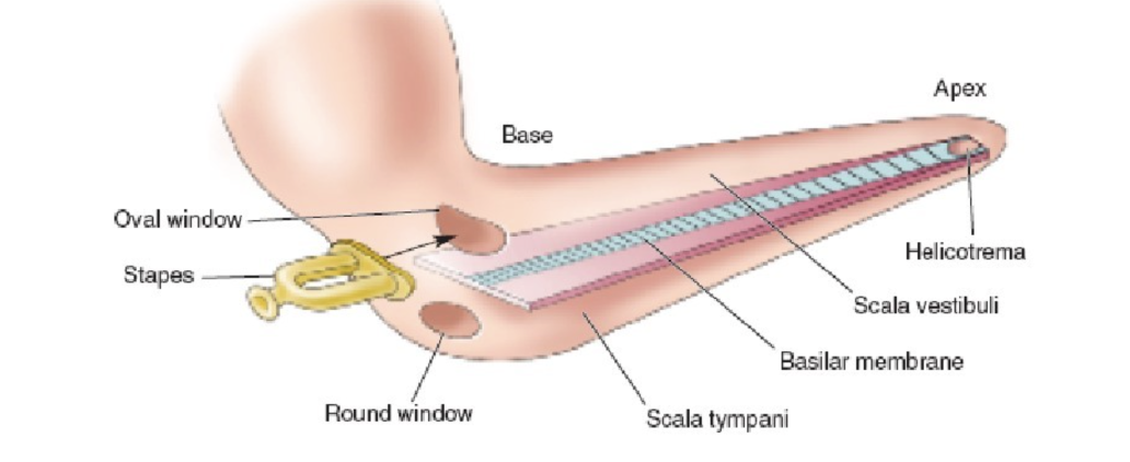
base of stapes in oval window
parts of the inner ear and their function
Structure | Function |
|---|---|
Cochlea | Converts sound to neural signals |
Basilar membrane | Frequency separation (tonotopy) |
Inner hair cells | Primary sensory receptors for sound |
Outer hair cells | Amplify sound, sharpen tuning |
Semicircular canals | Detect rotational head movement |
Utricle | Detect horizontal linear acceleration & head tilt |
Saccule | Detect vertical linear acceleration & head tilt |
Vestibular nerve | Sends balance signals to brain |
Auditory nerve | Sends sound information to brain |
Attenuation reflex muscles
protective mechanism in the middle ear that reduces the transmission of loud sounds to the inner ear
controlled by two tiny muscles:
tensor tympani muscle
attached near the ossicles
tenses the tympanic membrane and reduces vibrations from self-generated sounds
stapedius muscle
attached near the oval window
pulls the stapes away from the oval window
primary muscle responsible for this reflex
reduces transmission of low-frequency loud sounds
protects the cochlea from damage
Pressure amplification in the middle ear and why it is needed
Needed:
air is easy to move, fluid is not, hence the pressure needs to be amplified to move the fluid in the inner ear
2 mechanisms:
the oval window is much smaller than the tympanic membrane. Thus the force is funnelled to a smaller area, increasing pressure
the ossicles act like a lever system (mechanical advantage)
coverts large, low-force movements of the eardrum into small, high-force movements of the stapes
the tympanic membrane moves a lot but with little force
the stapes moves only 1/10 as much, but pushes the oval window with much greater force
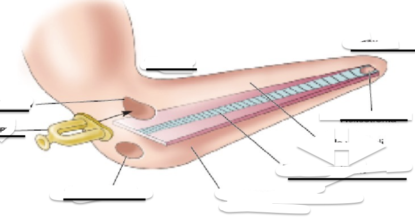
Name the following in the cochlea uncoiled underlined:
Explain the Basilar membrane → base and apex and attributes corresponding to them
base:
narrow, stiff
high frequency sound
20 kHz
sound that produces max vibration
150 micrometers wide
apex:
wide, floppy
low frequency sound
20Hz
500 micrometers wide
other animals and their auditory bandwidth
cat and dog → 20 to 40k
bat is higher frequency than humans to around 160k
elephant and mole is lower frequencies → 0-100
Describe the tonotopy of the basilar membrane and the auditory nerve fibers
axons of the auditory innervate throughout the basalar membrane
lowest points on the curves is where the soft sounds are
each axon has a preferred frequency
Name the following in the cochlea:
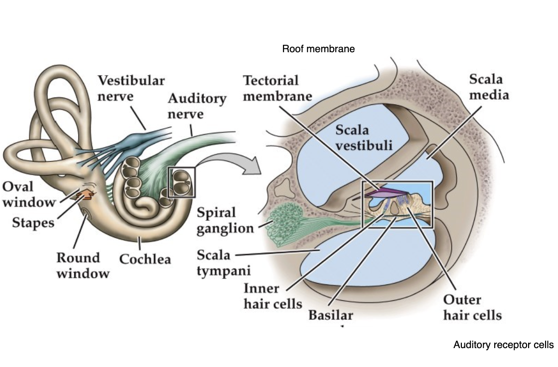
Explain the concentrations of ions within the cochlea: organ of corti and its 3 chambers
three chambers:
scala vestibuli (top chamber)
filled with perilymph (low K+m like normal extracellular fluid)
receives sound vibration from the oval window
2. Scala media (middle chamber)
Filled with endolymph (VERY high K⁺ ~150 mM — like intracellular fluid!)
This high potassium environment is essential for hair cell activation
Contains the Organ of Corti, where sound transduction happens
3. Scala tympani (bottom chamber)
Also filled with perilymph
Connects to the round window, which releases pressure
Fluid | Location | K⁺ Concentration | Purpose |
|---|---|---|---|
Endolymph | Scala media | High K⁺ (~150 mM) | Drives K⁺ into hair cell stereocilia during sound transduction |
Perilymph | Scala vestibuli & tympani | Low K⁺ (~7 mM) | Surrounds the hair cell bodies |
Arrangement of inner and outer hair cells
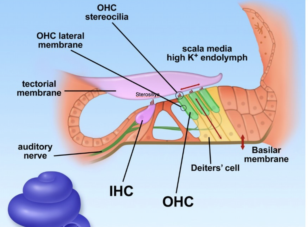
Inner hair cells (IHCs) → the true sensory receptors
Outer hair cells (OHCs) → the amplifiers
Tectorial membrane → the structure stereocilia push against
Basilar membrane → vibrates with sound
Endolymph → high-K⁺ fluid bathing stereocilia
Perilymph (below basilar membrane, not labeled) → low-K⁺ fluid bathing the hair cell bodies
what are stereocilia
tiny, hair-like projections on top of inner and outer hair cells in the cochlea
bundles of stiff, rod-like structures arranged in rows of increasing height
they are not true cilia → actin-filled microvilli that function as mechanical sensors for sound
make them rigid
What they do?
covert mechanical vibration → neural signals
Explain what mechano (acoustical) transduction is
coverting mechanical sound energy into neural (electrical) signals
Describe the process of mechano-acoustical transduction
sound waves vibrate the basilar membrane
floor of organ of corti moves up and down
different frequencies vibrate different locations (tonotopy)
shearing motion bends stereocilia
tectorial membrane stays relatively still
hair cells are sheared between them
stereocilia (multiple) bend toward or away from the tallest sterocilium (one)
tip links stretch → ion channels open
on top of the stereocilia are mechanically gated channels
when stereocilia bend toward the tallest tip, the tiny protein “tip links” pull open ion ion channels
K+ rushes in from the endolymph
usually high in K+
when channels open→ k+ flows into sterocilia → hair cells depolarizes
depolarization triggers neurotransmitter release
at the base of the hair cell:
voltage-gated Ca2+ channels open
hair cell releases glutamate onto the auditory nerve
auditory nerve fires action potentials to the brain
the signal now travels through:
cochlear nerve → cochlear nuclei → superior olive → inferior colliculus → thalamus (MGN) → auditory cortex
Hair cells do not fire action potentials, what do they do?
use graded potentials, and rely on mechanical movement to open their ion channels directly
Why does negative displacement only allow for little hyperpolarization compared to positive displacement of the hair cells that allow for a lot of depolarization
Most of the hair cells channels are already closed when your not pushing them, so negative displacement does not do much as theres not a lot more to close
as for hyperpolarization, there is a lot of change
the more you push, the more channels you open
Outer hair cells act as motors, how?
process is called cochlear amplifier
overall: OHCs change their length when they depolarize, this motion boosts basilar membrane vibration, making hearing more sensitive and precise
the wavelength sent along the basilar membrane is amplified (large wavelength)
why soft sounds can be heard
How this works:
inner hair cells (send info to brain), OHCs have a motor protein called prestin in their membrane
prestin makes the OHC physically contract and expand when the cell’s voltage changes
When OHC depolarizes (k+ enters from endolymph):
prestin proteins shrink
the entire OHC contracts
when the OHC hyperpolarizes, it lengthens
when the OHCs contract:
pull the basilar membrane upward
boost the vibration at the exact spot
makes the inner hair cells bend more
send more signals to the brain
sharper freq tuning
why humans can hear soft sounds
The motor protein in OHC
prestin
Two types of hearing loss
conductive hearing loss
sensorineural hearing loss
conductive hearing loss (what is it and causes)
→ vibration impeded from reaching inner ear (middle ear)
causes:
wax
otitis media
behind the eardrum becomes infected and filled with fluid, usually due to bacteria or virus
otosclerosis
when stapes get melded together with cochlear bone
cannot vibrate
Treatments:
antibiotics
poke hole with tube to drain puss out
sensorineural hearing loss (what is it and causes)
→ neural processing compromised (inner ear)
causes:
occupational deafness
due to jobs having loud noises available
presbycusis
damage of hair cells at the base of the cochlea
antibiotic ootoxicity
damages hair cells, antibiotics that end with mycin
acoustic neuroma (vestibular schwannoma)
tumour that presses up against the auditory nerve axon that prevents AP being transmitted
in vestibular system and pushes against the auditory nerve
benign
causes dizziness due to systems nearby
grows on schwann cells that myleinate PNS
TOW: how do antigens and antibodies work
antigen is the “target” the immune system recognizes
antibodies recognize antigen, bind, and neutralize, or cause cell lysis
some B cells become memory cells, and later enables a faster, stronger response upon re-exposure
TWO: Direct and indirect method of immunofluorescence
Direct method
the primary antibody (anti-a or b) be fluorescently tagged
antibody binds straight to the antigen
signal detected
fast → weak signal
Indirect method:
more efficient
different primary antibodies have the same tail regions → a single tagged secondary antibody can serve as an all-purpose labeller
allows for amplification → several tagged secondary antibodies can bind to the same primary antibody tail
many fluorescent signals
TWO: how has immunofluorescence revealed damage caused by loud sound exposure
loud noise damages the synapses between IHCs and auditory nerve fibres
immunofluorescence labels specific proteins → can see where damage occurs
labelled:
synaptic ribbon protein (CTBP2)
found in IHCs
help IHCs release neurotransmitter quickly
missing = causes synapse loss
heavy neurofilament protein
structural proteins in axons
labelling marks auditory nerve fibers (afferent axons)
missing or disrupted = nerve damage or degeneration
control:
lots of synaptic ribbons and heavy neurofilament proteins
after 1 day post exposure
loss of synaptic ribbons and disrupted neurofilament protein