5 - transport in humans
1/34
There's no tags or description
Looks like no tags are added yet.
Name | Mastery | Learn | Test | Matching | Spaced |
|---|
No study sessions yet.
35 Terms
advantage of a double circulatory system
ensures that blood is pumped at low pressure to the lungs so that blood can be properly oxygenated and the oxygenated blood can be pumped out at high pressure to all body cells at a fast rate
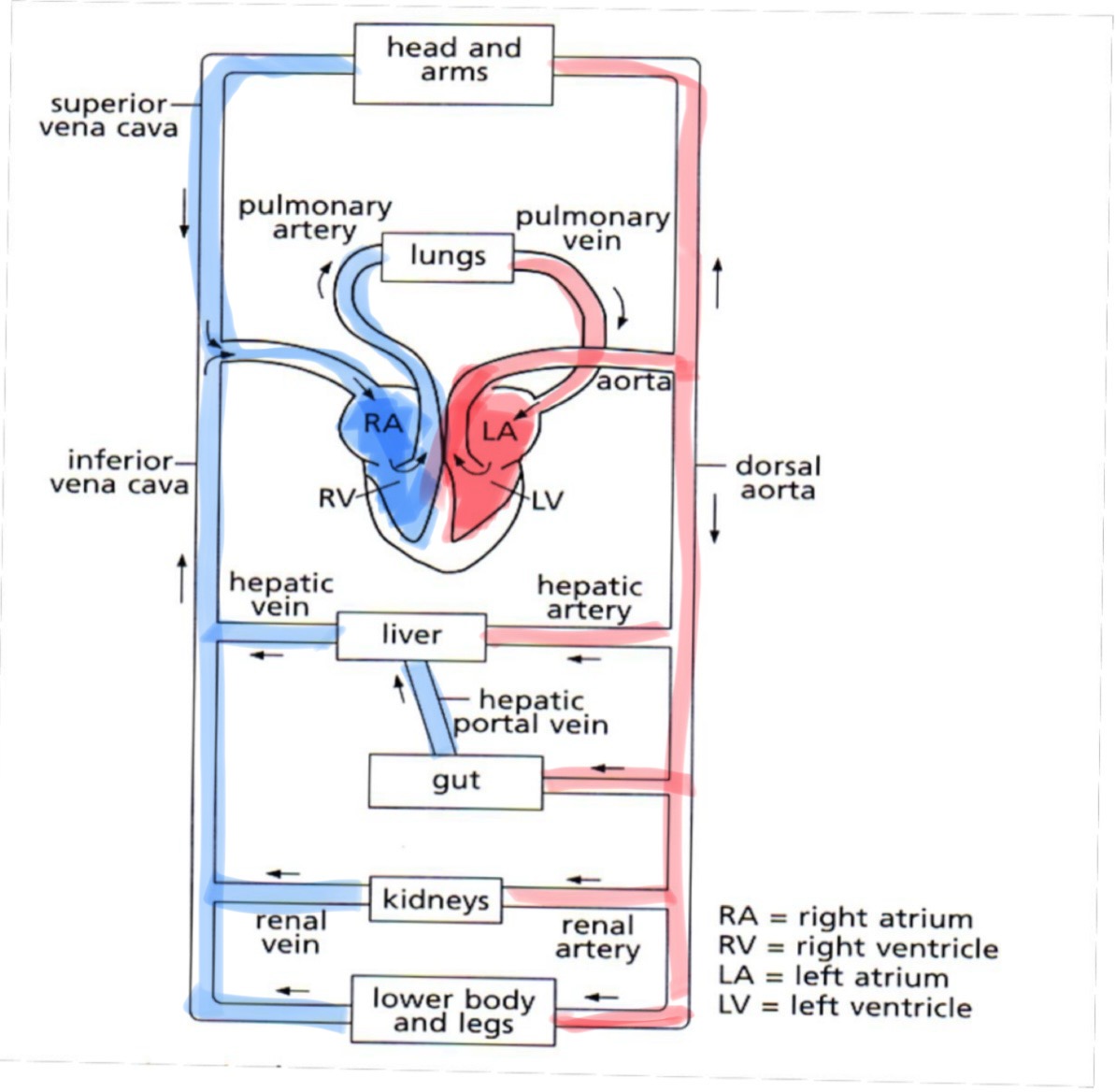
pulmonary circulation
carries oxygenated blood from lungs to the heart and deoxygenated blood from heart to the lungs, gaseous exchange happens in the lungs
systemic circulation
carries oxygenated blood from heart to rest of the body and deoxygenated blood from the rest of the body to the heart, gaseous exchange happens at the cells of the body
hepatic blood cells are connected to what
liver
renal blood vessels are connected to what
kidneys
4 components of blood
plasma
red blood cells (eruthrocytes)
white blood cells (leukocytes)
platelets
2 functions of blood
to transport oxygen, food substances, waste, hormones, heat from one body part to another
to protect the body by preventing the entry of foreign bodies and fighting infections
plasma consists of 90% water, what is rhe 10% of dissolved substances
mineral ions
digested food substances
metabolic waste products
plasma proteins
hormones
why do rbc have haemoglobin
haemoglobin binds reversibly to oxygen to form oxyhaemoglobin which helps to transport oxygen in the rbc around the body
why do rbc have no nucleus
more space available for more haemoglobin to be pack inside the cell cytoplasm and more oxygen to be transported
why do rbc have a circular and biconcave shape
increases surface are to volume ratio of the cell thus allowing quicker diffusion of oxygen in and out of the cell
why does rbc have an elastic and flexible cell surface membeane
cell can change its shape while squeezing through the small capillaries
properties and functions of lymphocytes
lymphocytes are round in shape and have a large round nucleus, they produce antibodies that protect our bodies against diseases by 1. destroying bacteria by attaching to them and causing the bacteria surface membrane to rupture 2. cause bacteria to agglutinate so that they can be easily digested by phagocytes 3. neutralise the toxins produced by bacteria 4. attach to viruses and prevent them from binding to the host cell
properties and functions phagocytes
phagocytes are more irregular in shape and have multi-lobed/bean-shaped nuclei, with more granular cytoplasm. they can move out of leaky capillaries to carry out phagocytosis
properties and functions of platelets (thrombocytes)
not true cells but fragments of larger bone marrow cells called megakaryocytes, they play an important role in blood clotting by forming “sticky plugs” at wound sites
importance of blood clotting
prevents excessive loss of blood
prevents further entry of pathogens though the wound
mechanism of blood clotting
damaged tissue and platelets → thrombokinase (enzyme)
prothrombin, inactive -(thrombokinase and calcium ions)→ thrombin, active
fibrinogen -(thrombin, enzyme)→ fibrin threads to form a mesh to trap blood cells
definition of agglutination
clumping of red blood cells which is fatal and occurs when anti-A antibodies bind with antigen A on red blood cells
universal donor
O
universal receipient
AB
the 3 main categories of blood vessels
arteries - bring blood away from heart at higher pressure
veins - bring blood away at a relatively lower pressure
capillaries - blood vessels with 1-cell thick walls
3 layers of wall in arteries and veins
innermost - endothelium
middle - smooth muscle tissue and elastic fibres
outer - connective tissue like collagen fibres
describe arteries
blood vessels that carry oxygenated blood away from the heart (except pulmonary artery), blood flow is rapid and under high pressure, thick elastic walls allow the arteries to stretch and recoil to push the blood along it in spurts, maintaining high pressure of blood flowing through the artery.
what do arteries branch to form
arteries branch to form smaller arteries called arterioles, to form many tiny blood vessels called capillaries
describe veins
blood vessels that carry deoxygenated blood towards the heart (except pulmonary vein), blood flow in vein is slow and at low pressure, they have relatively thinner muscular and elastic walls as the pressure of the blood flowing through it is at low pressure, semilunar valves are present to prevent the back-flow of blood in the vein and ensure blood flows in one direction in the vein only. veins are located between large muscles of body, the contraction of these skeletal muscles squeeze the veins and push the blood along, up towards the heart.
describe capillaries
microscopic blood vessels that connect the arteries to veins and are found between cells of most tissues, capillary walls are only one-cell thick and are partially permeable so that certain substances are able to diffuse through their walls quickly, they branch repeatedly to provide a large surface area for the quick exchange of substances between blood and tissue cells
what does tissue fluid contain
dissolved food substances such as glucose and amino acids as well as waste products like urea and hydrogencarbonate ions, proteins are not found in tissue fluid as it is too large to pass through the gaps between the cells of capillary walls, phagocytes can squeeze through the gaps to enter tissue fluid but not rbc
“glucose and other substances diffuse directly from blood plasma into body cells surrounding the capillary” is accurate or not?
inaccurate as tissue fluid exists as a medium between blood plasma and body cells
why are the walls of ventricles thicker and more muscular than walls of atria?
the walls of ventricle contract to pump blood to the rest of the body, so the walls are thicker and more muscular to exert a higher pressure, ensuring blood reaches all areas of the body, however atria only pump blood to the ventricles which is a short distance, so the walls are thinner and less muscular as blood can be pumped at a lower pressure
atrial systole
muscles of atrial wall contract, further increasing atrial pressure,
bicuspid valve is already open to allow blood flow,
blood therefore flows from left atrium to left ventricle, down a pressure gradient,
as aortic pressure is higher than ventricular pressure, semilunar valve is closed to prevent backflow of blood from aorta into left ventricle
ventricular systole
ventricular pressure rises such that it is higher than aortic pressure,
semilunar valves open to allow blood to flow through,
blood flows from left ventricle into aorta
meanwhile, atrium undergoes diastole and begins to be filled with blood, bicuspid valve closes to prevent backflow of blood from ventricle to atrium (Dub)
ventricular diastole
ventricle pressure decreases until lower than aortic pressure,
semilunar valves close to prevent backflow of blood from aorta into left ventricle (Lub)
ventricular pressure decreases further until it is lower than atrial pressure
bicuspid valve opens to allow blood to flow
blood therefore flows from left atrium to left ventricle, down the pressure gradient
coronary heart disease
coronary arteries that supply oxygen and nutrients to the heart muscles become narrowed
and eventually blocked by a build-up of fatty deposits containing cholesterol, resulting in coronary heart disease.
the blood supply to part of the heart muscle is cut off,
as a result heart muscle cells are deprived of oxygen and nutrients, cannot respire and dies.
part of the heart is unable to contract, causing a heart attack
causes of coronary heart disease
diet high in cholesterol and saturated animal fats
lack of exercise - sedentary lifestyles increase the risk
stress and smoking
increases blood pressure
narrows artery walls through build up of plague
preventive measures against coronary heart disease
diet - reduce in take of cholesterol and saturated animals
regular physical exercise
strengthens heart
maintains elasticity of arterial walls
manage stress and avoid smoking