Shoulder X-Ray and MRI
1/83
There's no tags or description
Looks like no tags are added yet.
Name | Mastery | Learn | Test | Matching | Spaced | Call with Kai |
|---|
No analytics yet
Send a link to your students to track their progress
84 Terms
Shoulder trauma, dislocation, and suspected GHJ OA, ACJ OA, or ACJ separation
When is a shoulder x-ray indicated?
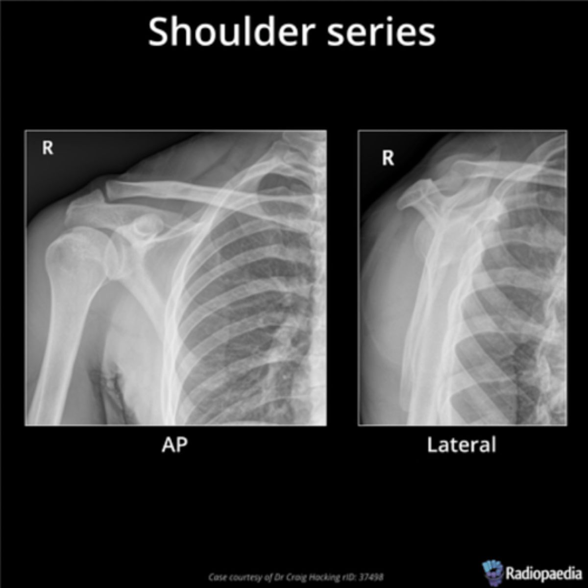
AP (frontal/coronal), lateral (sagittal), Y-view/scapular view (allows you to see the orientation of the head of the humerus in the glenoid), and axial (transverse)
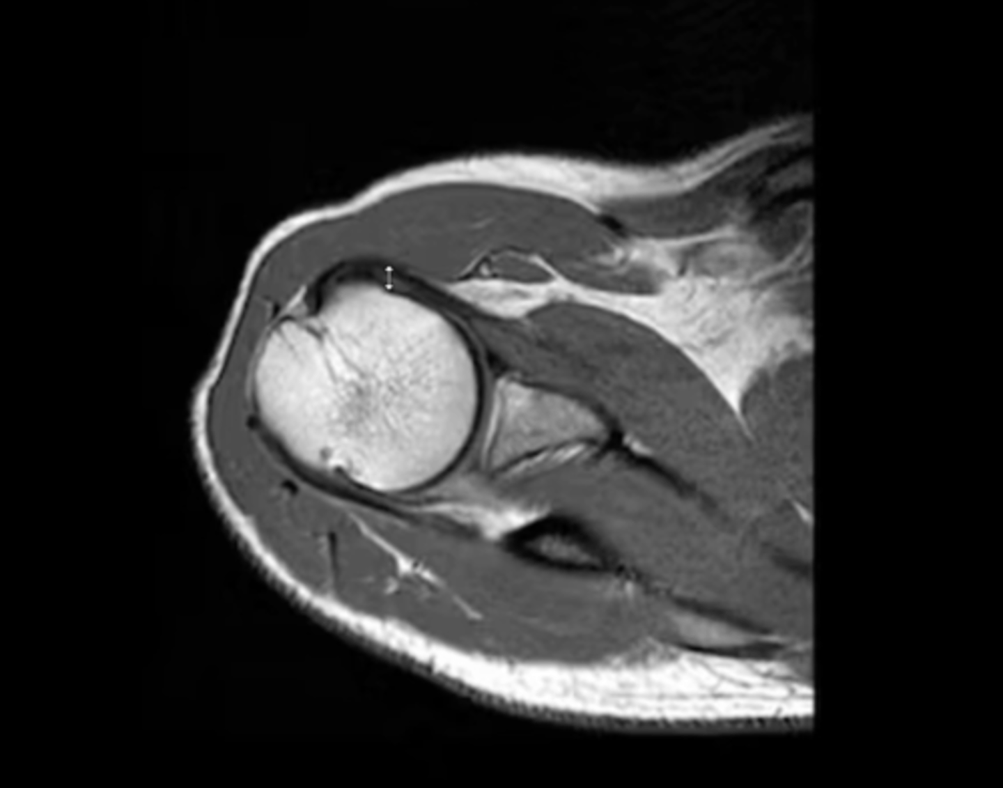
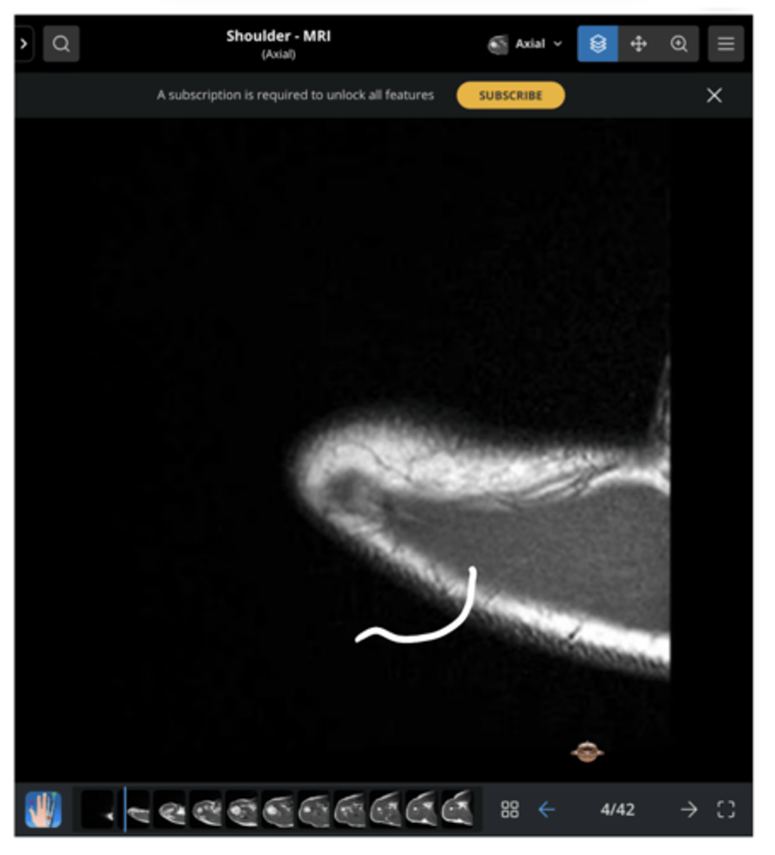
NOTE: this picture is an AXIAL view of the shoulder
Which types of x-ray views are common at the shoulder?
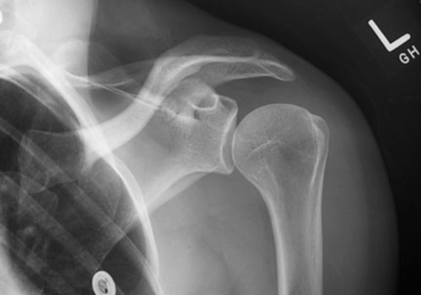
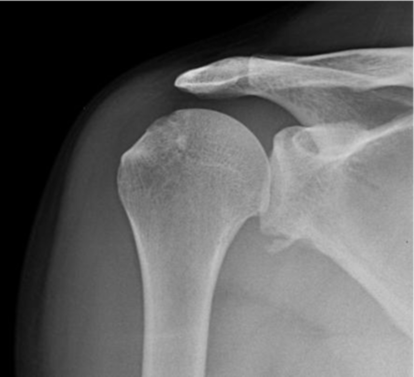
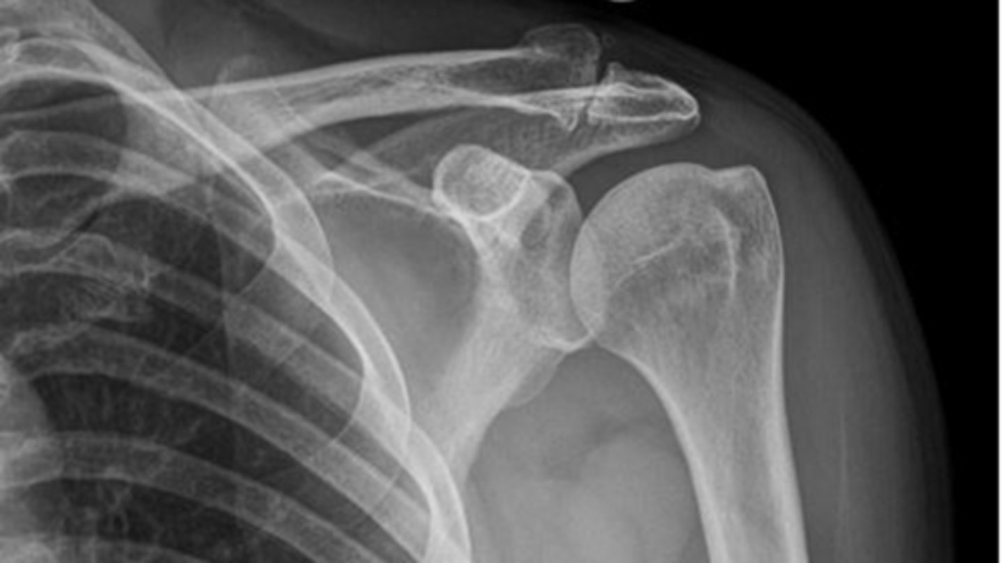
TRUE!!--we see healthy space b/w the clavicle and acromion, the humerus and the acromion, and b/w the glenoid and humerus...also, there is no sclerosing or osteophytes
True or false: this is a healthy GHJ
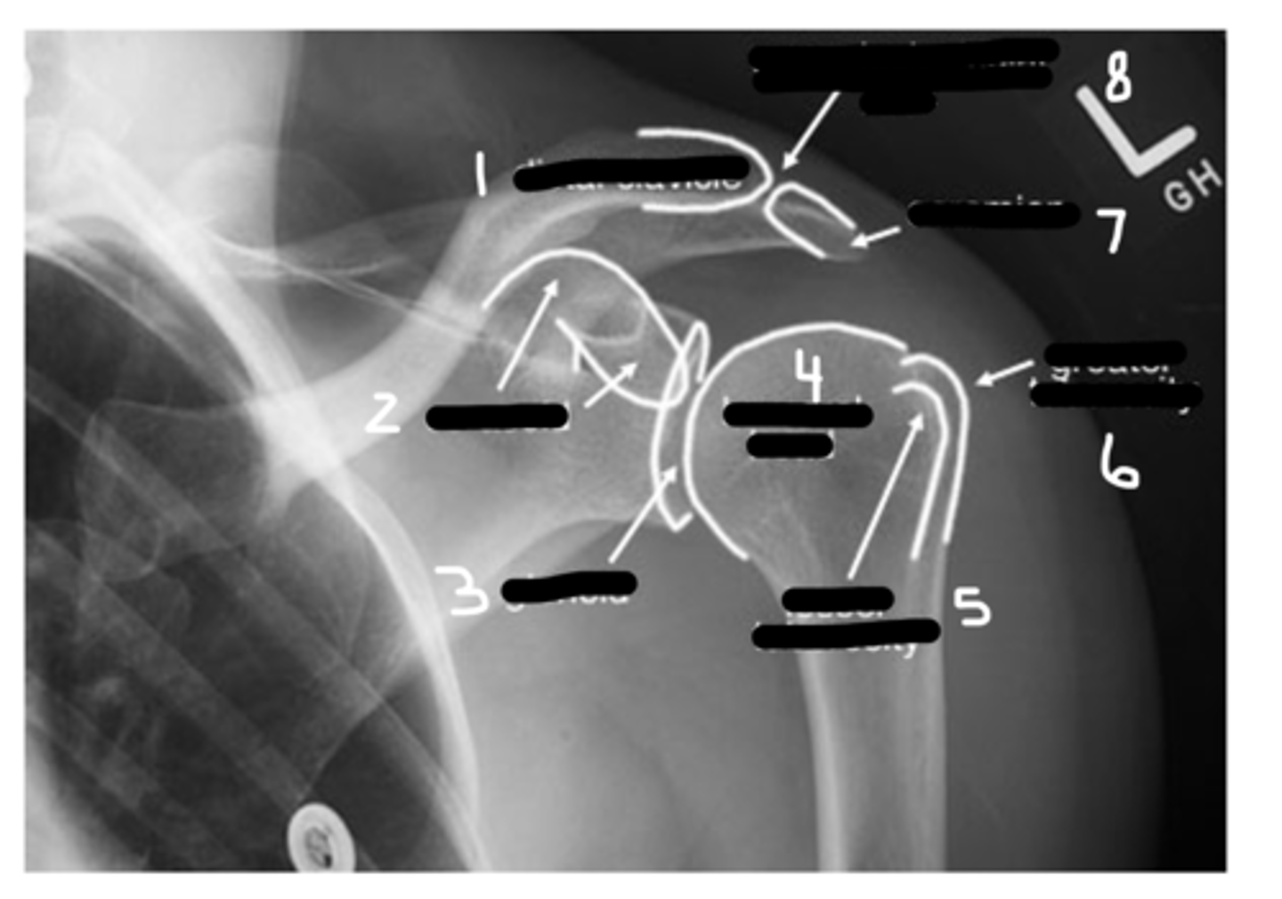
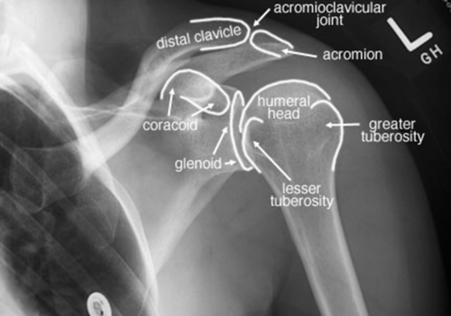
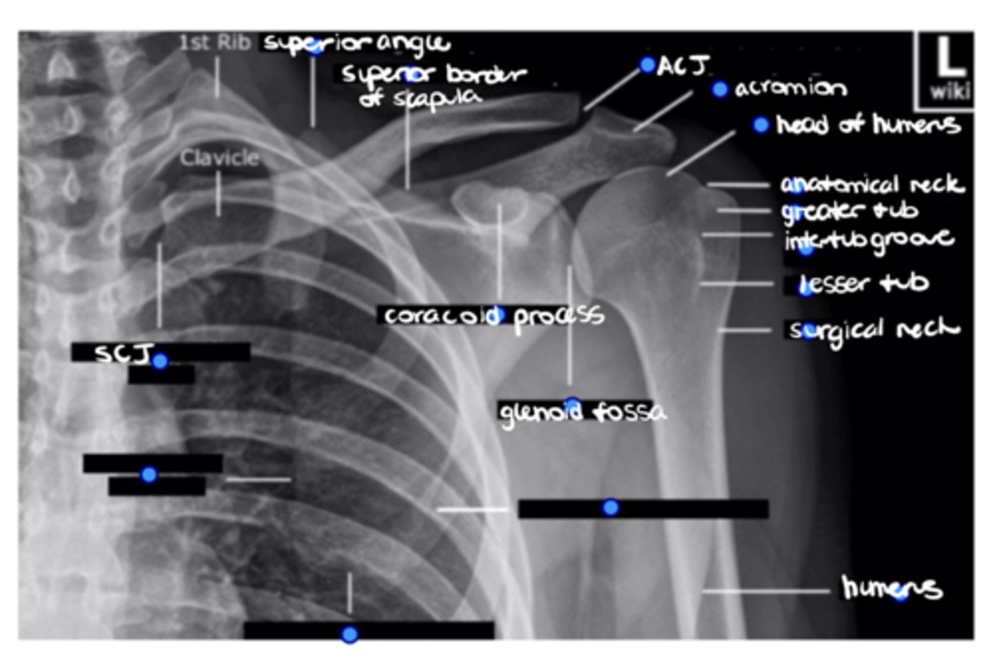
1. Distal clavicle
2. Coracoid
3. Glenoid
4. Head of humerus
5. Lesser tuberosity
6. Greater tuberosity
7. Clavicle
8. AC joint
Label this image.
INTERNAL, because the lesser tuberosity is positioned medially vs. in line with the greater tuberosity, as seen in external rotation
Is this shoulder in EXTERNAL or INTERNAL rotation? Why?
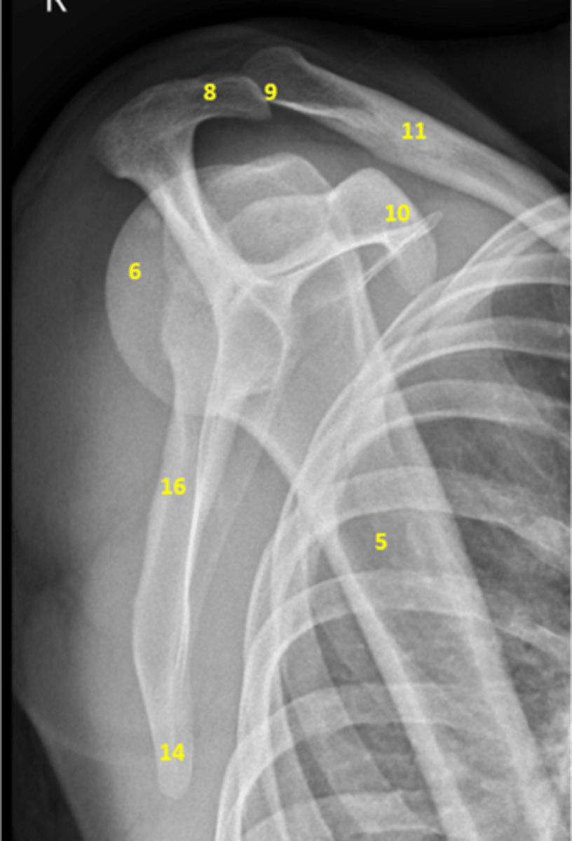
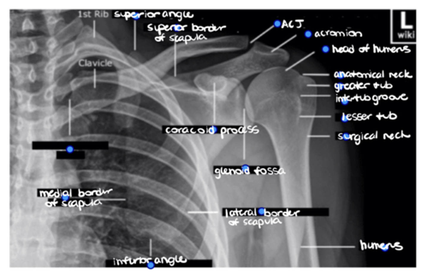
View: Y-view
5. Humeral shaft
6. Head of the humerus
8. Acromion
9. Distal end of the clavicle
10. Coracoid process
11. Clavicular shaft
14. Inferior angle of the scapula
16. Medial border of the scapula
Label this image. Bonus: which type of view is this?
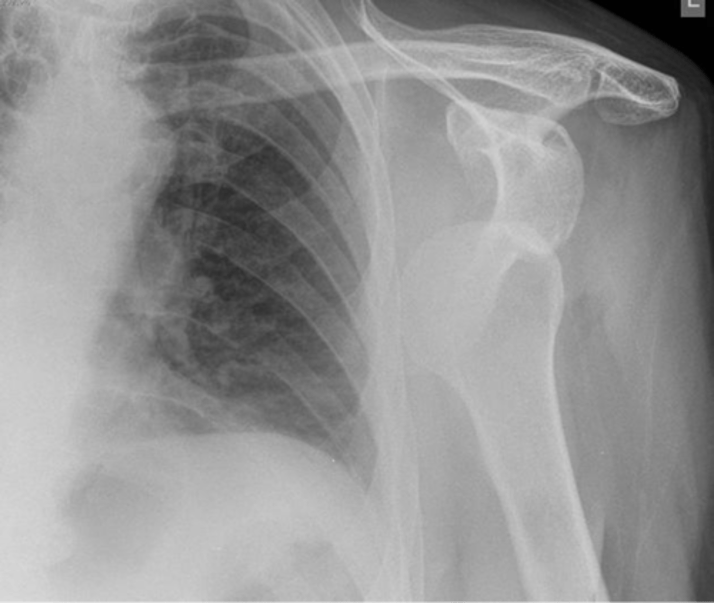
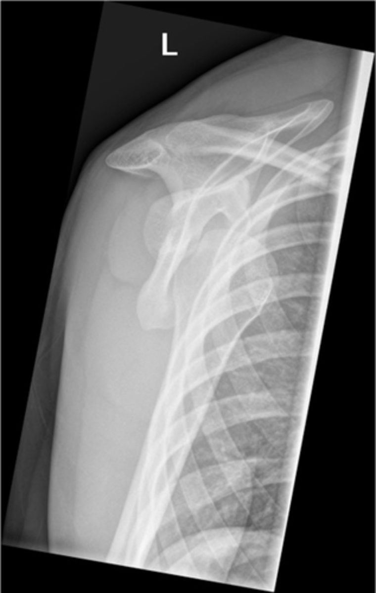
Condition: anterior GHJ dislocation
REALLY important to know BEFORE you relocate the humerus into the GHJ to know where the fracture(s) is and the integrity of the neurovascular structures
What is this image depicting? In an ideal scenario, determining this diagnosis is important before what? Why?
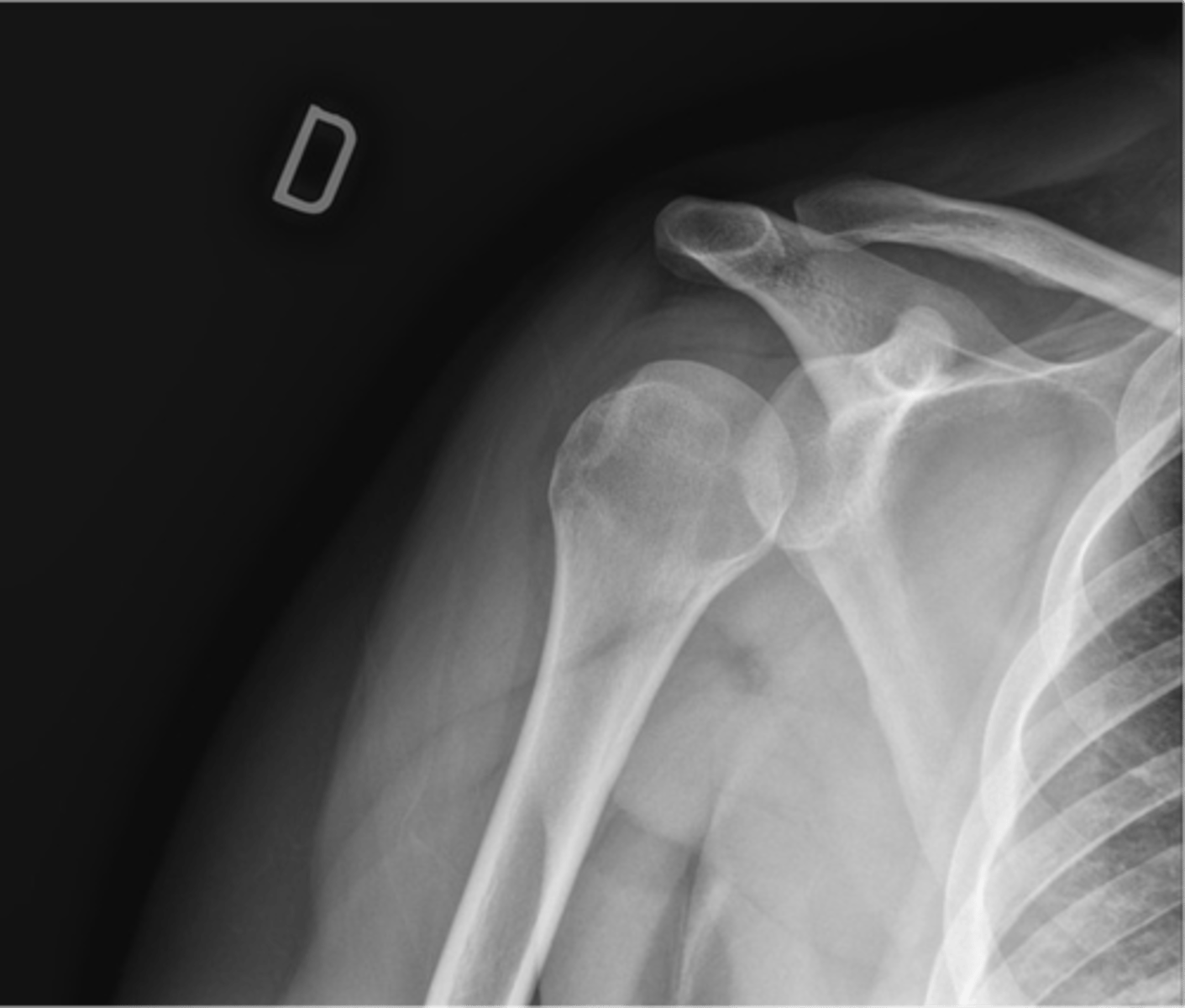
Condition: anterior GHJ dislocation via a Y-view -- makes dislocations MUCH more obvious
What is this image depicting? What view is it?
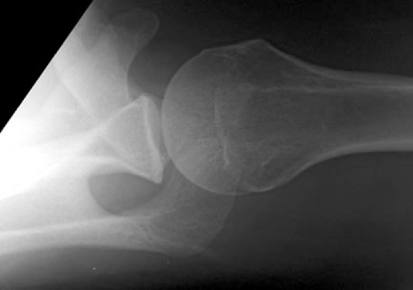
Bony Bankart Lesion (part of the glenoid fossa chips off)
What is this image depicting?
Hill-Sachs Lesion (humerus catches the bony glenoid rim as it moves towards the dislocated position --> compression fracture of the head of the humerus)
What is this image depicting?
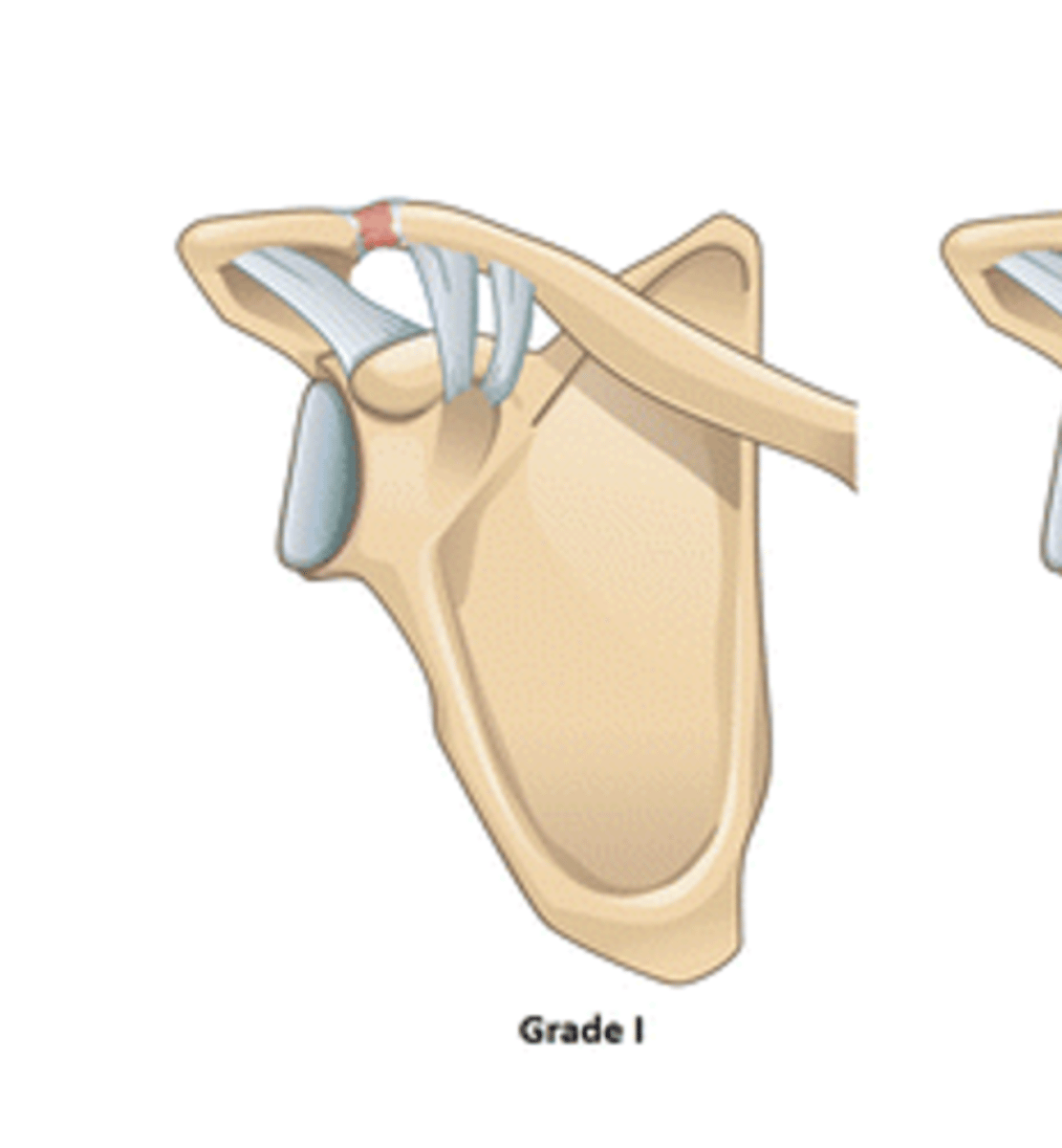
No ligamentous injury --> disruption of collagen fibers of the AC ligament + pain + swelling
According to the Rockwood Classification System, a grade I ACJ separation entails: __________
Complete tear of the AC ligament --> separation of the clavicle and acromion = MOST PAINFUL!!
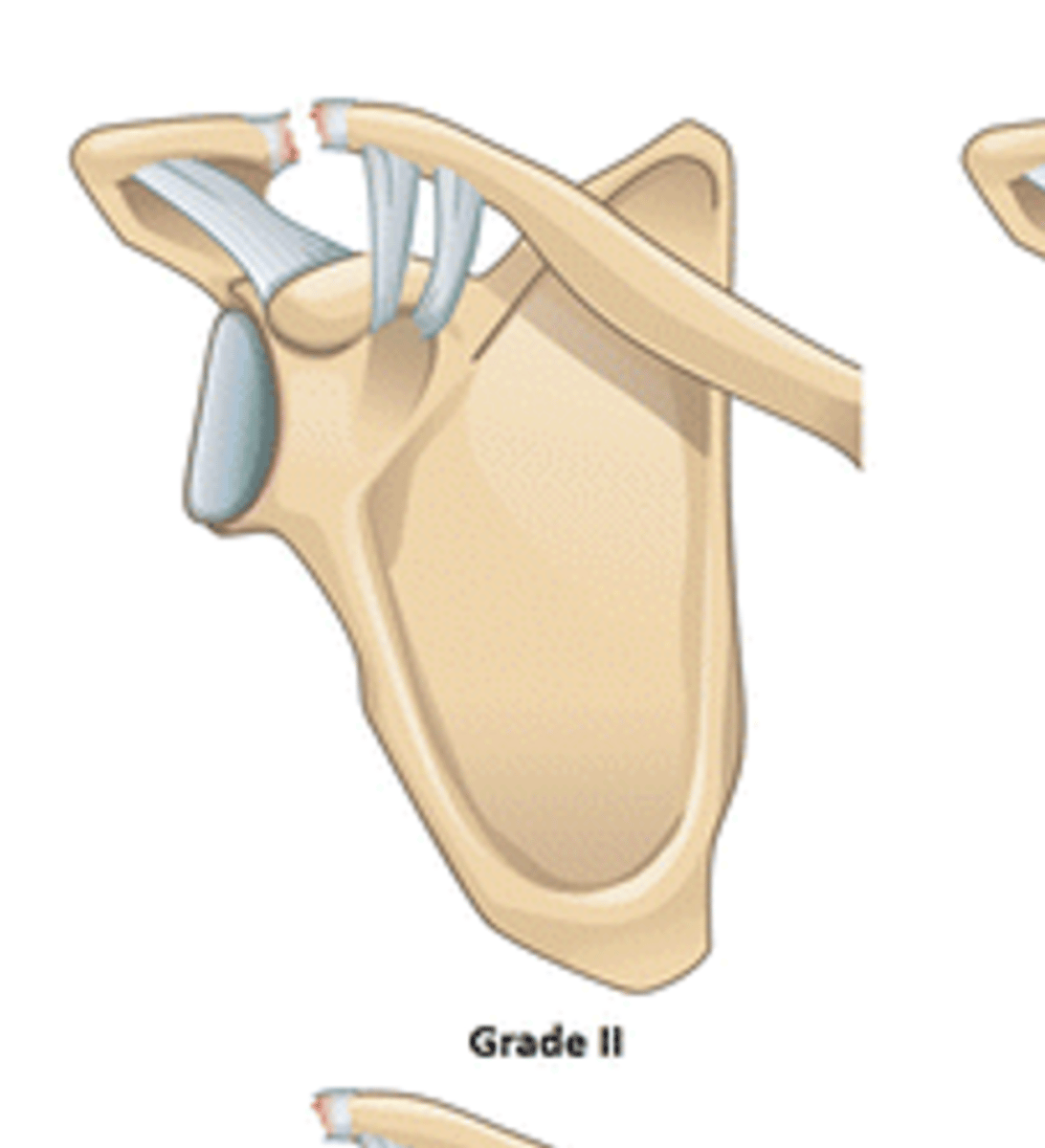
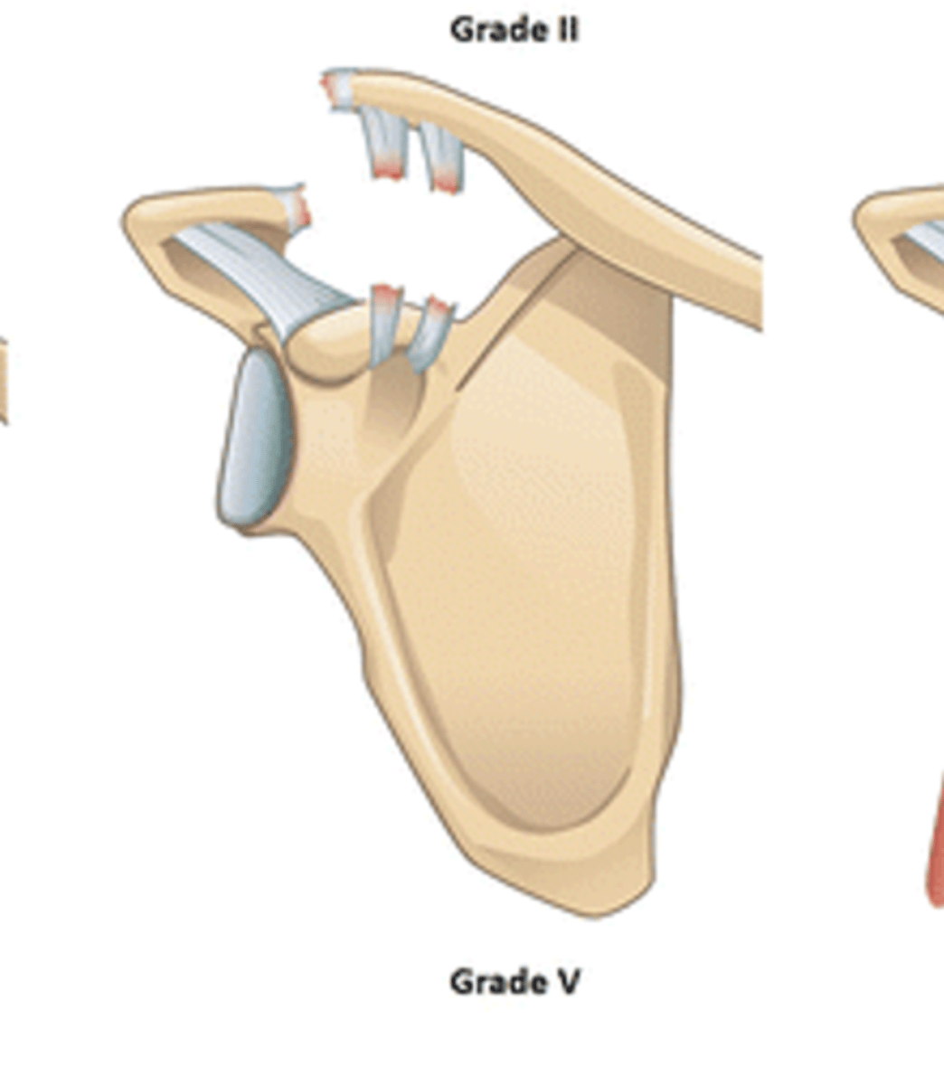
According to the Rockwood Classification System, a grade II ACJ separation entails: __________. What is unique about it?
Rupture of the AC AND CC ligaments w/ detachment of the deltoid and upper trap and separation of the ACJ
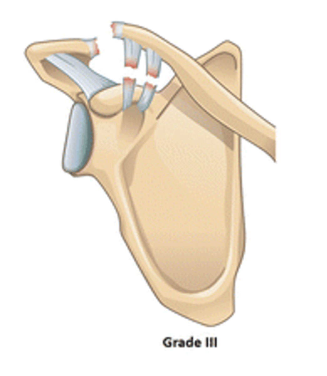
According to the Rockwood Classification System, a grade III ACJ separation entails: __________
Posterior displacement of the clavicle
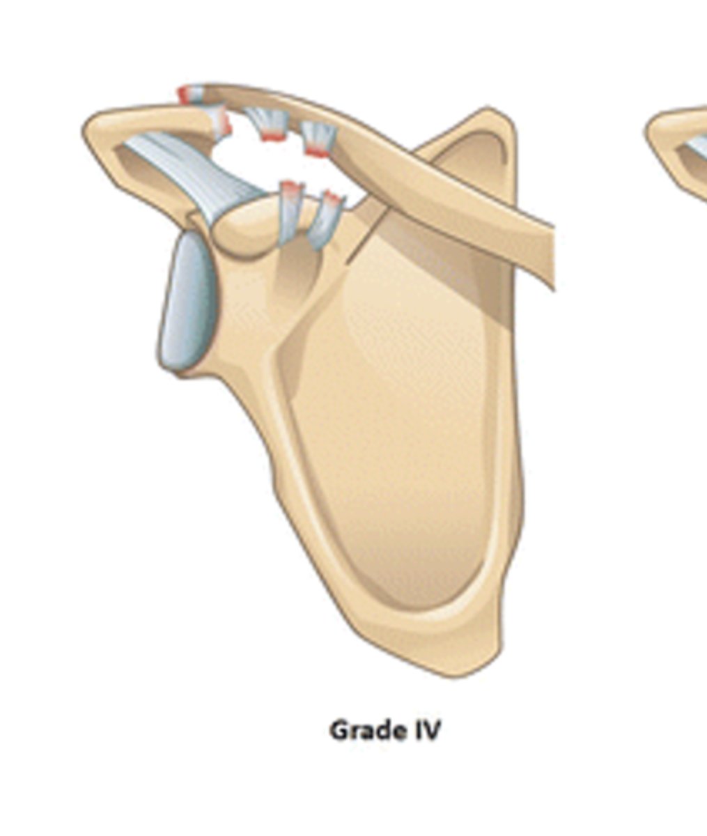
According to the Rockwood Classification System, a grade IV ACJ separation entails: __________
Superior displacement of the clavicle
According to the Rockwood Classification System, a grade V ACJ separation entails: __________
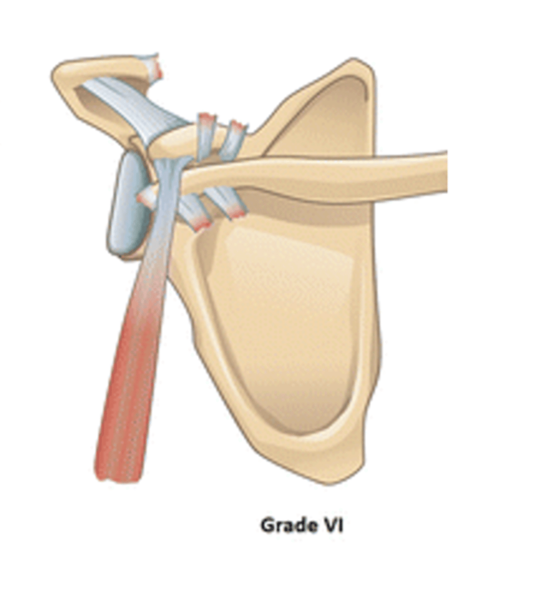
Inferior displacement of the clavicle under the coracoid process
According to the Rockwood Classification System, a grade VI ACJ separation entails: __________
Grade II -- normal to palpate a little step-up deformity at this time, but does NOT require surgical repair
Which grade ACJ separation would this be considered?
Grade III -- acromion migrated WELL below the clavicle and the AC and CC ligaments are both torn
Which grade ACJ separation would this be considered?
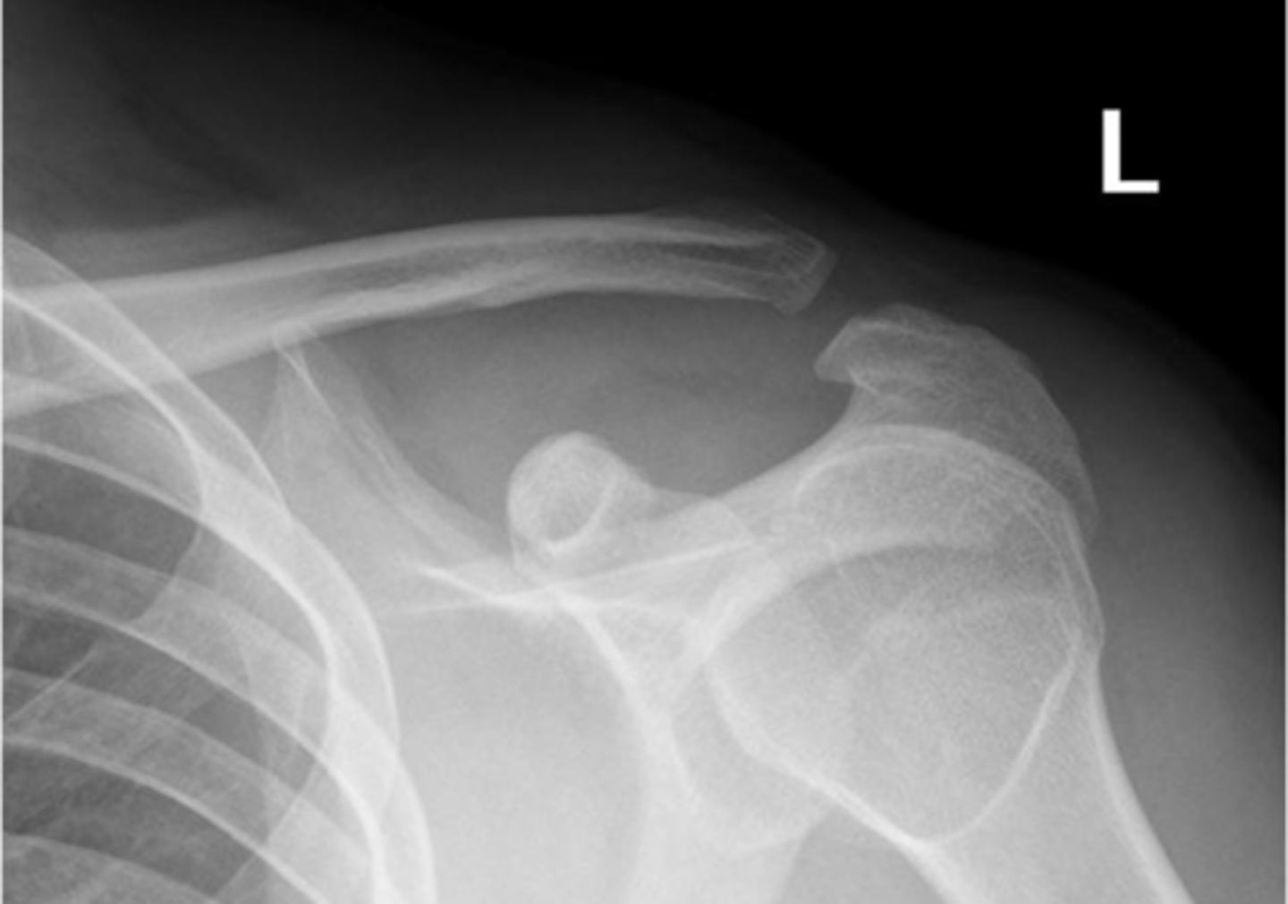
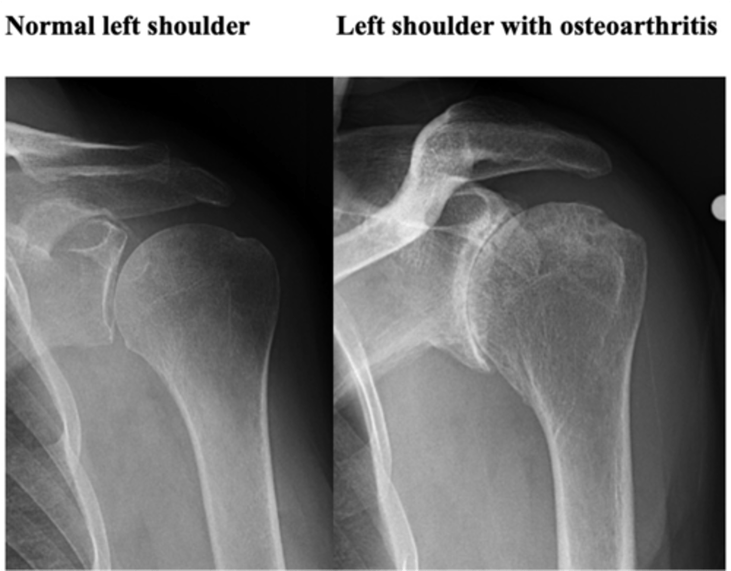
ACJ OA -- A (alignment) = osteophyte formation on either side of the acromion, C (cartilage space) = loss of AC joint space
What is this image depicting? How do you know?
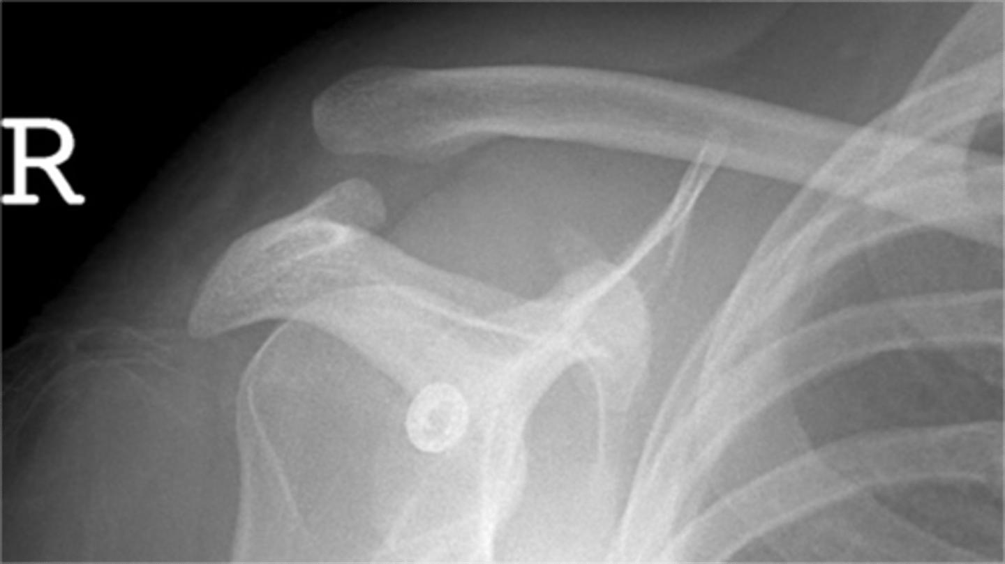
ACJ OA
What is this image depicting?
1. A (alignment) = osteophytes
2. B (bone density) = sclerosis
3. C (cartilage space) = loss of space
Which 3 MAIN things are you looking for when it comes to classifying OA?
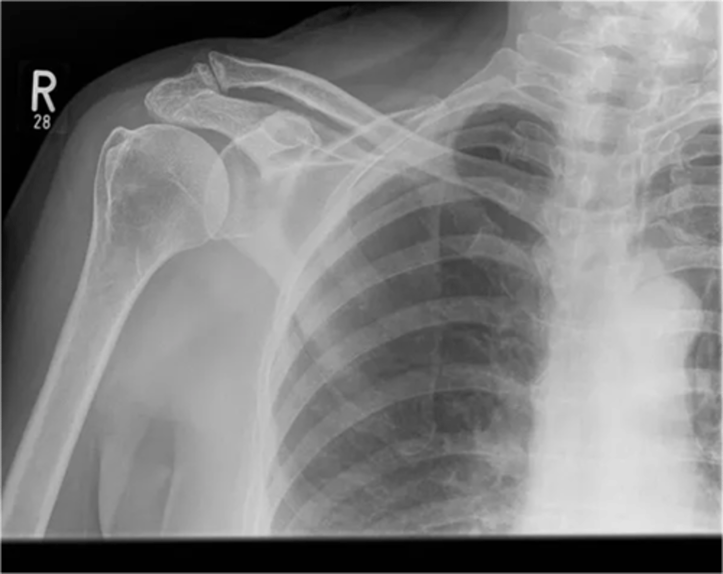
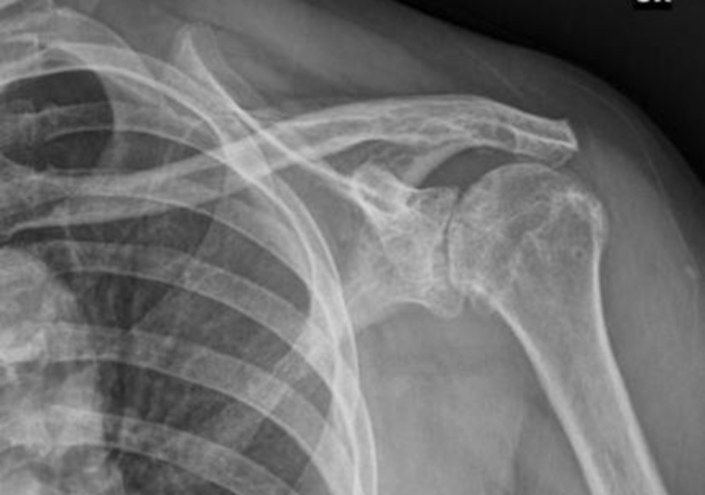
GHJ OA w/ cystic changes of the glenoid fossa, which essentially means the bone tissue is starting to wear away --> concern when it comes to TSA
What is this image depicting? Why does it matter?
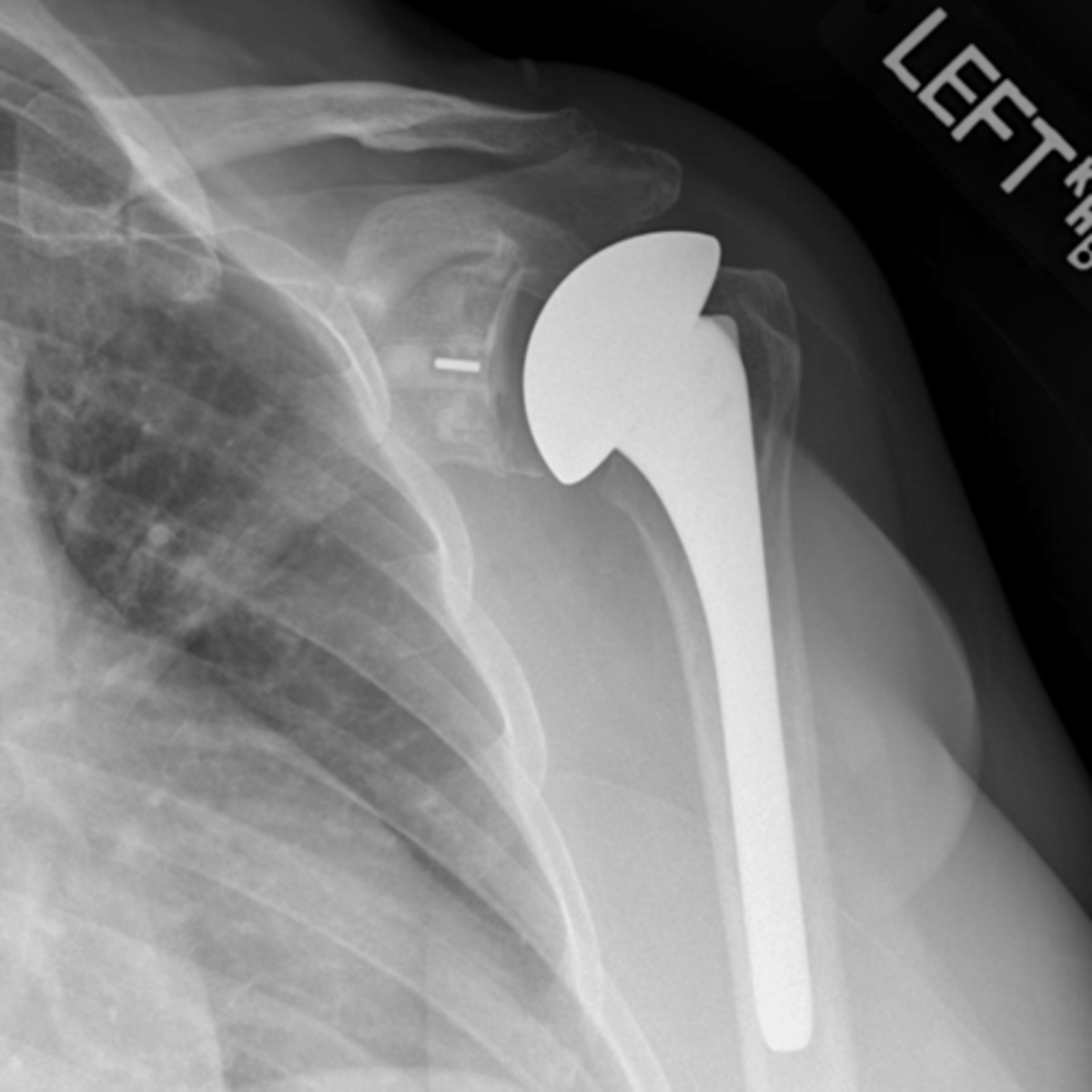
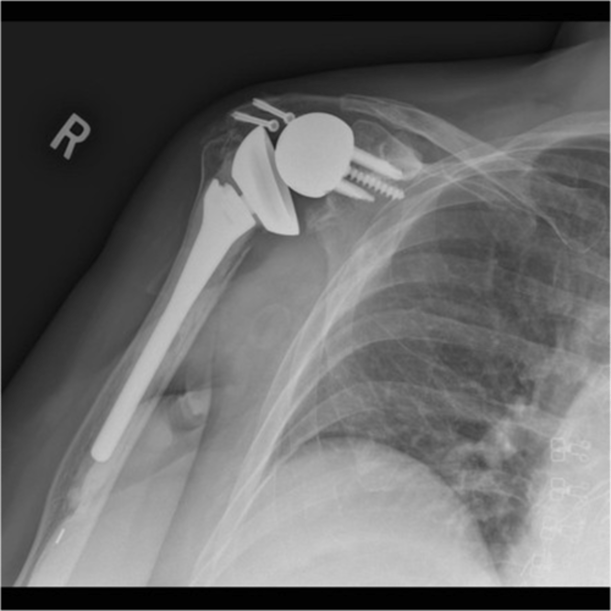
TSA
What is this image depicting?
RTSA
What is this image depicting?
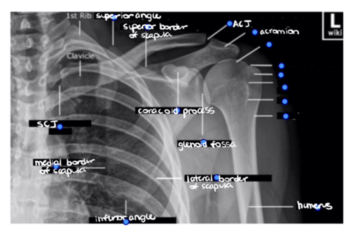
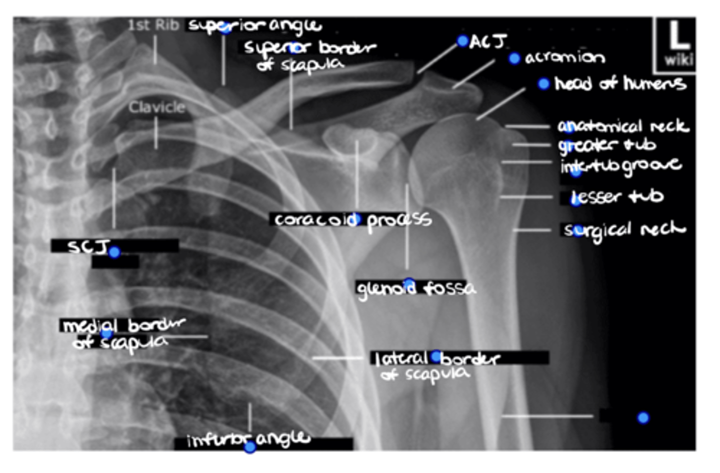
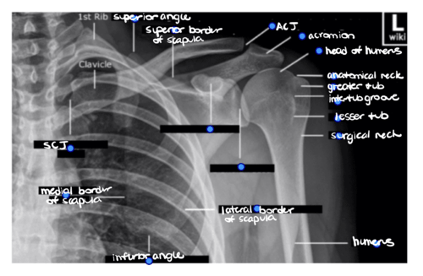
Superior angle of the scapula
ID the missing structure.
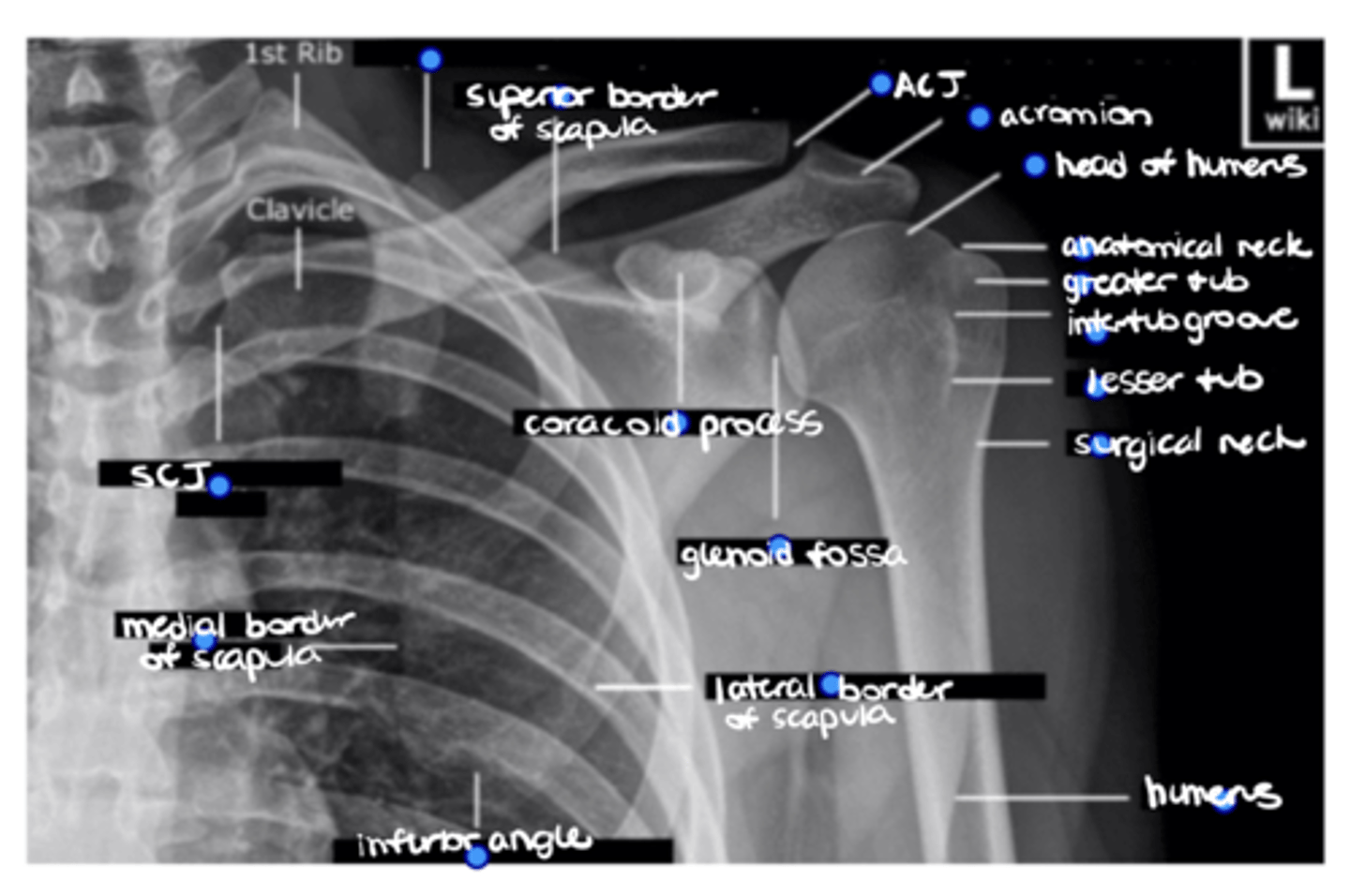
Superior border of the scapula
ID the missing structure.
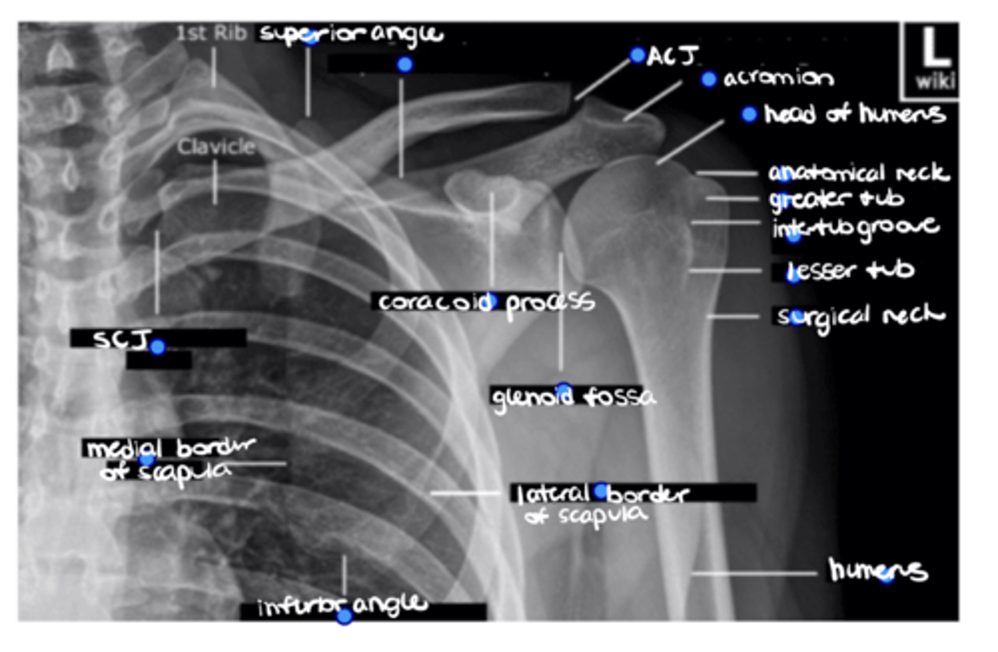
ACJ
ID the missing structure.
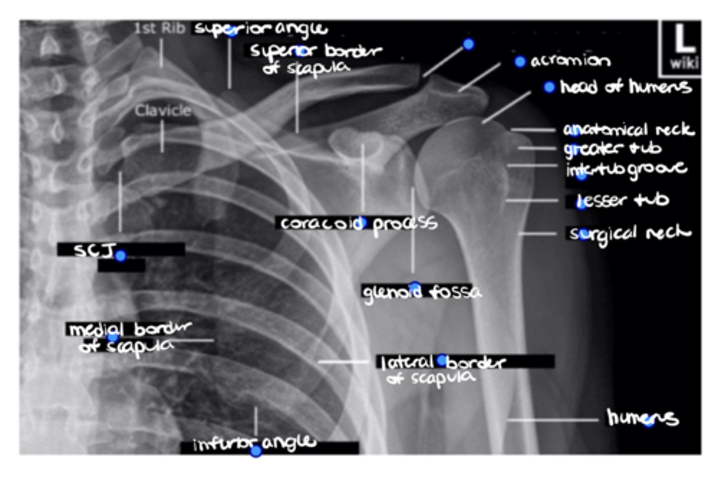
Acromion
ID the missing structure.
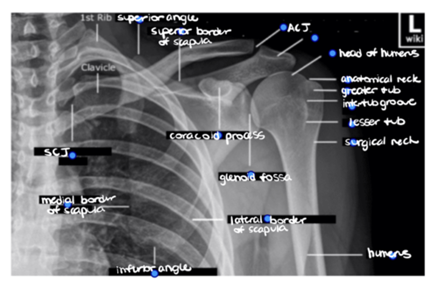
1. Head of humerus
2. Anatomical neck
3. Greater tuberosity
4. Intertubercular groove
5. Lesser tuberosity
6. Surgical neck
ID the 6 missing structures.
Shaft of the humerus
ID the missing structure.
1. Coracoid process
2. Glenoid fossa
ID the 2 missing structures.
1. Lateral border of the scapula
2. Inferior angle of the scapula
3. Medial border of the scapula
ID the 3 missing structures.
SCJ
ID the missing structure.
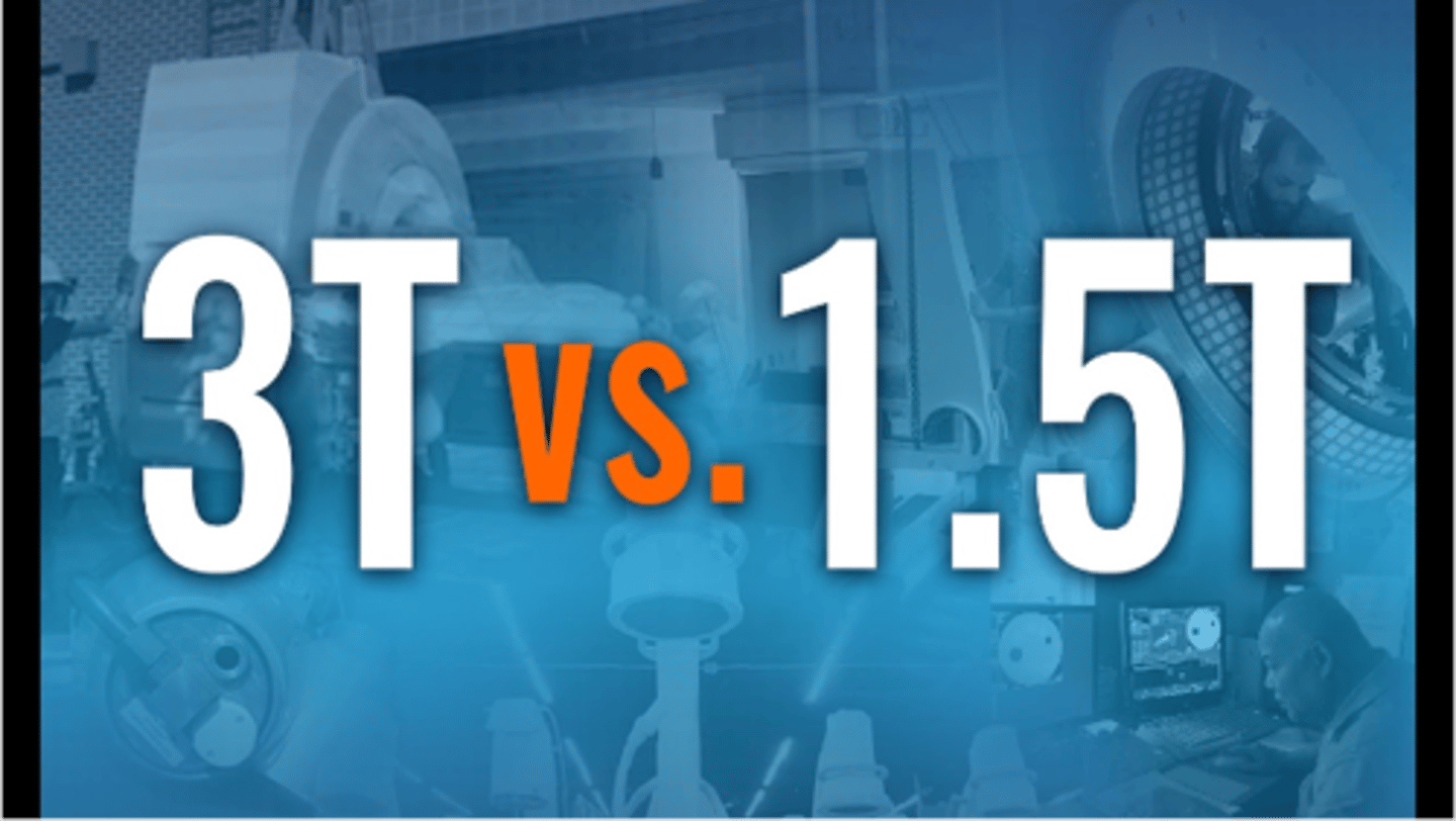
Both are measurements of magnet strength in MRIs -- 1.5T is VERY common and might be a better option for some people due to hardware, artifacts, etc. vs. 3.0T is MUCH more $$$ due to better resolution
What is the difference between 3T and 1.5T MRI machines?
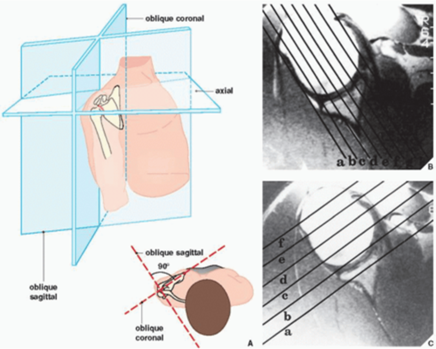
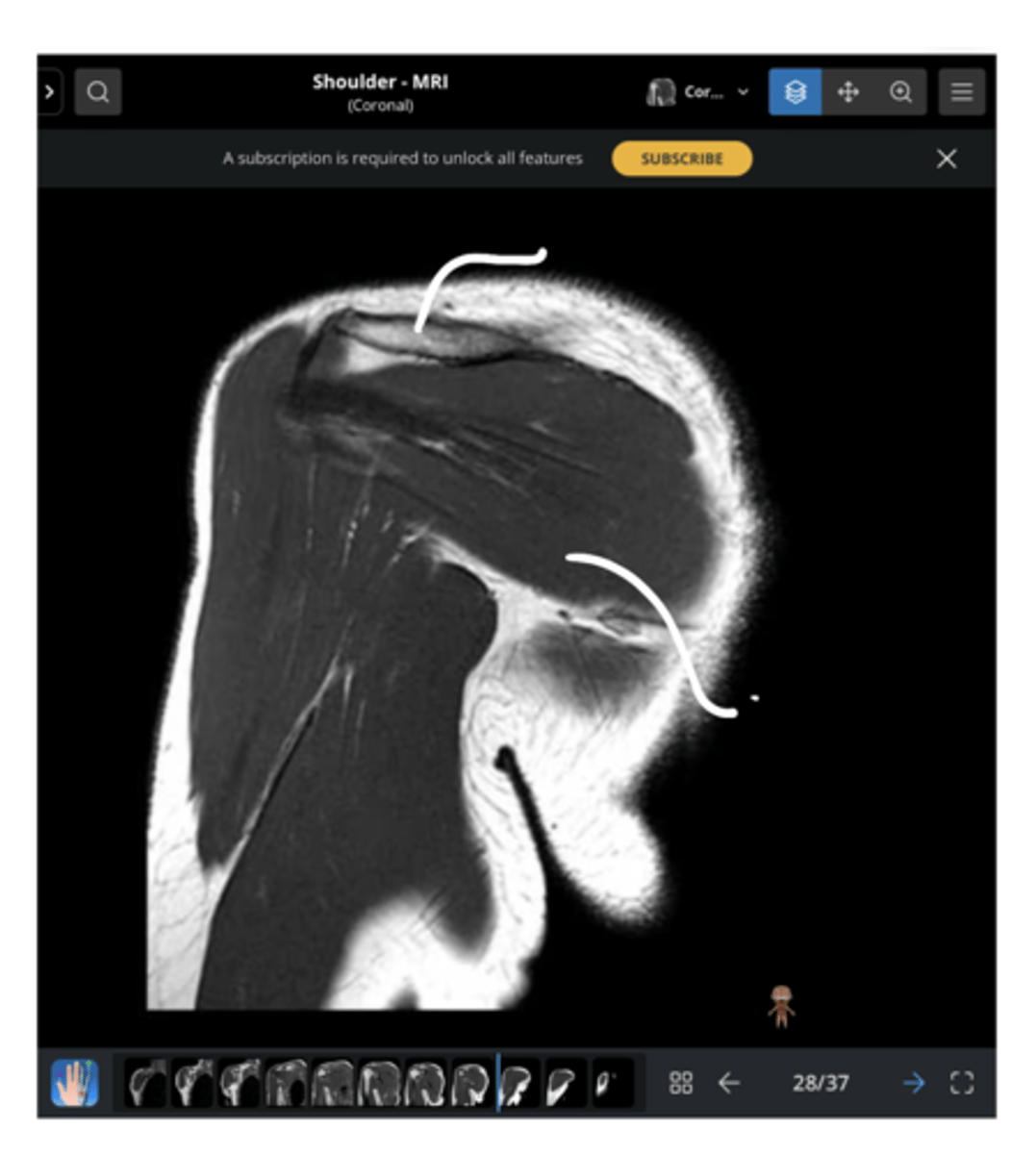
1. Sagittal = slices outside --> in
2. Coronal = slices w/ a traditional AP view
3. Axial = slices looking down
Explain the difference between each of the MRI slices.
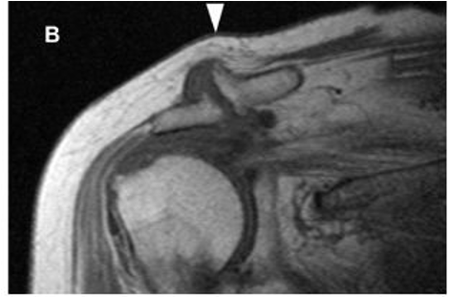
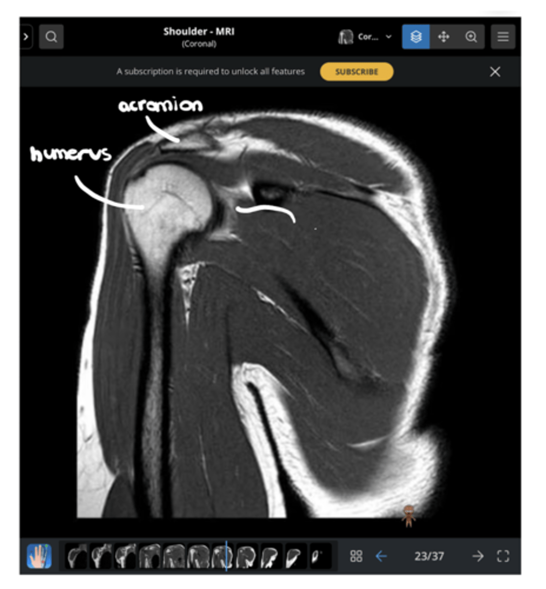
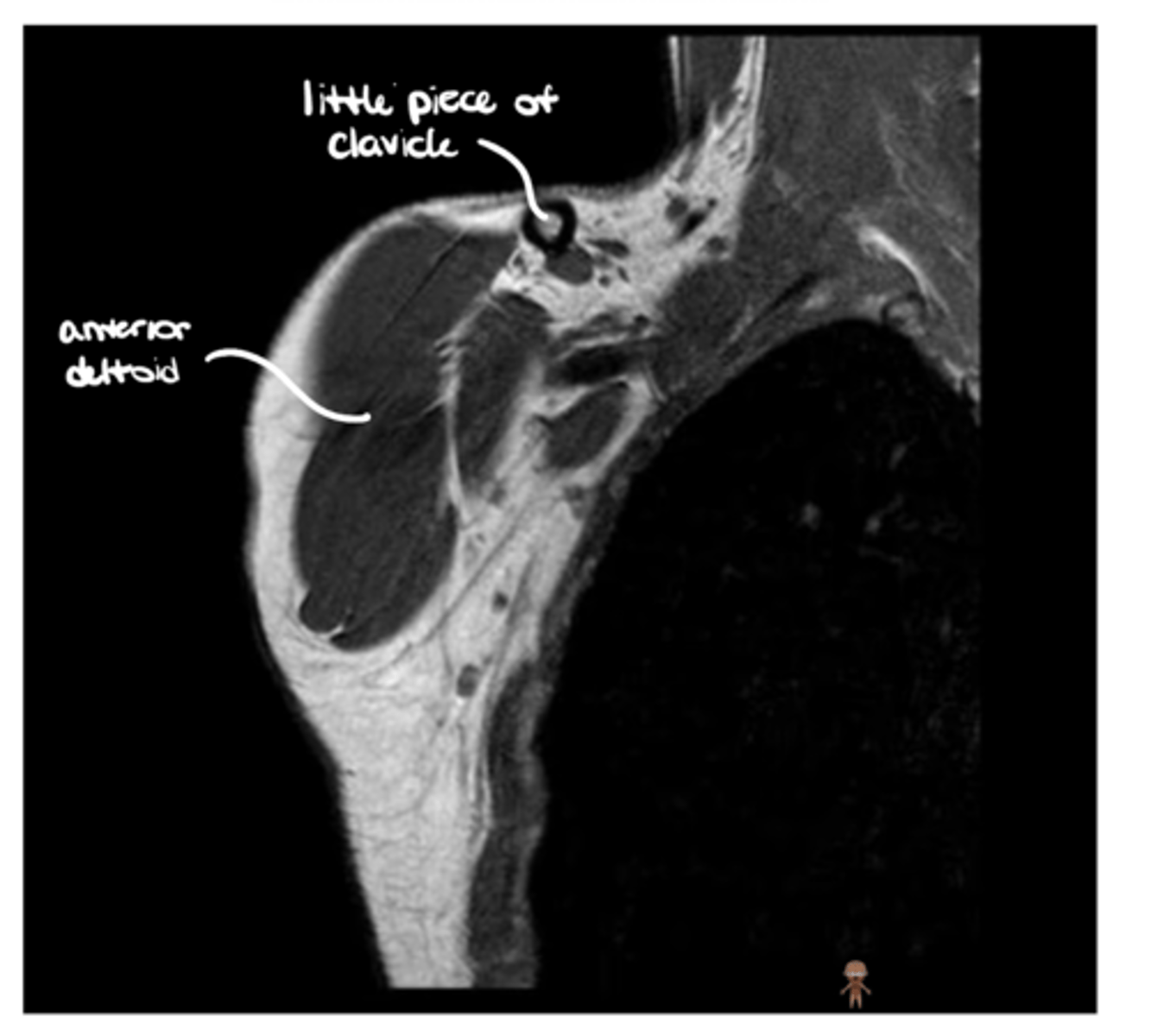
Anterior coronal view of the shoulder -- key to setting this image up is finding the coracoid process, which is NOT present on the back side
What type of view is this? Anterior vs. posterior? How do you know?
Posterior coronal view of the shoulder -- now, instead of seeing the supraspinatus, we are seeing the infraspinatus
What type of view is this? Anterior vs. posterior? How do you know?
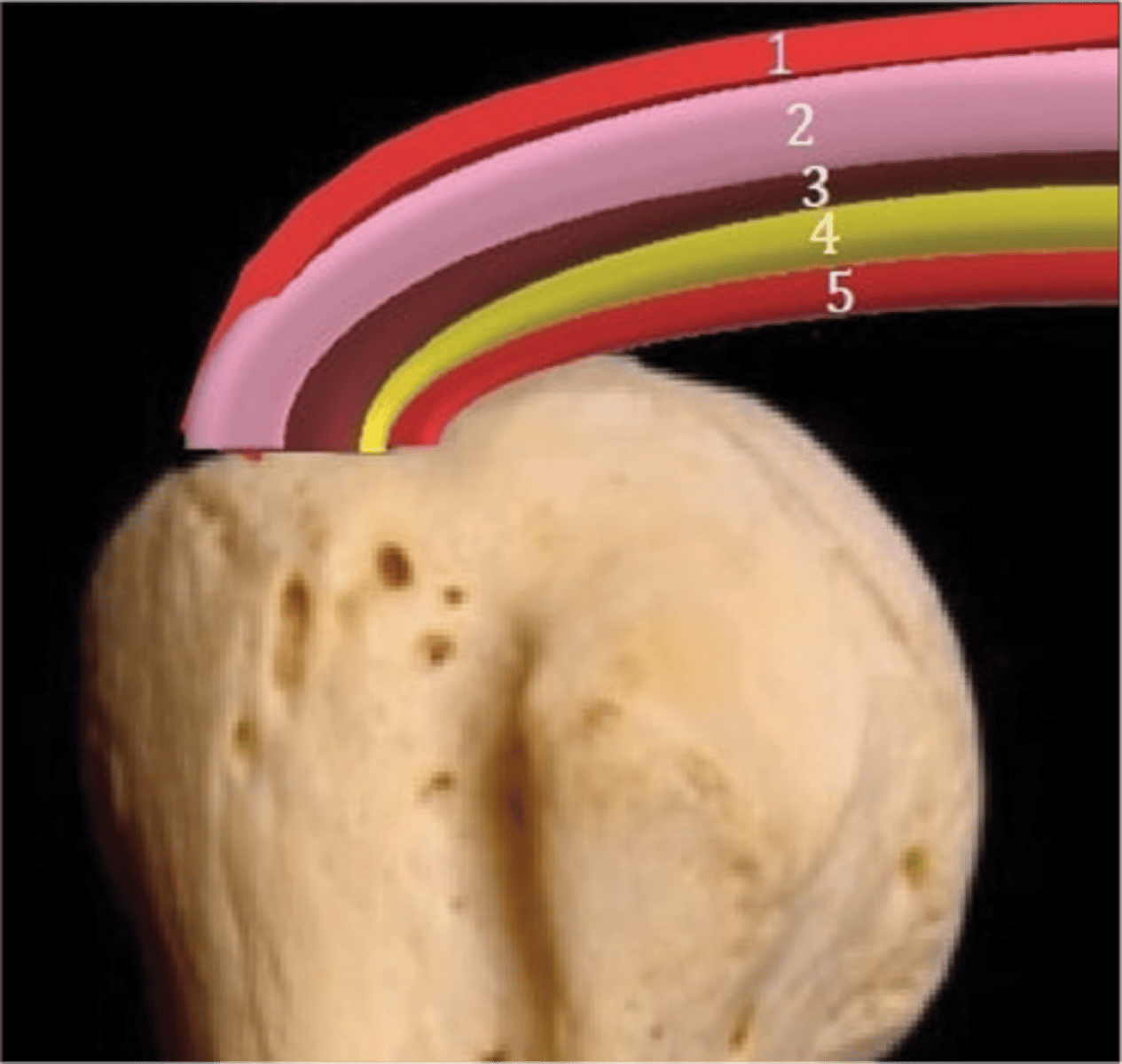
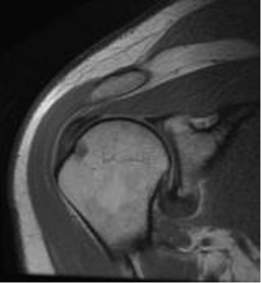
Sagittal view of the anterior, middle, and posterior deltoid (from left to right)
What type of view is this?
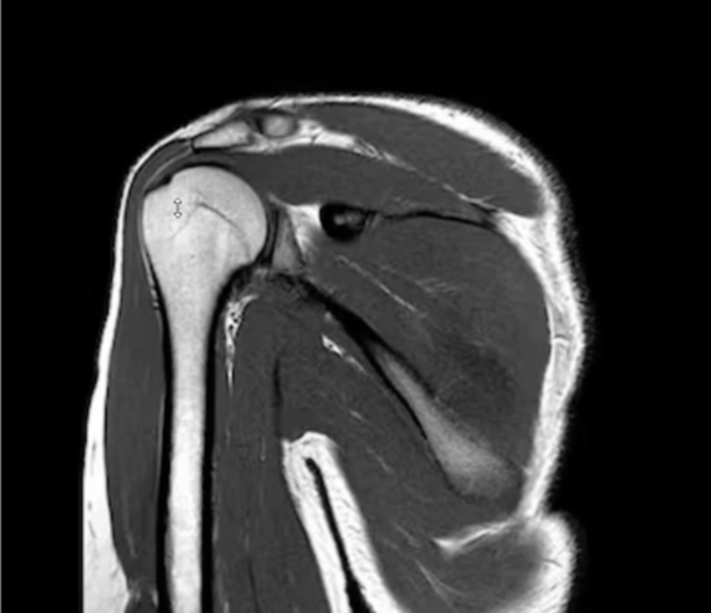
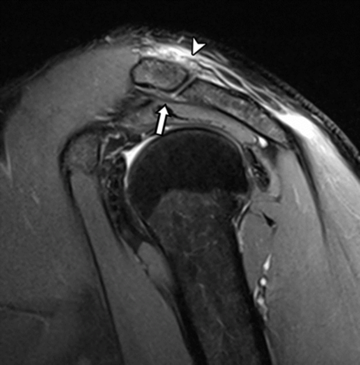
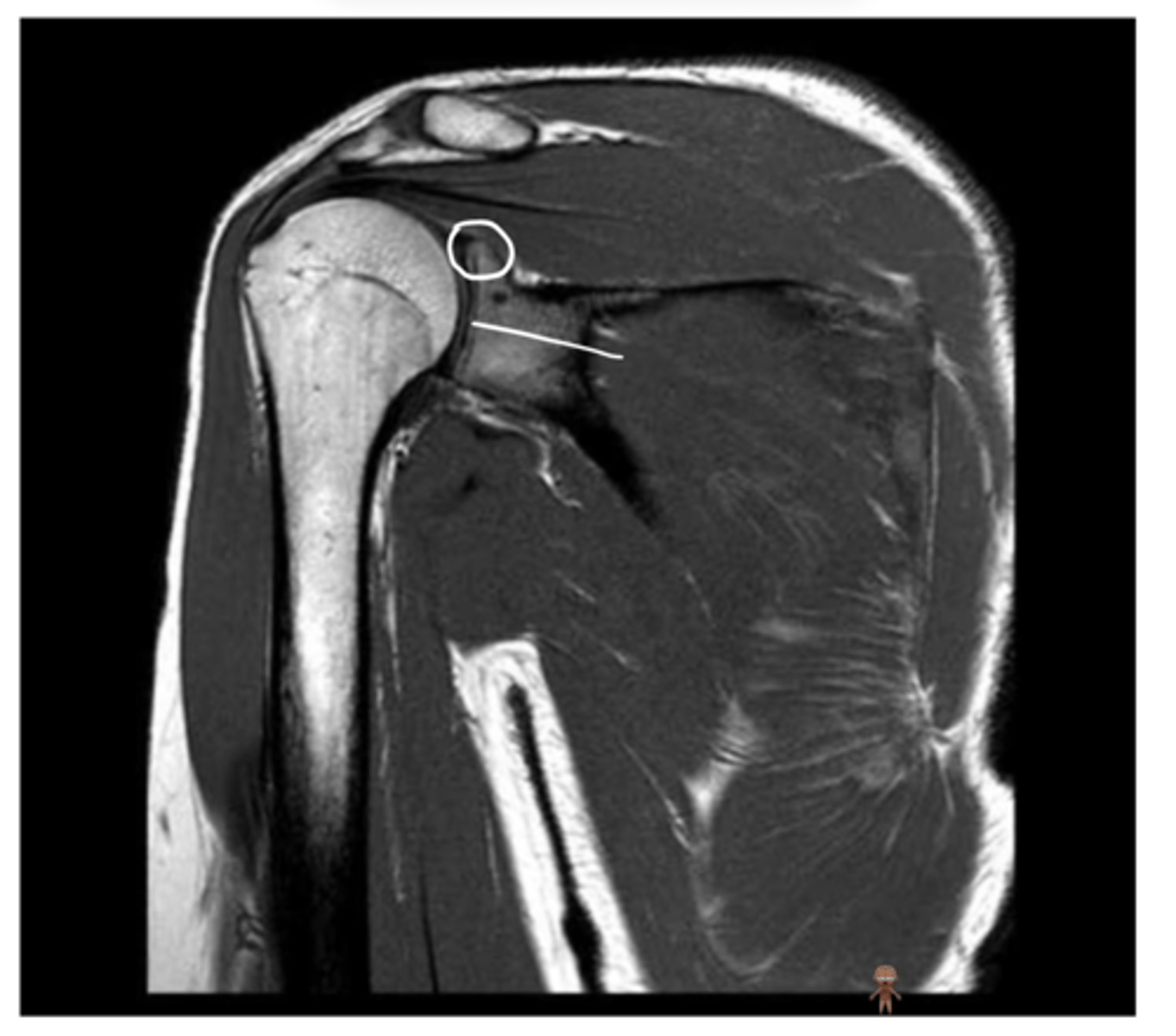
Right where the pointer is -- this is where the supraspinatus attaches to the humerus
If there was a supraspinatus tear, where would you see it? Why?
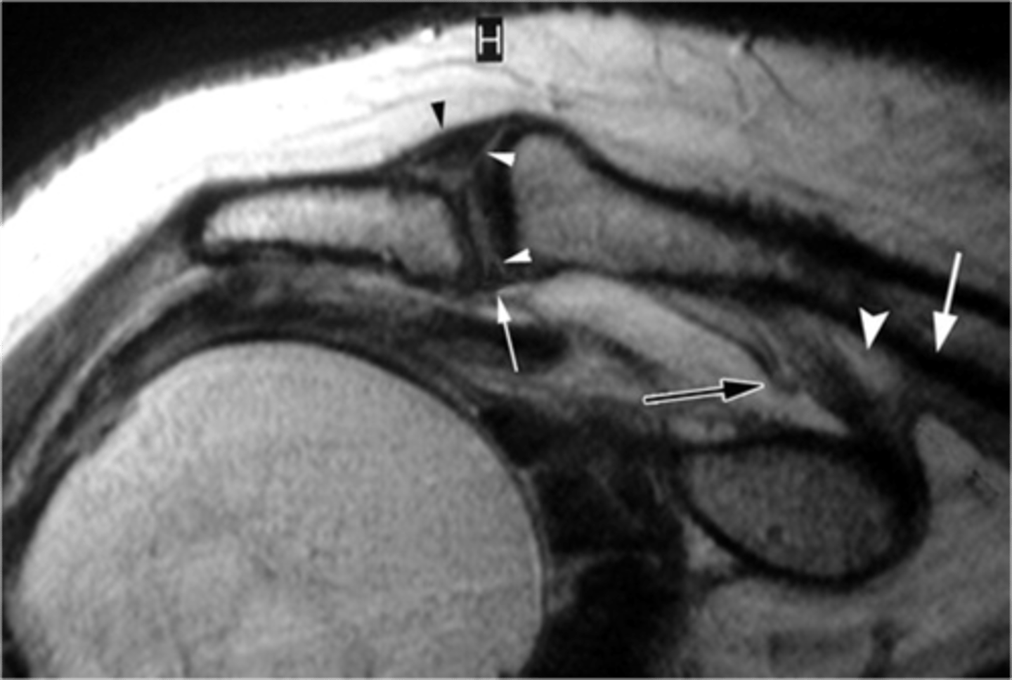
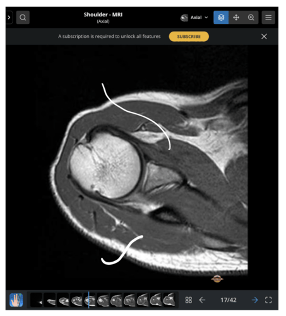
AC joint, coracoid, short head of biceps (attaching to the coracoid), long head of biceps (making its way to attach to the superior glenoid labrum), anterior deltoid, and posterior deltoid
Which structures are present in this image as we work our way inwards towards the glenoid fossa and labrum?
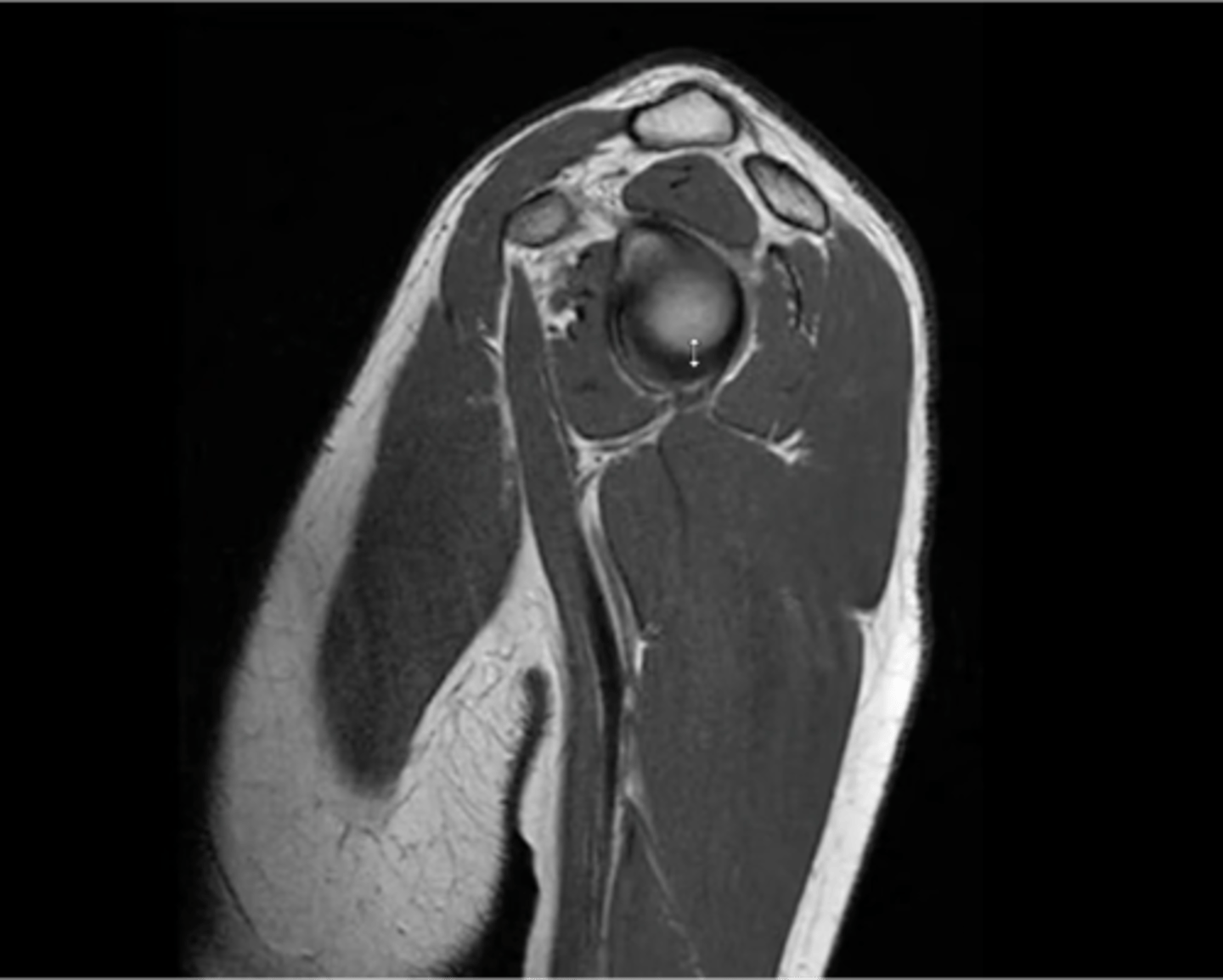
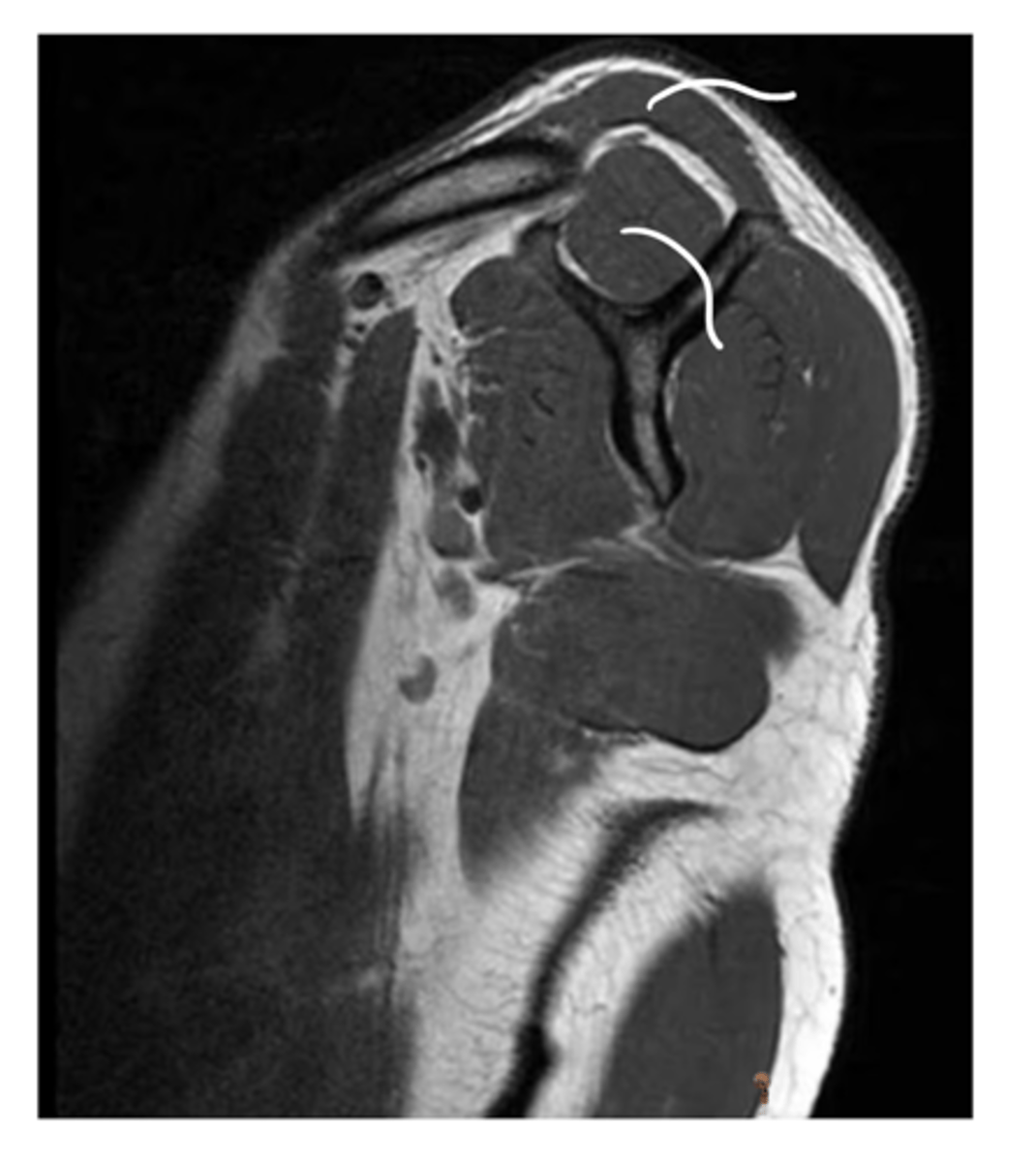
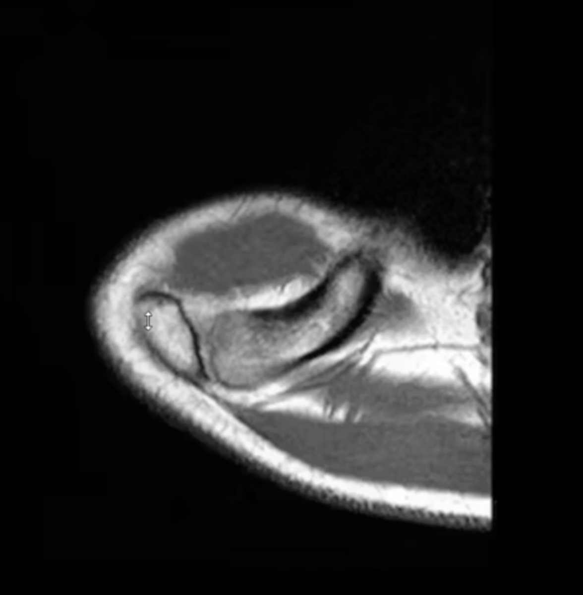
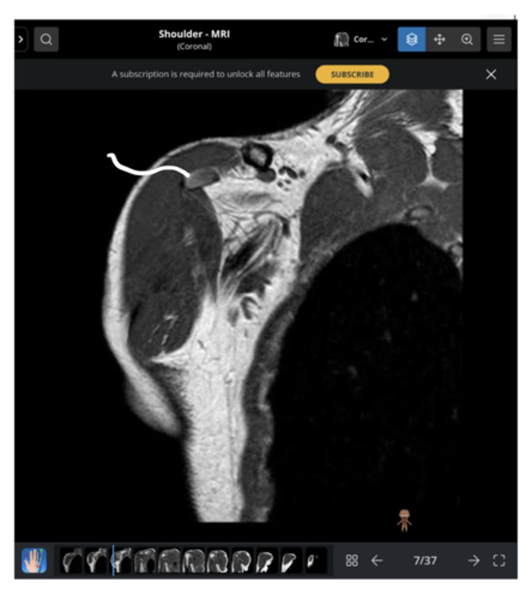
An axial view of the AC joint
What is this image depicting?
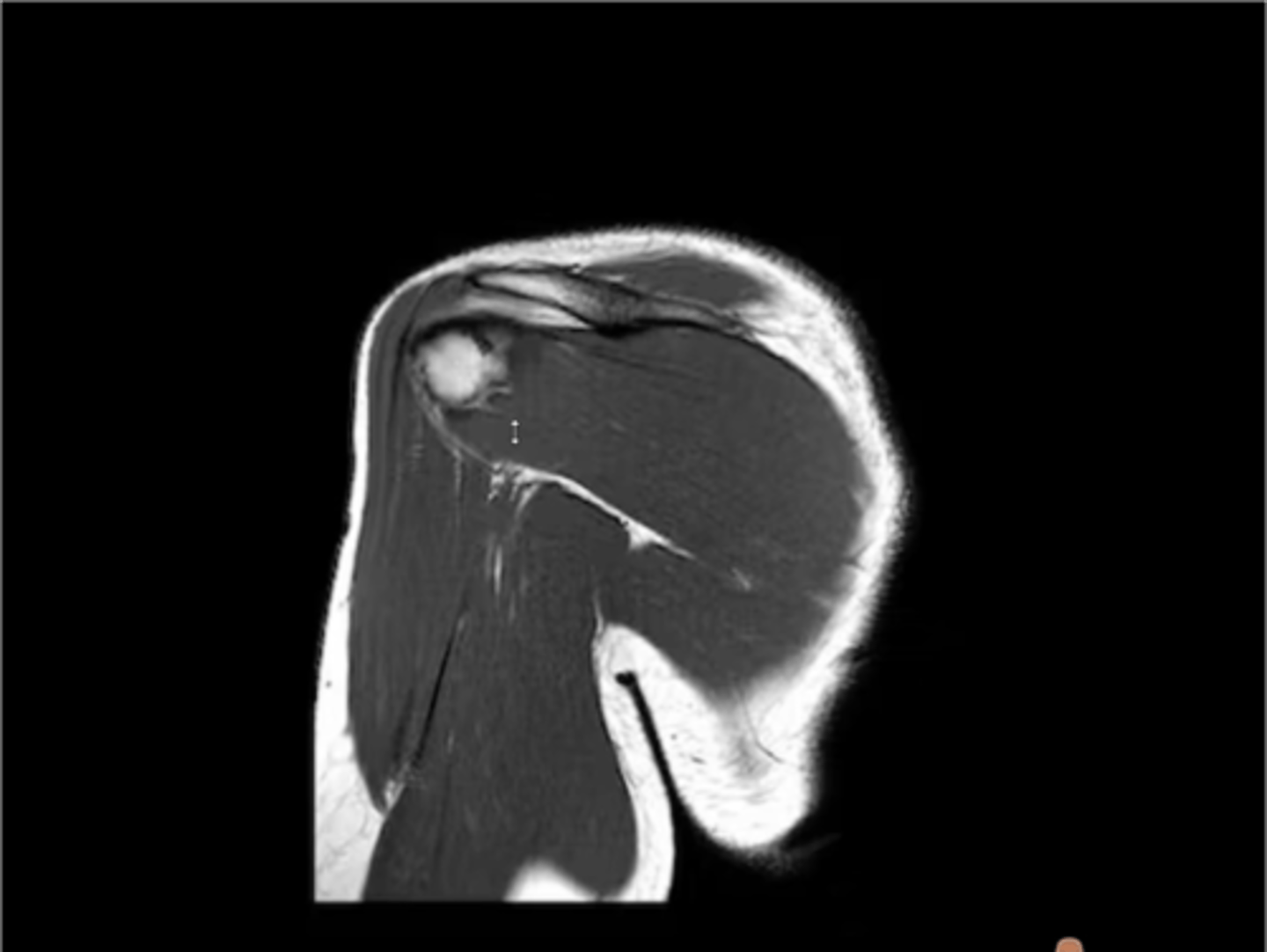
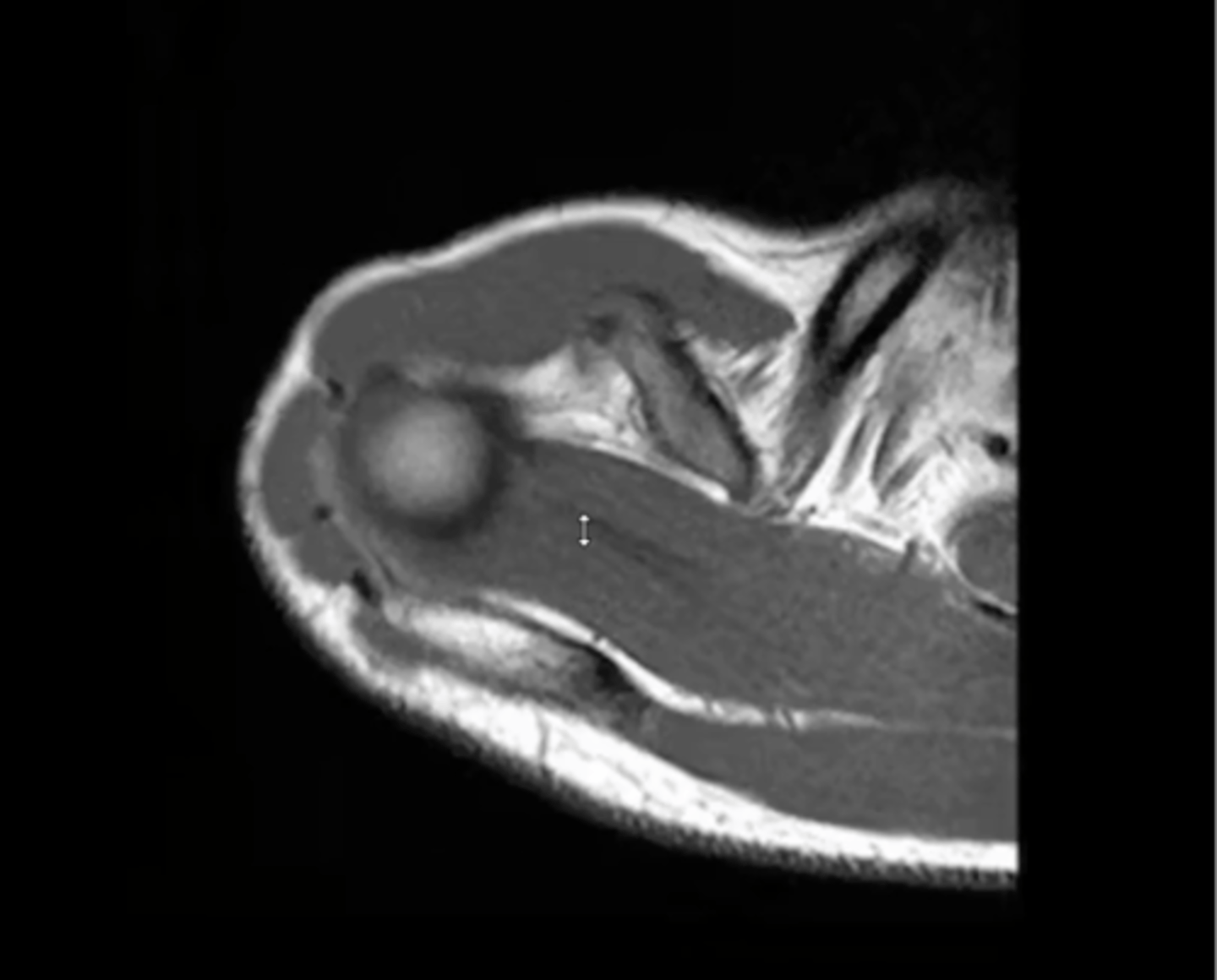
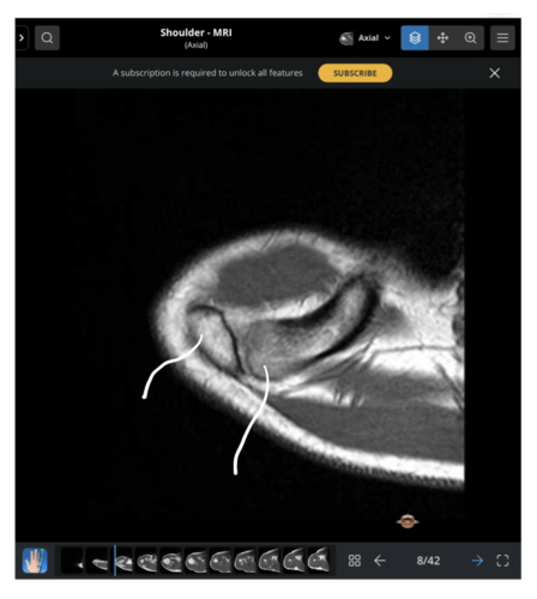
An axial view of the supraspinatus muscle belly
What is this image depicting?
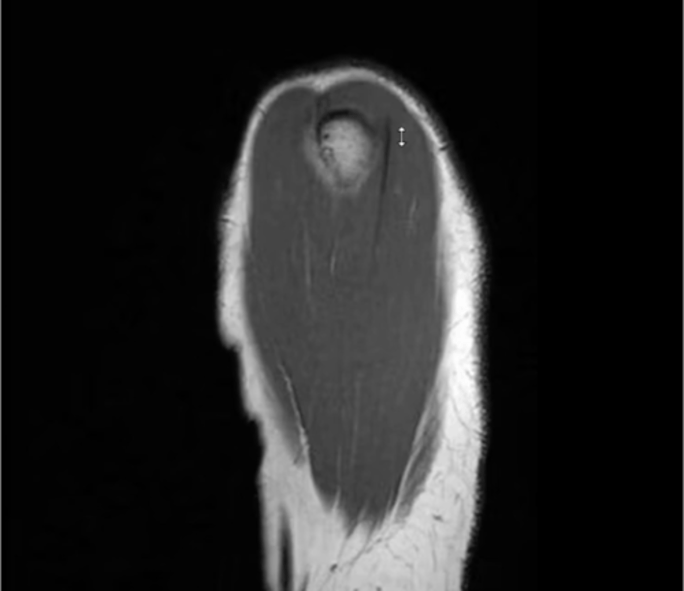
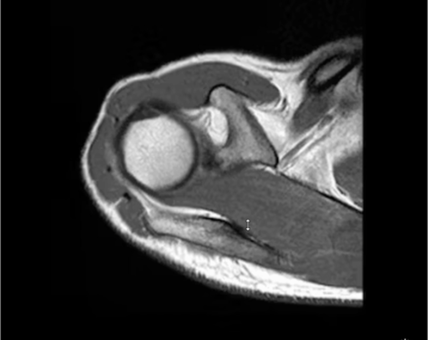
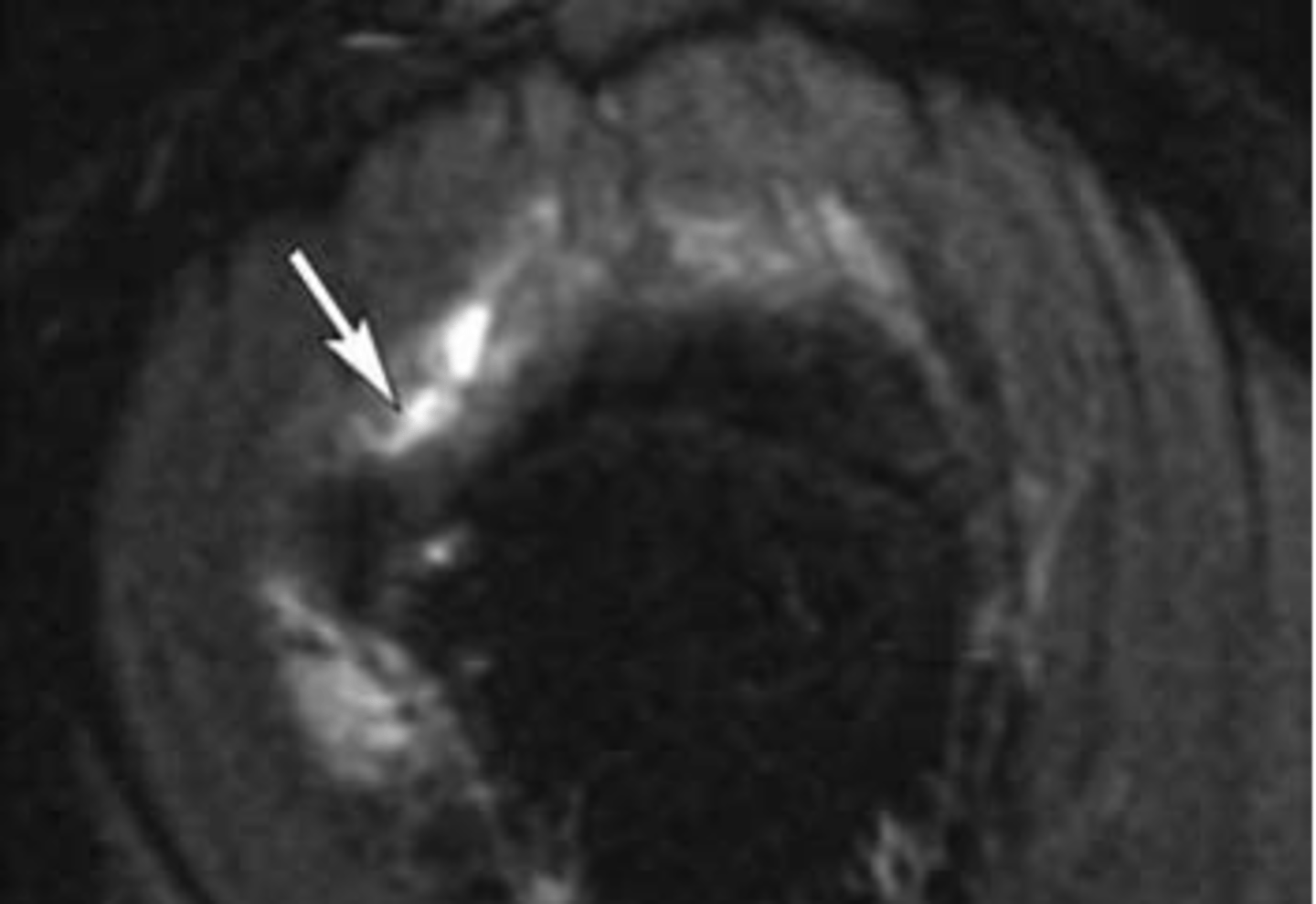
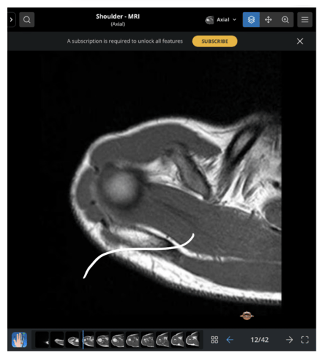
An axial view of the supraspinatus tendon fibers wrapping around the head of the humerus
What is this image depicting?
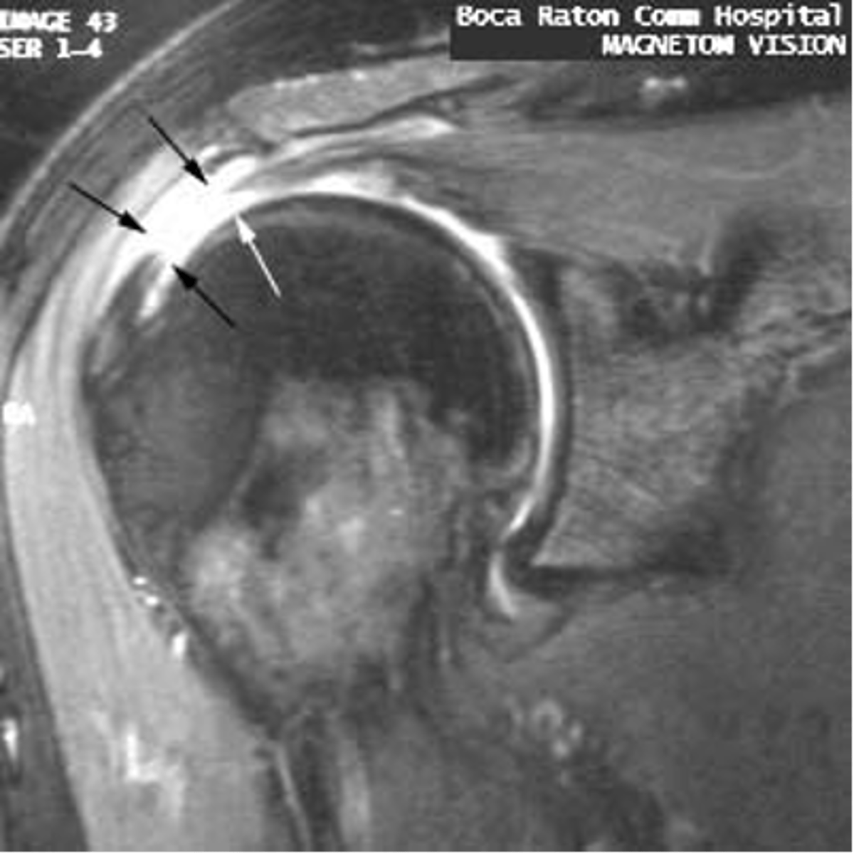
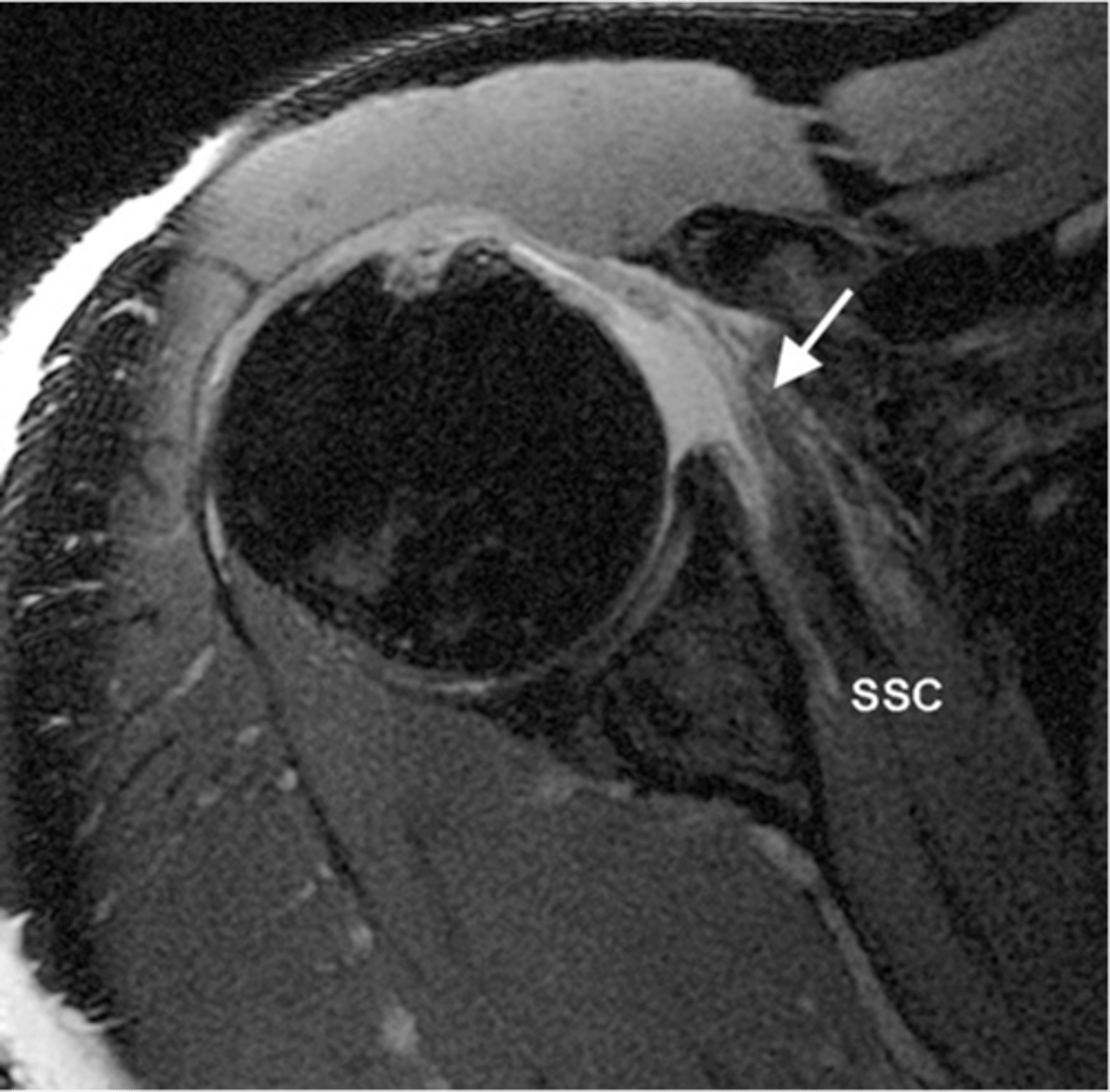
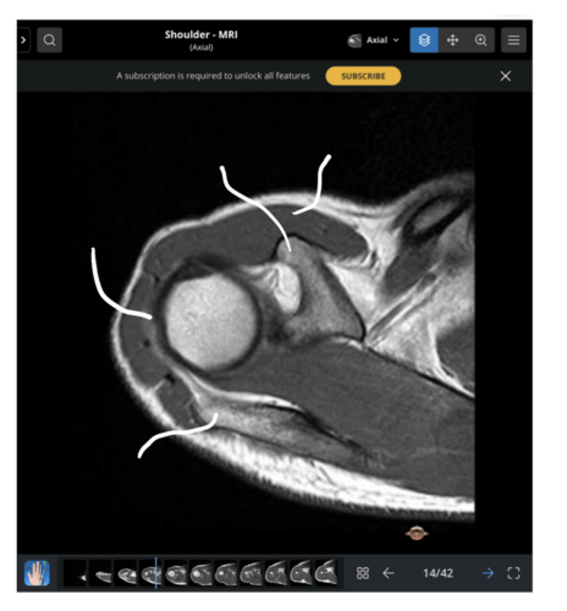
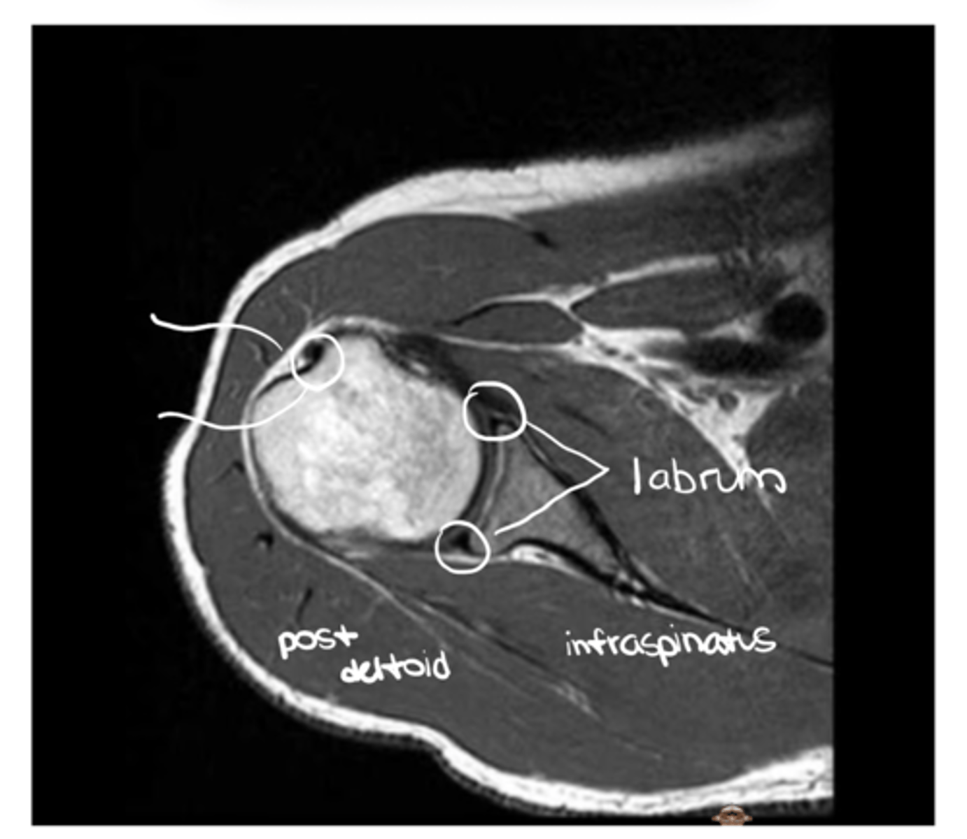
An axial view of the subscapularis tendon wrapping around the anterior portion of the humerus & the infraspinatus tendon on the back side
What is this image depicting?
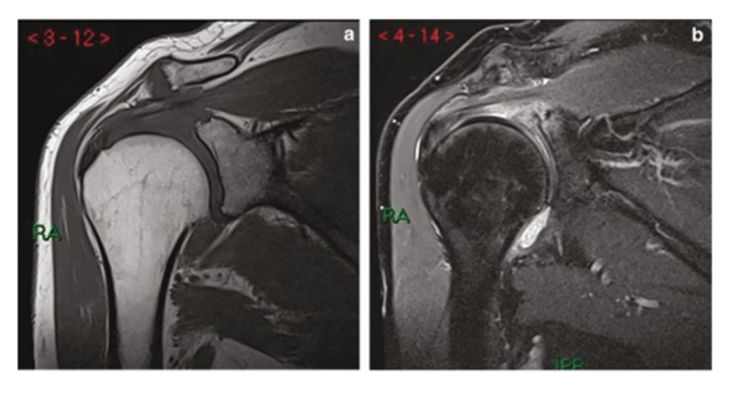
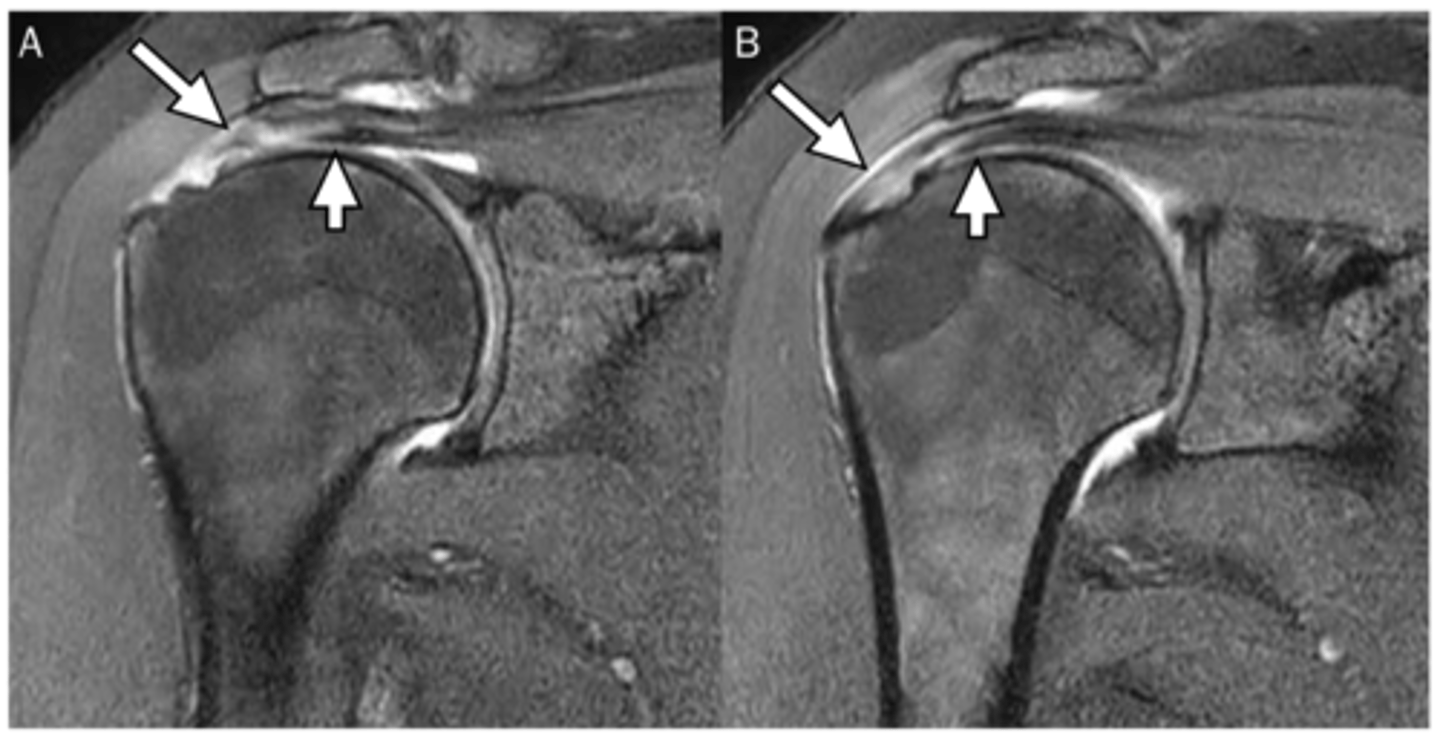
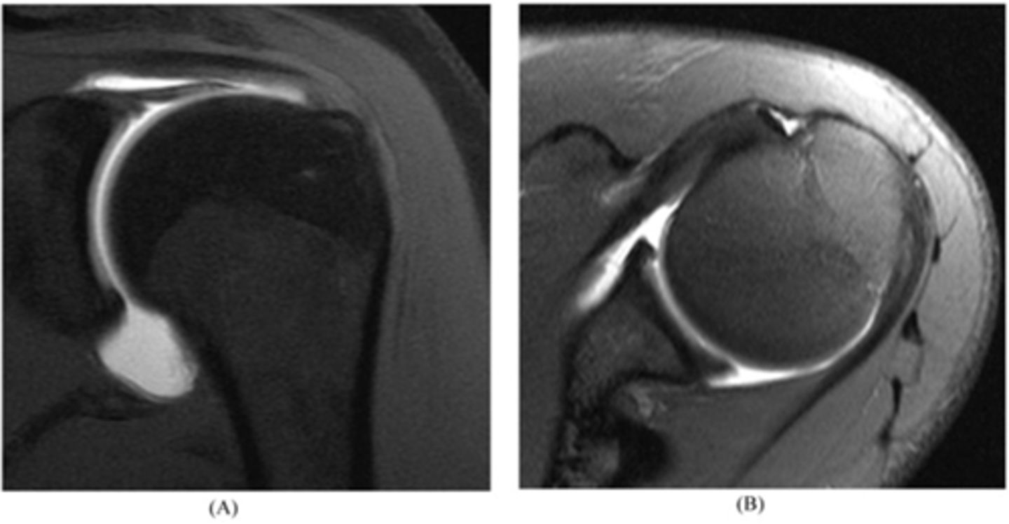
Coronal view of an AC joint separation (grade II sprain) -- an MRI allows us to see the torn ligaments, fluid accumulation, swelling, and get a good sense of the edge of the joint to see how healthy the cartilage is
What is this image depicting? Why is an MRI helpful vs. an x-ray?
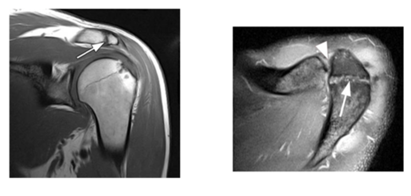
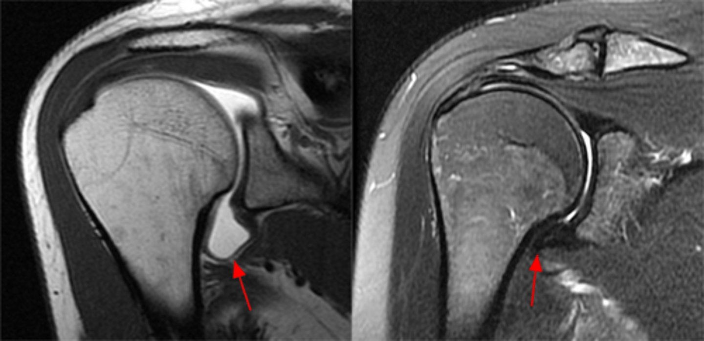
Sagittal view of an AC joint separation (grade I-II) -- torn ligaments (sitting on top of the ACJ) buckle and get wavey (when they should be straight and tight normally) and inflammation is also present on the bottom side of the joint
What is this image depicting? How do you know?
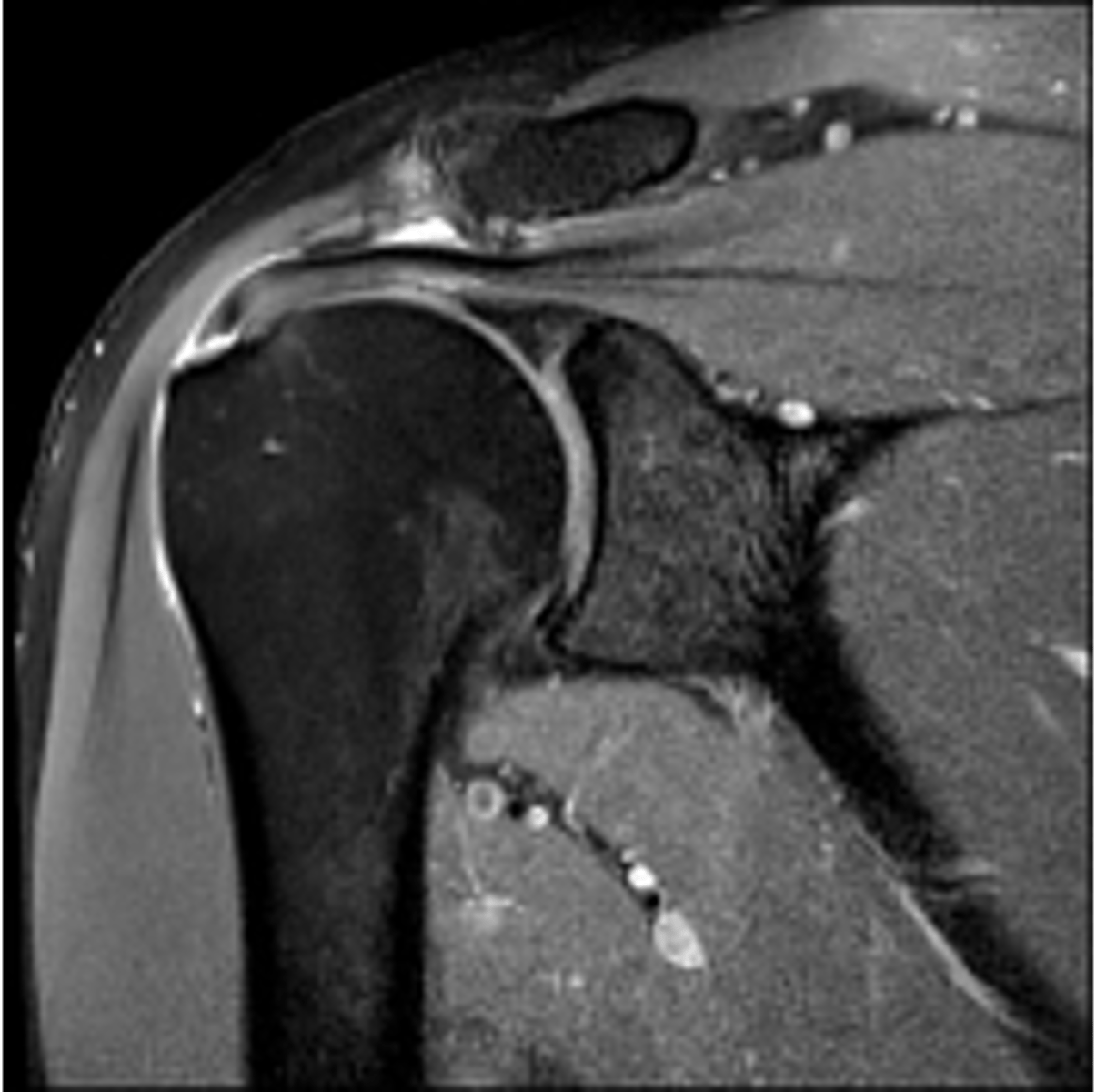
AC joint OA w/ thick, fibrous fluid accumulating and eating away at the joint (note: with OA, hypertrophy, joint surface changes, and bone edema are common)
What is this image depicting at the ACJ?
AC joint OA progression w/ roughed surfaces, hypertrophy, and lytic changes (bone has almost been eaten away due to the damage)
What is this image depicting at the ACJ?
Lytic/arthritic changes in the ACJ --> hypertrophy on the inferior side --> compression on the rotator cuff --> increases the likelihood of a rotator cuff tear (i.e., the supraspinatus falls victim to bone spurs, swelling, hypertrophy, etc.)
Case: a 73-year-old swimmer fell on a boat in rough weather. There was expansion of the ACJ, impingement of the supraspinatus, erosions of articular surfaces, and subdeltoid hematoma (chronic).
Which changes may occur as a result of this accident?

Os Acromiale, a disorder that involves lack of fusion of the acromion (i.e., the growth plate did NOT fuse --> mobile acromion --> encroachment and impingement on the rotator cuff tendons)
What are these images depicting at the ACJ?
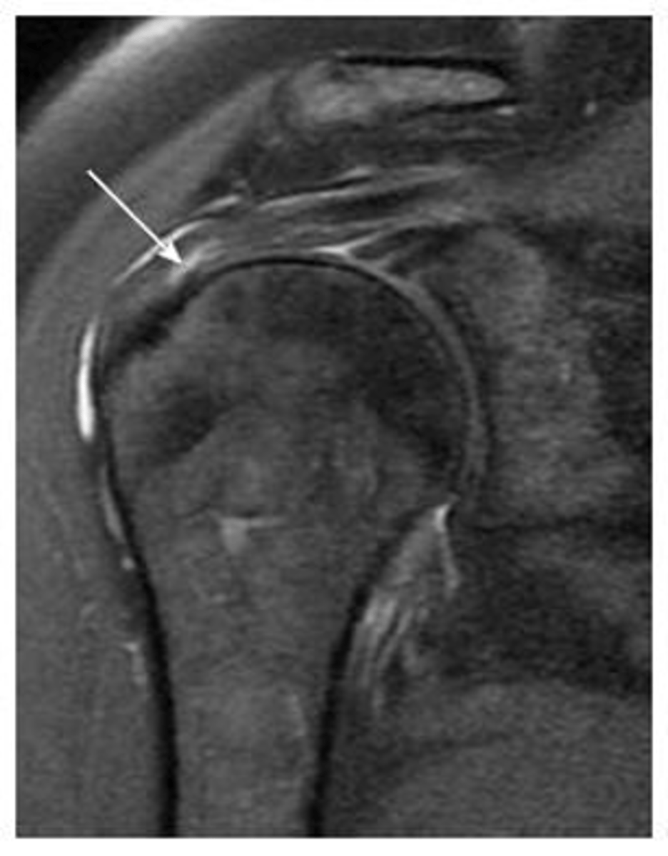
Rotator cuff tendinopathy -- image involves signal changes but the tendon is NOT yet torn
What is this image depicting w/ the rotator cuff? How do you know?
Partial tear of the rotator cuff -- focal area of disruption
What is this image depicting w/ the rotator cuff? How do you know?
Partial rotator cuff tear -- focal area of disruption
What is this image depicting w/ the rotator cuff? How do you know?
Full thickness rotator cuff tear -- 100% of the fibers are involved
What is this image depicting w/ the rotator cuff? How do you know?
This system allows us to classify partial tears of the rotator cuff:
1 = Bursal-sided tear
3 = Intra-substance tear (in the middle of the tendon itself)
5 = Articular-sided tear
Note: we can also identify "delamination," where these fibers separate from one another, which would also be considered a partial tear
Why is understanding this image important?
Left image: bursal-sided tear w/ SOME fibers still intact
RIght image: bursal sided AND intrasubstance tears
NOTE: our concern is these partial tears will progress to full tears
What are these images depicting w/ the rotator cuff? What is our concern?
Full-thickness tear!!
- fluid (area of tear) in white
- retraction in red
- bucking in green
What is this image depicting w/ the rotator cuff? (hint: it is a T2 image)

Complete thickness tear of the supraspinatus w/ major bucking and retraction as the muscle pulls the tendon back
What are these images depicting w/ the rotator cuff?
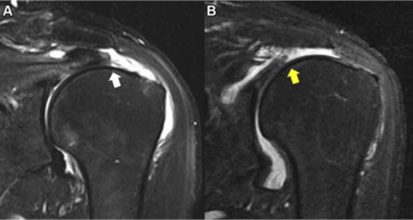
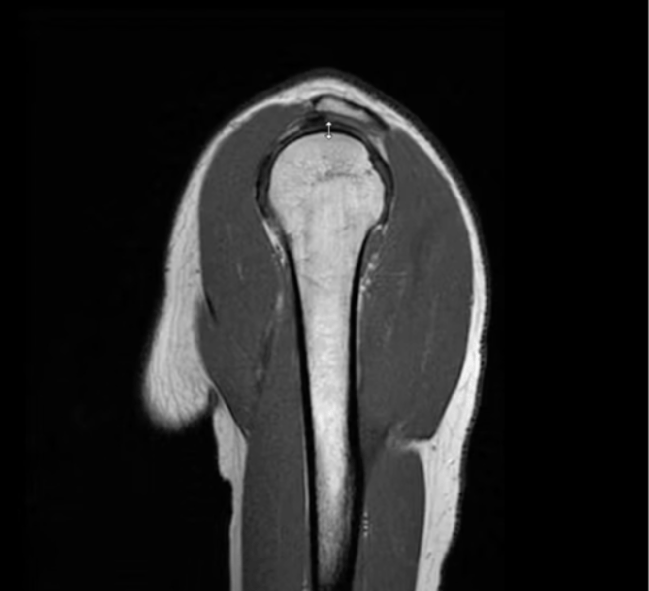
Supraspinatus tendon
What is this image calling out in white on the left and with a star on the right?
Axial view of the shoulder w/ atrophy of the subscapularis -- fat is depicted in white in the middle of the muscle, which is a tell-tale sign of disuse, atrophy, neurological impairments, or tendon tears
What is this image depicting? How do you know?
MORE fatty atrophy and infiltrate -- this is a prognostic factor for patients! (i.e., we DON'T want to repair a cuff that already has a ton of fatty infiltrate)
What is this image depicting? Why is it important?
Adhesive capsulitis (frozen shoulder) -- axillary recess can get fibrose, thick, and larger than normal
Note: typically, MRIs should NOT be ordered for adhesive capsulitis because they're not necessary
What is this image depicting? How do you know?
**ALTHOUGH THIS IS IN THE ADHESIVE CAPSULITIS SECTION, DR. C SAID THAT THIS IS A CRAPPY EXAMPLE AND IS ACTUALLY A NORMAL AXILLARY RECESS THAT IS BEING HIGHLIGHTED BY THE DYE IN THE MRI ARTHROGRAM**
Note: normally, you DON'T see the axillary recess very easily, but in this image, the dye makes it clear as day
What is this image depicting?
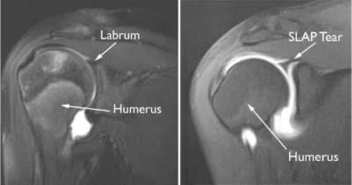
1. When there is a labral tear, the dye will make its way into that region, as depicted in this image
2. ADHESIVE CAPSULITIS IS NOT PRESENT -- THIS IS A NORMAL PRESENTATION DUE TO THE DYE IN THE RECESS
Two questions for you on this one: why do we know there a labral pathology present? (1) and why is this NOT adhesive capsulitis? (2)
THEN, that would be considered full-blown adhesive capsulitis
Essentially, if there IS dye, it's always going to look like there's something going on with the axillary recessive. What about if there was NO dye?
Labral tears -- dye is creeping into the labral region
What are these images depicting? How do you know?
1. Posterior coronal view
2. NO CORACOID
3. Posterior deltoid
1. What view is this image?
2. How do you know?
3. Which structure is being pointed to?

1. Posterior coronal view
2. NO CORACOID -- spine of the scapula
3. Superior = spine of the scapula, inferior = infraspinatus
1. What view is this image?
2. How do you know?
3. Which structures are being pointed to?
1. Posterior coronal view
2. Glenoid
1. What view is this image?
2. Which structure is being pointed to?
1. Anterior
2. Coracoid
1. What view is this image?
2. Which structure is being pointed to?
1. Lateral deltoid
2. Acromion
3. Clavicle
4. Supraspinatus footprint
5. Glenoid
Working our way around clockwise, which structures are being pointed to?
1. Anterior deltoid
2. Middle deltoid
3. Posterior deltoid
From left to right, which structures are being pointed to?
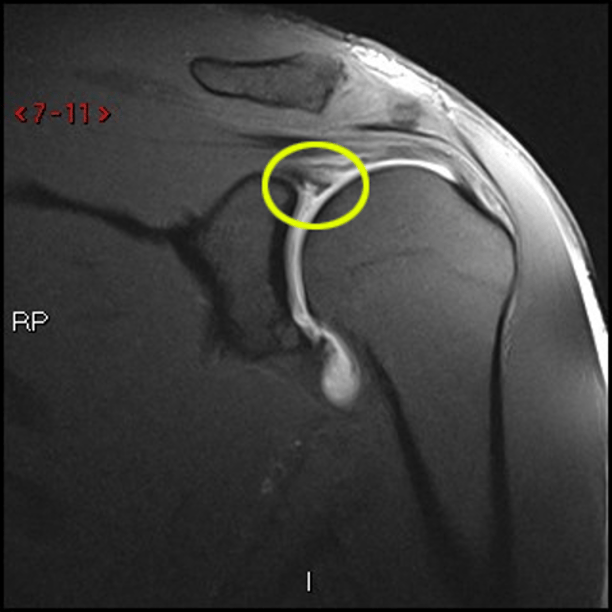
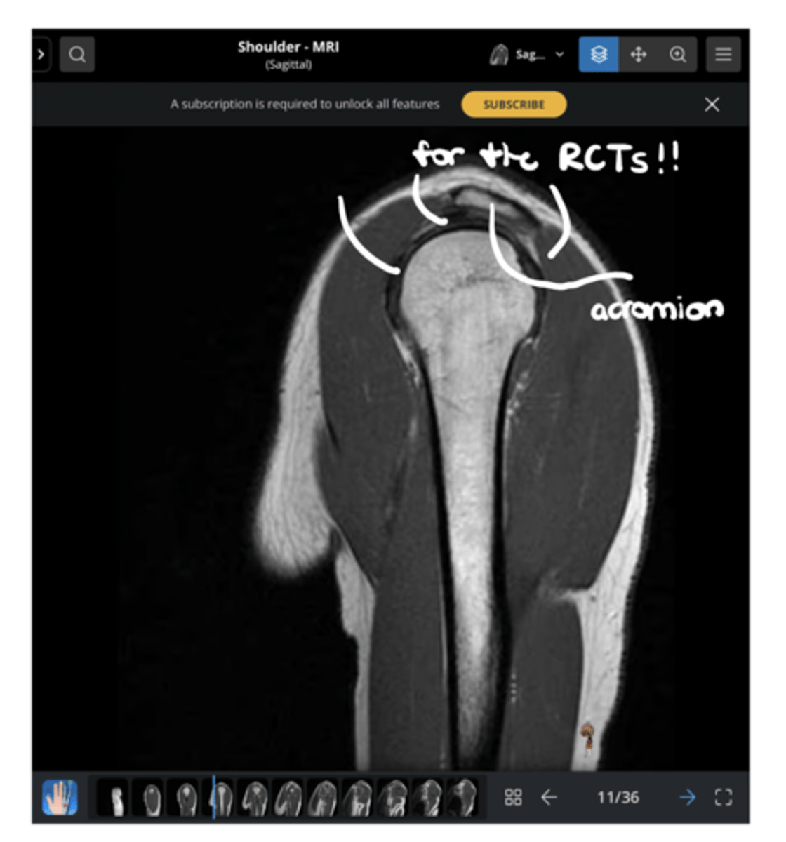
Footprints!!
What is the missing word in this image: "________ for the RCTs!!"
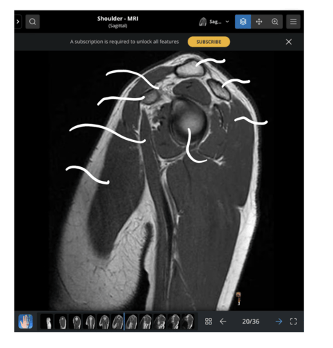
1. Anterior deltoid
2. SH biceps
3. Coracoid
4. LH biceps (making its way to attach to the superior glenoid labrum)
5. Clavicle
6. Acromion
7. Posterior deltoid
8. Glenoid fossa & labrum
Working our way clockwise, which structures are being pointed to in this image?
Upper trap
Which structure is being pointed to in this image?
1. Acromion
2. Clavicle
From left to right, which structures are being pointed to in this image?
Supraspinatus
Which structure is being pointed to in this image?
1. Scapula
2. Supraspinatus tendon fibers (wrapping around the humerus)
3. Coracoid
4. Anterior deltoid
Working our way clockwise, which structures are being pointed to in this image?
1. Subscapularis (superiorly)
2. Infraspinatus (inferiorly)
Which structures are being pointed to in this image?
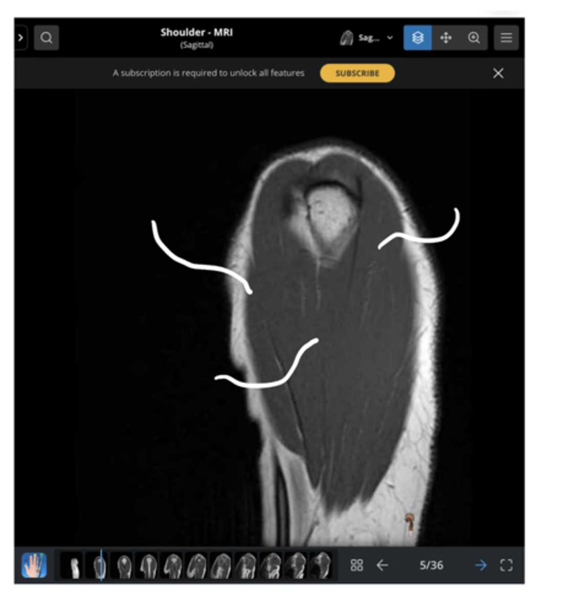
1. Anterior coronal view
2. Lung space (this is not as easily visualized from a posterior view due to the movement of the scapula on the thorax while laying down)
Note: usually, a great way to tell is by the coracoid process, but it is NOT visible this superficially
1. Which view is this?
2. How do we know -- from what we can see here?
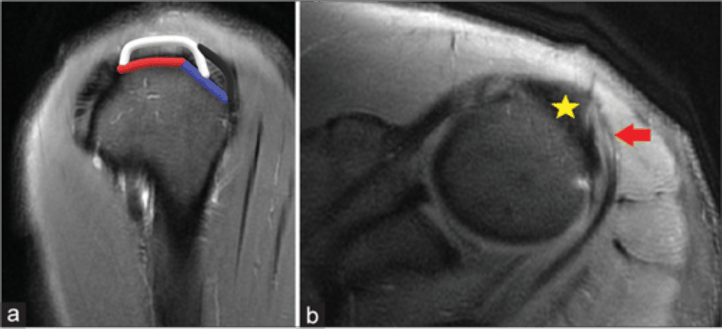
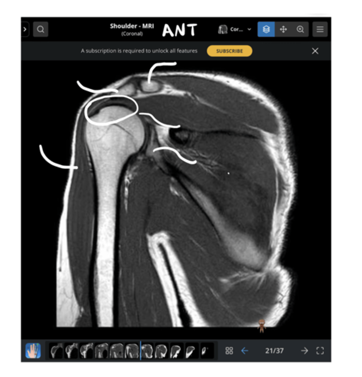
Circle = labral triangle
Pointer = articular cartilage
What has been circled and what has been pointed to in this image?
1. AC joint sprain
2. RTC tear (the worst versions will involve retraction and atrophy)
3. Adhesive capsulitis (axillary recess will enlarge)
4. Labral pathology (with dye -- NOT well diagnosed)
What are the MAIN pathologies can we see in a CORONAL view of the shoulder?
Circle = LH biceps
Pointer = transverse humeral ligament
What has been circled and what has been pointed to in this image?
1. Upper trap (superiorly)
2. Supraspinatus (inferiorly)
Which structures are being pointed to in this image?