ANAT 100 - Module 2
1/101
There's no tags or description
Looks like no tags are added yet.
Name | Mastery | Learn | Test | Matching | Spaced | Call with Kai |
|---|
No analytics yet
Send a link to your students to track their progress
102 Terms
5 main functions of the skeletal system
SUPPORT
- the skeleton provides framework that anchors soft organs
MOVEMENT
- skeletal muscles use bones as levers
PROTECTION
- protects our internal organs
STORAGE
- bones are storage for minerals and fat
HEMATOPOESIS
(production of red blood cells)
- blood cell formation happens in the marrow of bones
2 types of bone marrow
YELLOW MARROW
- stores fat
RED MARROW
- important for hematopoiesis (production of red blood cells)
Basic Composition of Bone
OUTER CORTICAL LAYER
- made of compact bone, hard and strong
INNER CANCELLOUS LAYER
- made up of spongy bone, porous and sponge-like
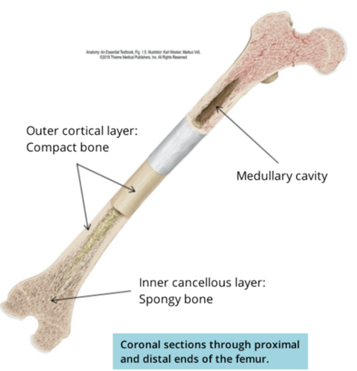
medullary cavity
- where blood cells are produced, found in the inner cavity of the bone

Types of Bones
FLAT
- ex. bones of the skull
IREGULAR
- ex. vertebrae
LONG
- ex. femur (longer than they are wide)
SHORT
- ex. bones of the ankle and wrist (long as they are wide)
SESAMOID
- ex. patella (kneecap)
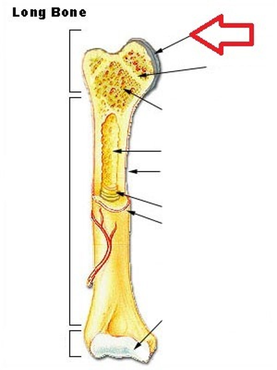
Structure of Long Bones
- most common bone in the body
EPIPHYSIS
- knobby enlarged region at the end that tendons and ligments attach on to
METAPHYSIS
- region between epiphysis and diaphysis (Middle)
DIAPHYSIS
-elongated cylindrical shaft
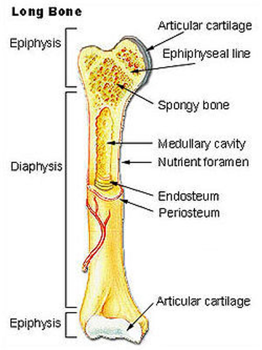
additional features of long bones: Articular cartilage
- covers the epiphysis
- reduces friction between joints and absorbs shock
- a hyaline cartilage because it lines the joint surface
additional features of long bones: periosteum
- a tough sheath of dense irregular connective tissue that covers the surface of bones
- protects the bone, and contains blood vessels and nerves that supply the bone
- it has cells responsible for forming new blood tissue
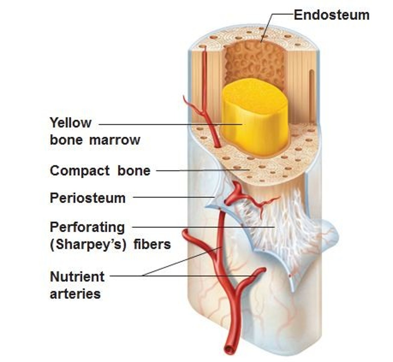
additional features of long bones: medullary cavity
- bone marrow that proceeds red blood cells (in the inner cancellous layer)
Axial Skeleton: Skull
2 sets of bones that make up the skull:
cranial bones
facial bones
Axial Skeleton: Skull: Cranial Bones
7 bones that form a roof (dome of the brain) and a floor (cranial base) creating the cranial cavity
- frontal
- temporal (2)
-sphenoid
- parietal (2)
- occipital
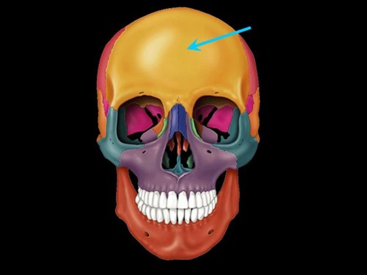
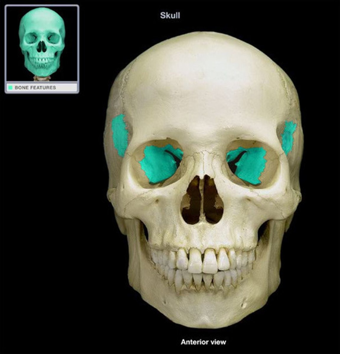
Cranial Bones: Frontal Bone
1 bone
- forms the forehead and roof of the orbits (eye sockets)
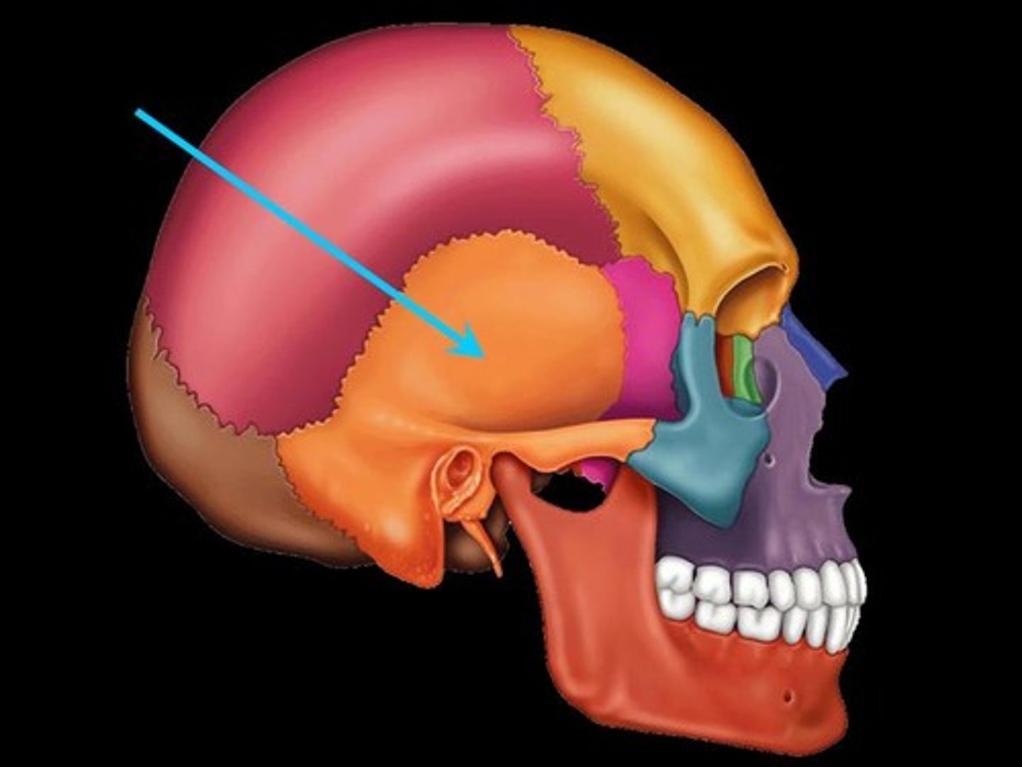
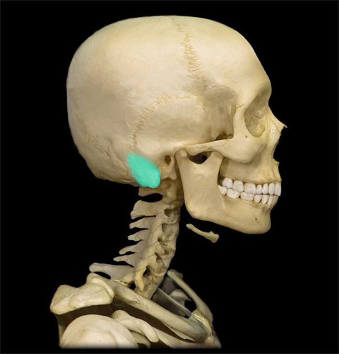
Cranial Bones: Temporal Bones
2 bones
features of the temporal bone:
- zygomatic process
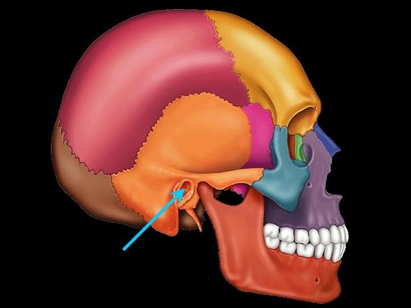
- external auditory meatus (ear hole)
- mastoid process
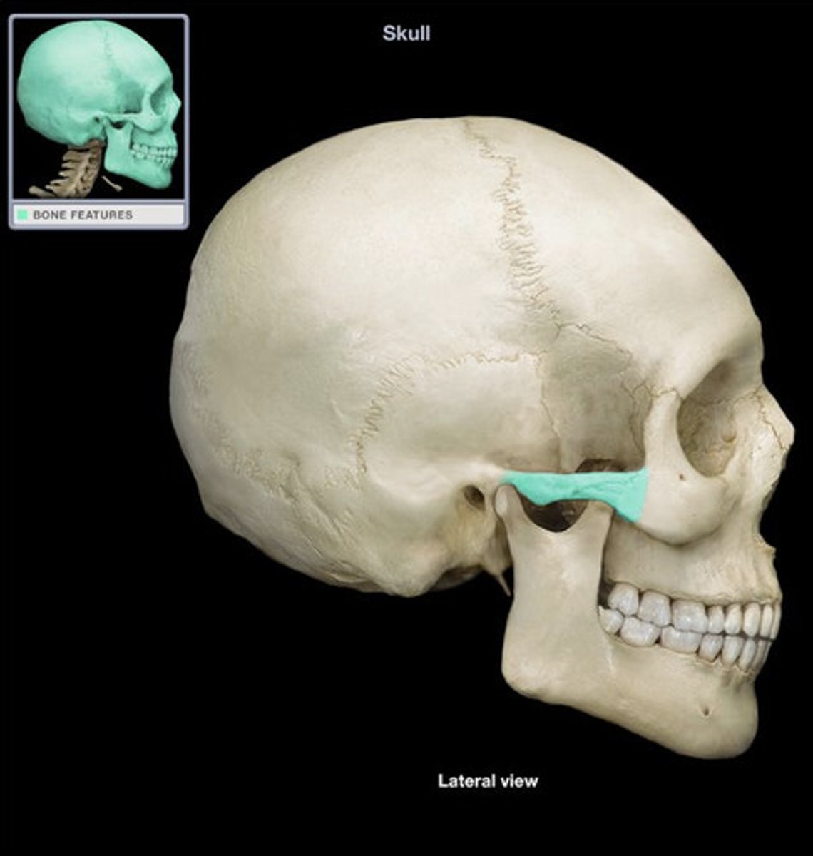
Temporal Bones: zygomatic process
Temporal Bones: External auditory meatus
Temporal Bones: Mastoid process
Cranial Bones: sphenoid bone
1 bone
- "keystone" of the skill because it joints the facial and cranial bones by attaching to almost every bone in the skull
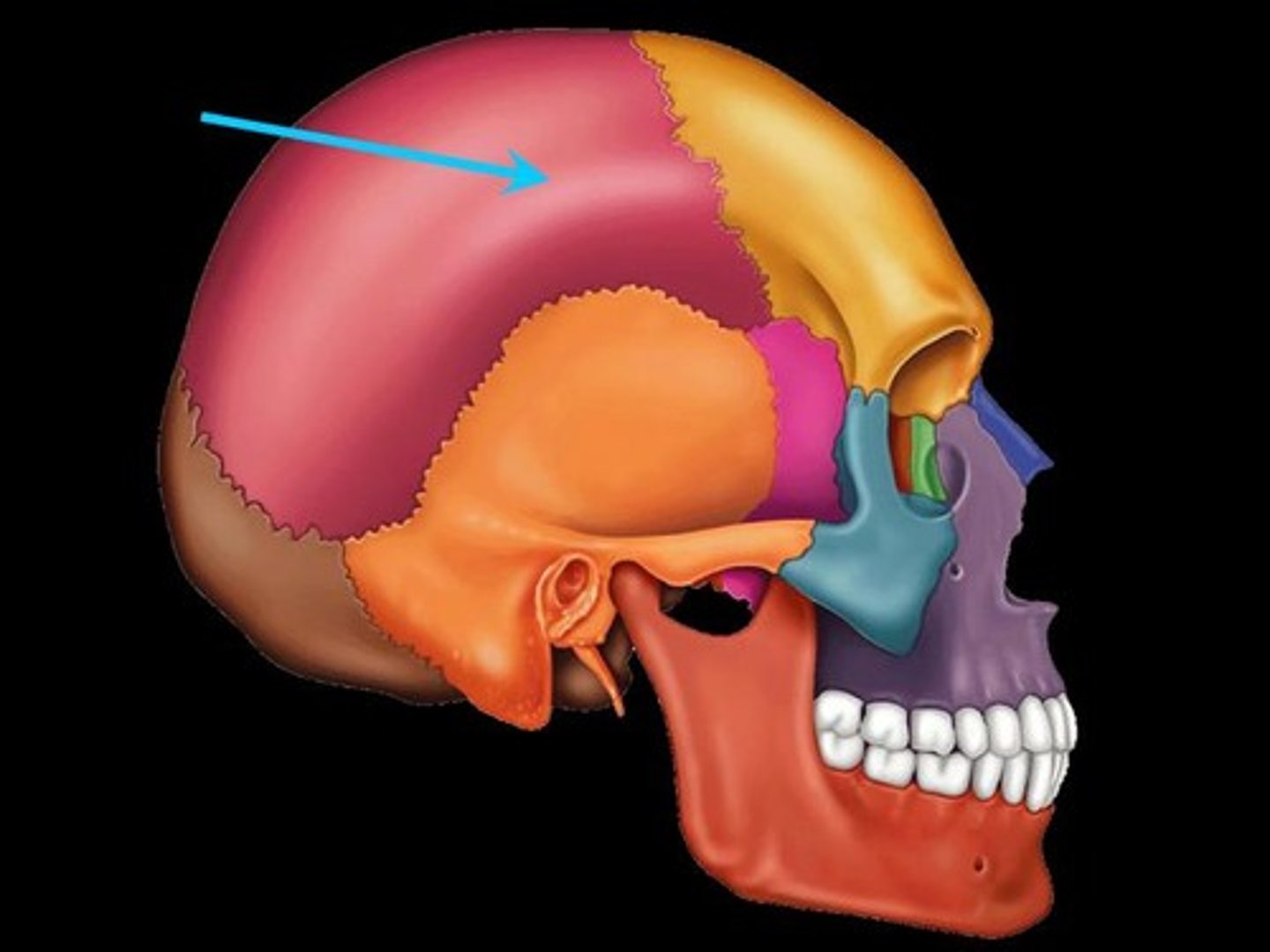
Cranial Bones: Parietal Bones
2 bones
- paired
- form the superior and lateral surfaces of skull (top and side)
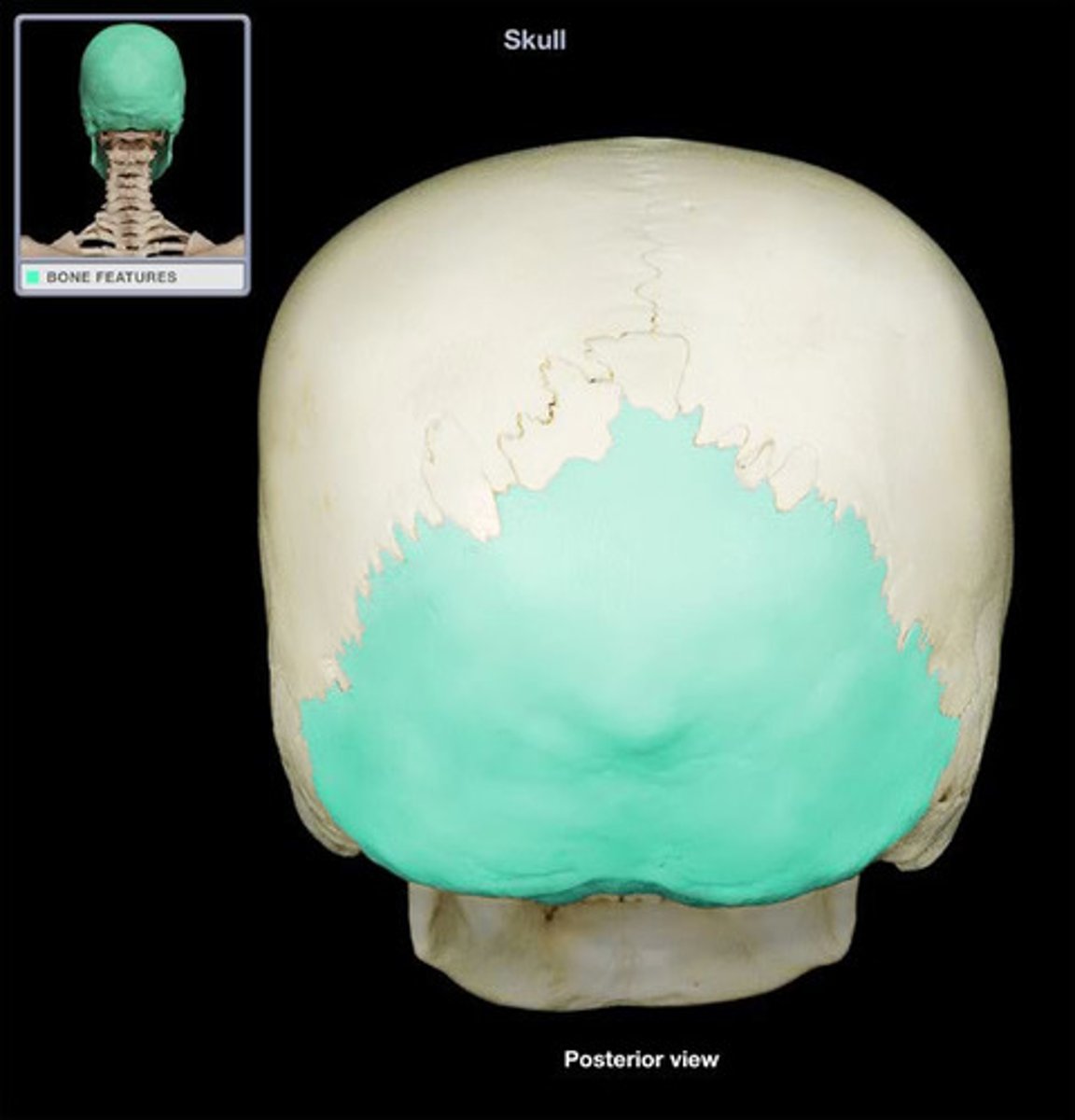
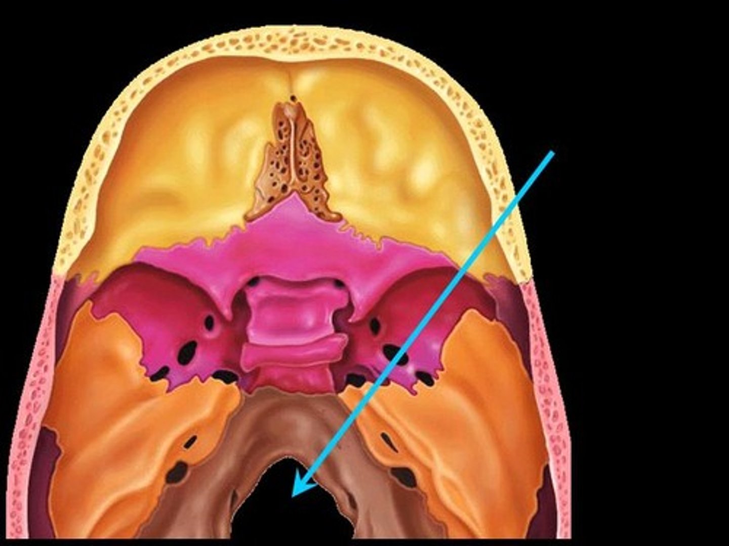
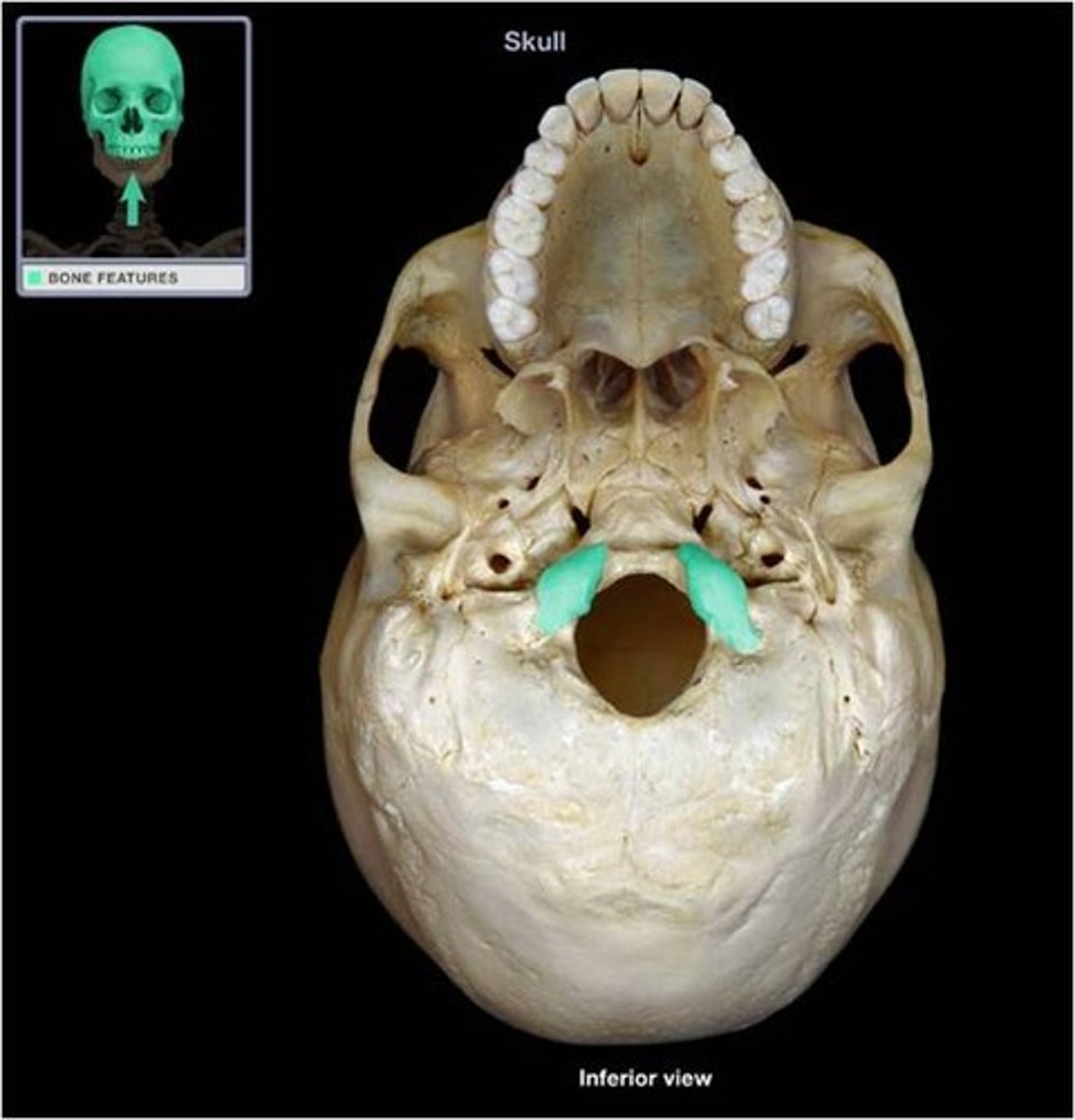
Cranial Bones: Occipital Bone
- forms posterior wall and base of skull
features:
- foramen magnum
- occipital condyles
Occipital bone: foramen magnum
- allows the spinal chord to exit the cranial cavity
Occipital Bone: Occipital condyles
- articulate with the first bone in the neck
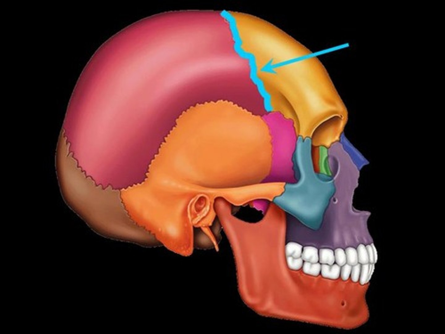
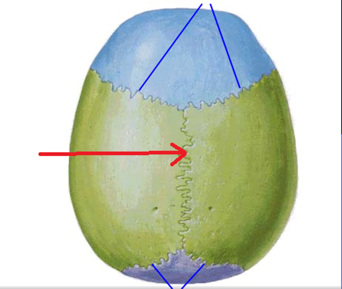
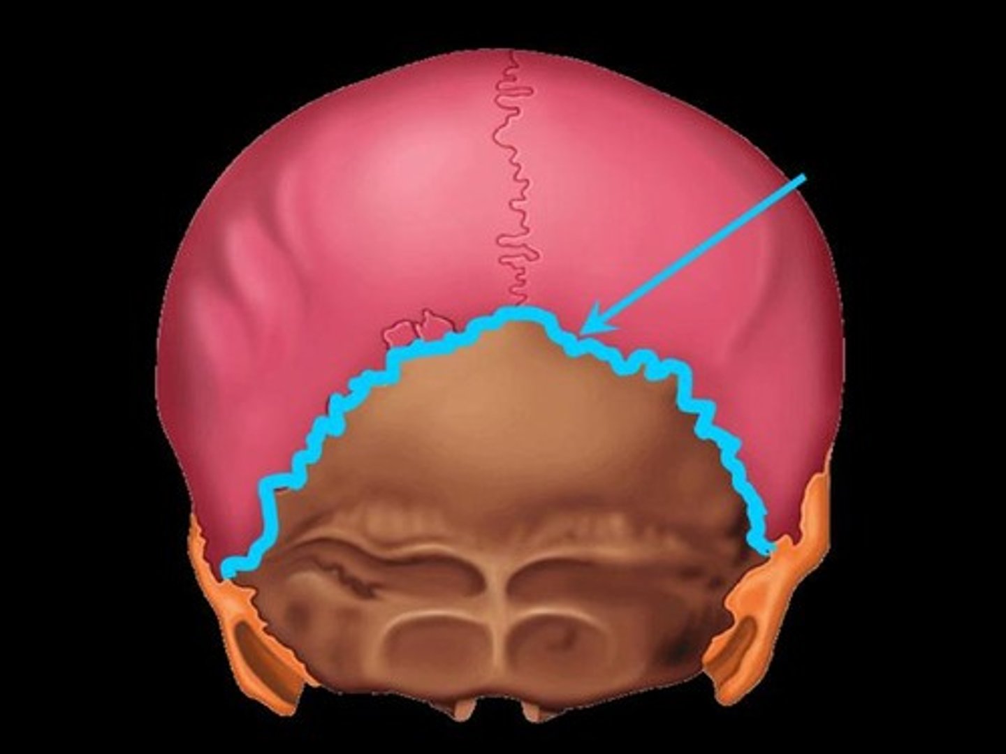
Sutures
- attachments between flat bones in the skull
coronal
sagittal
lambdoid
squamous
coronal suture
Junction between frontal and parietal bones
sagital suture
junction between 2 parietal bones
lambdoid suture
junction between occipital and parietal bones
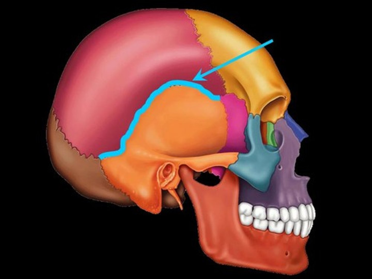
squamous suture
junction between temporal bone and parietal bones
cranial vault
- formed by frontal, parietal, and occipital bones
cranial base
3 fossa:
- anterior cranial fossa
- middle cranial fossa
- posterior cranial fossa
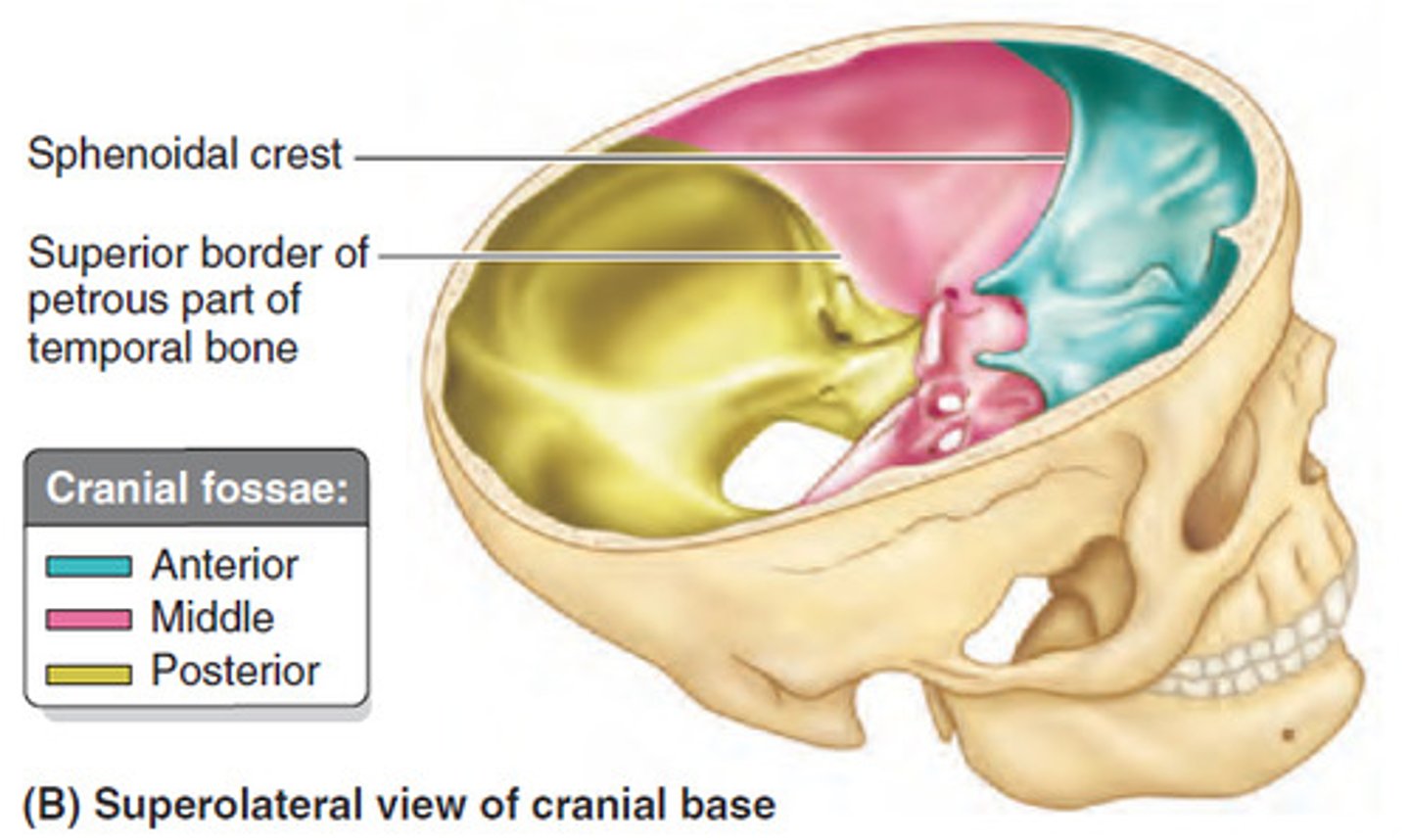
fossa
a depression in a bone
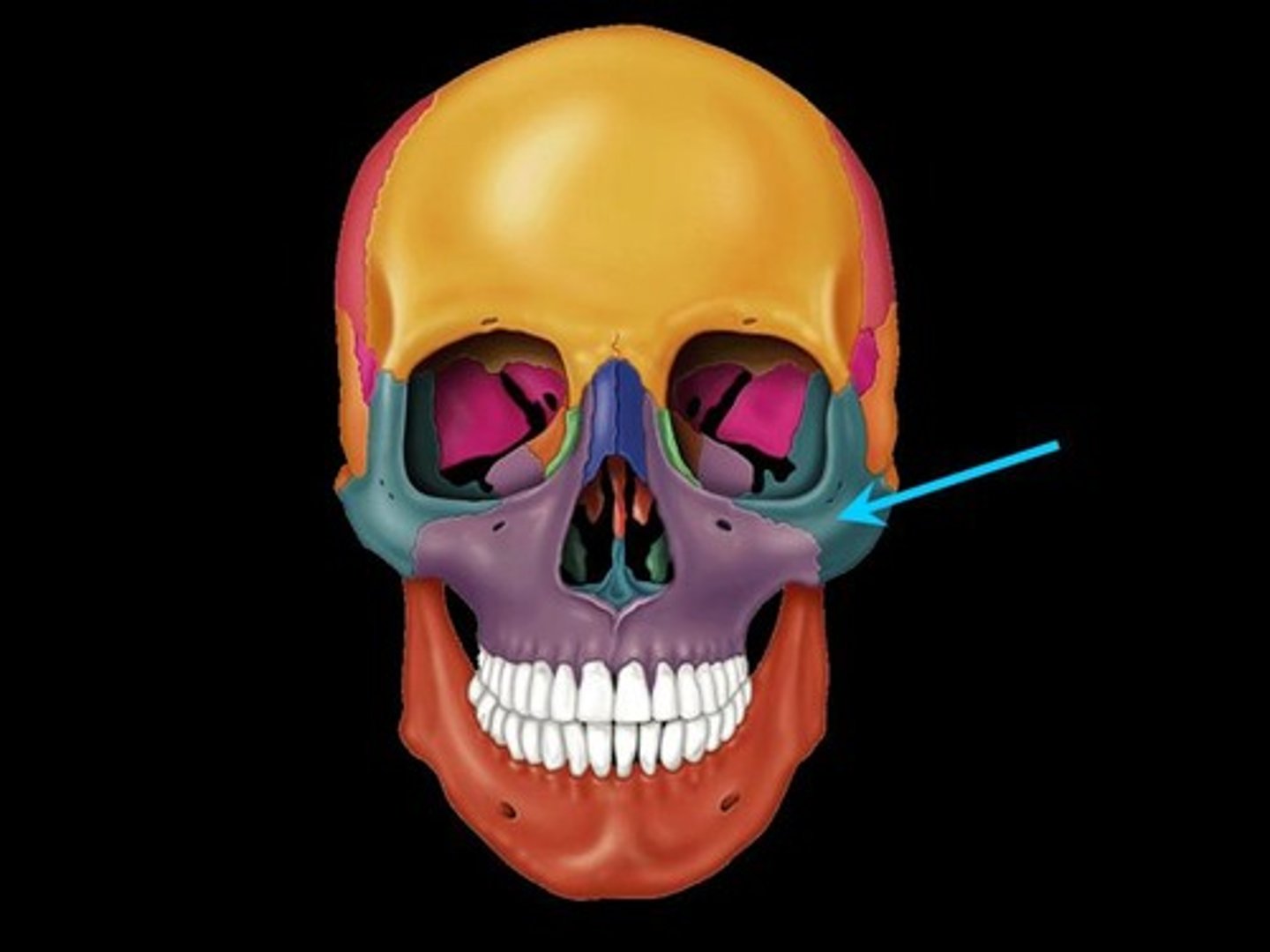
Skull: facial bones
7 bones to know:
- maxillary (2)
- nasal (2)
- zygomatic (2)
- mandible (1)
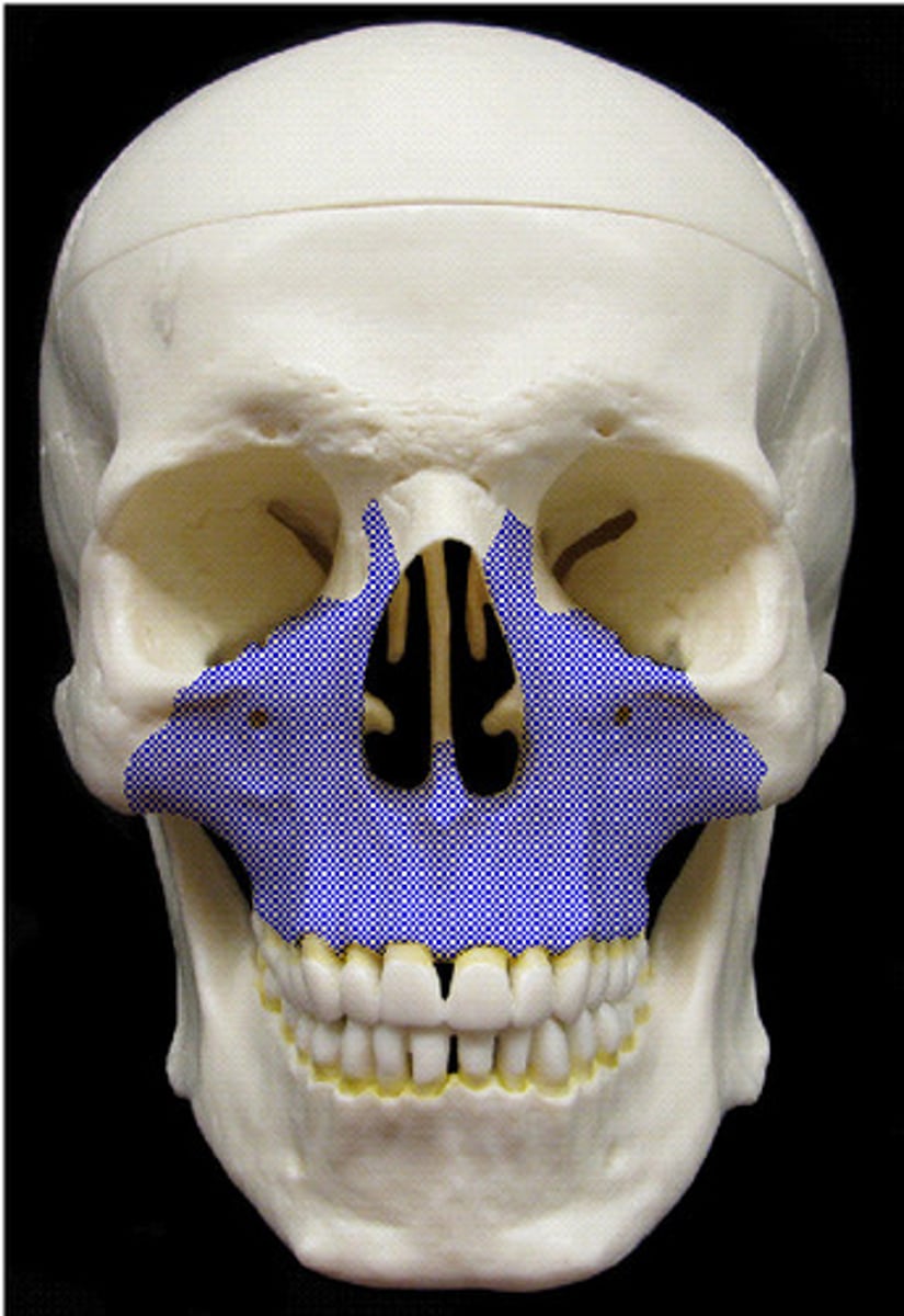
Facial Bones: Maxillary Bones
- paired (2)
- upper jaw bones
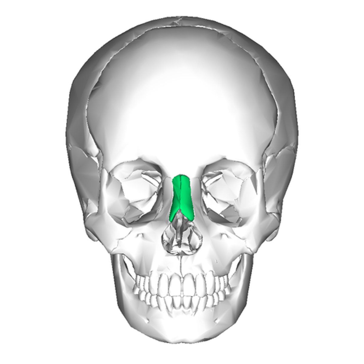
Facial Bones: Nasal Bones
- paired (2)
- articulate with the frontal bone
- form the bridge of the nose
Facial Bones: Zygomatic Bones
- paired (2)
- form the cheekbones
feature:
- the temporal process (which articulate with the zygomatic process of the temporal bone to form the zygomatic arch)
temporal process

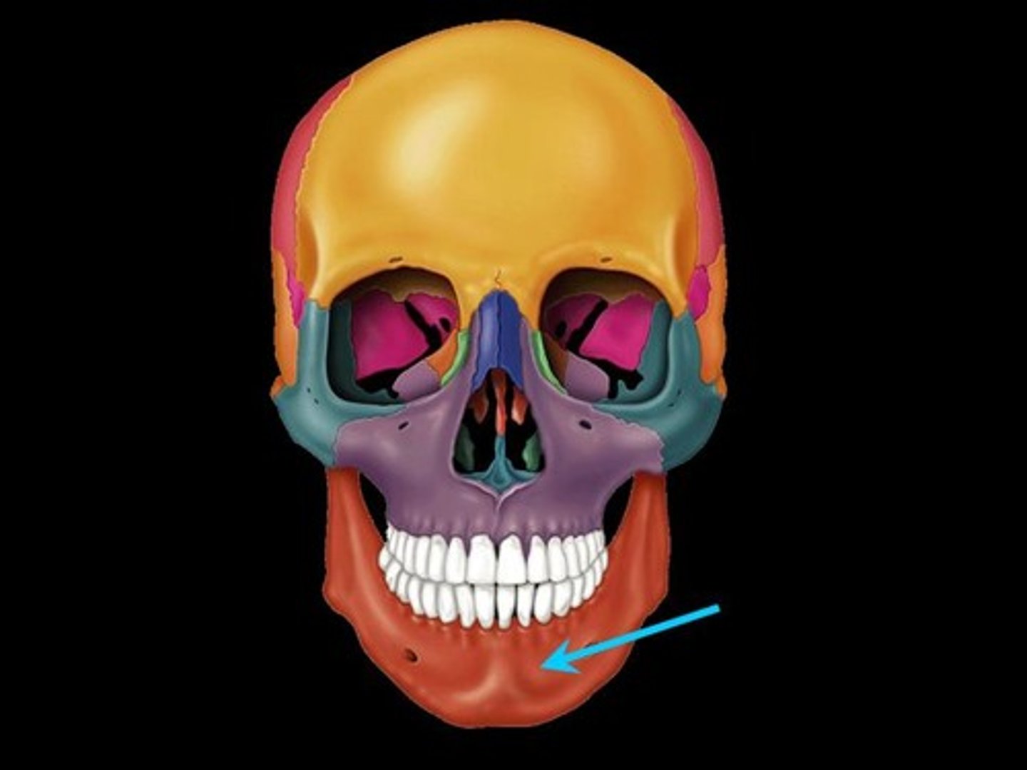
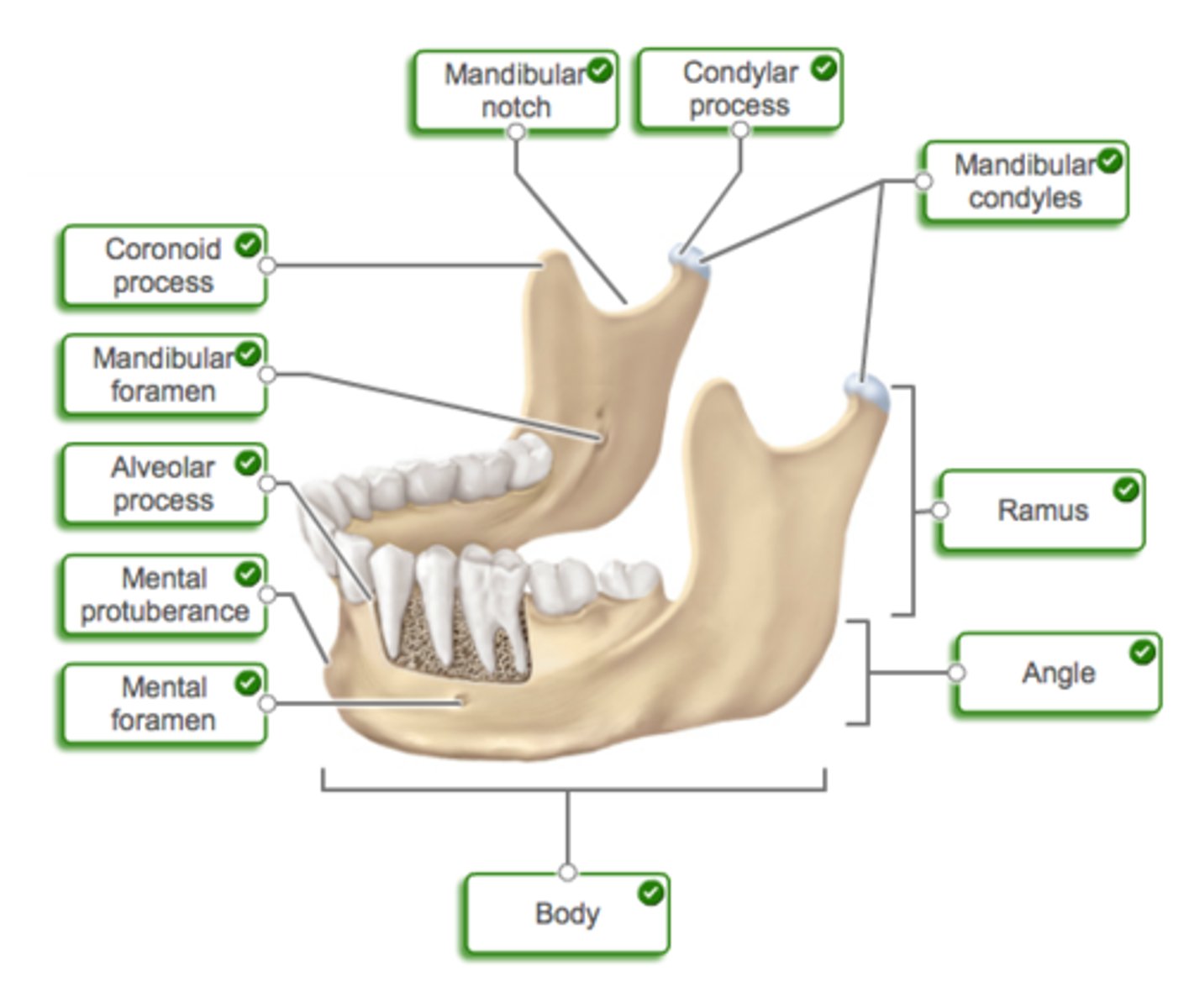
Facial Bones: Mandible Bone
- 1 bone
- forms the Lower jaw
- parts of the mantle: body, ramus, angle
labelled mandible bone
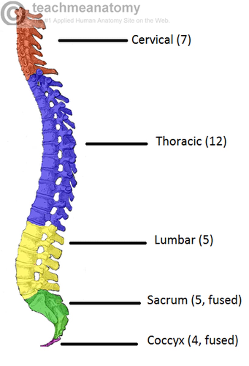
Axial Skeleton: Vertebral Column
- made up of 26 bones:
24 vertebrae
1 sacrum
1 coccyx
divisions of vertebral column
- cervical (7 vertebrae)
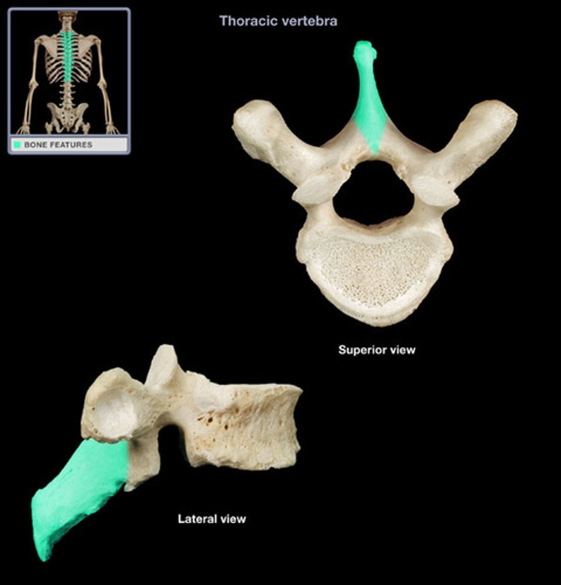
- thoracic (12 vertebrae)
- lumber (5 vertebrae)
- sacrum (5 fused vertebrae)
- coccyx (4 fused vertebrae)
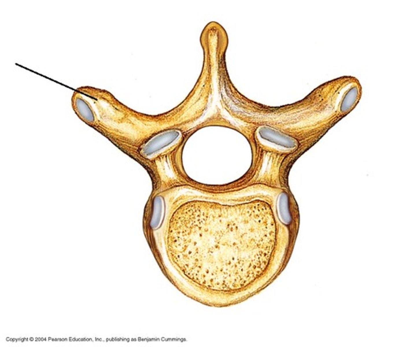
Basic Structure of vertebrae
BODY (anterior)
VERTEBRAL ARCH (posterior)
- extends into spinous and transverse processes
VERTEBRAL FORAMEN
- they all stack together to form the vertebral canal, which protects the spinal chord
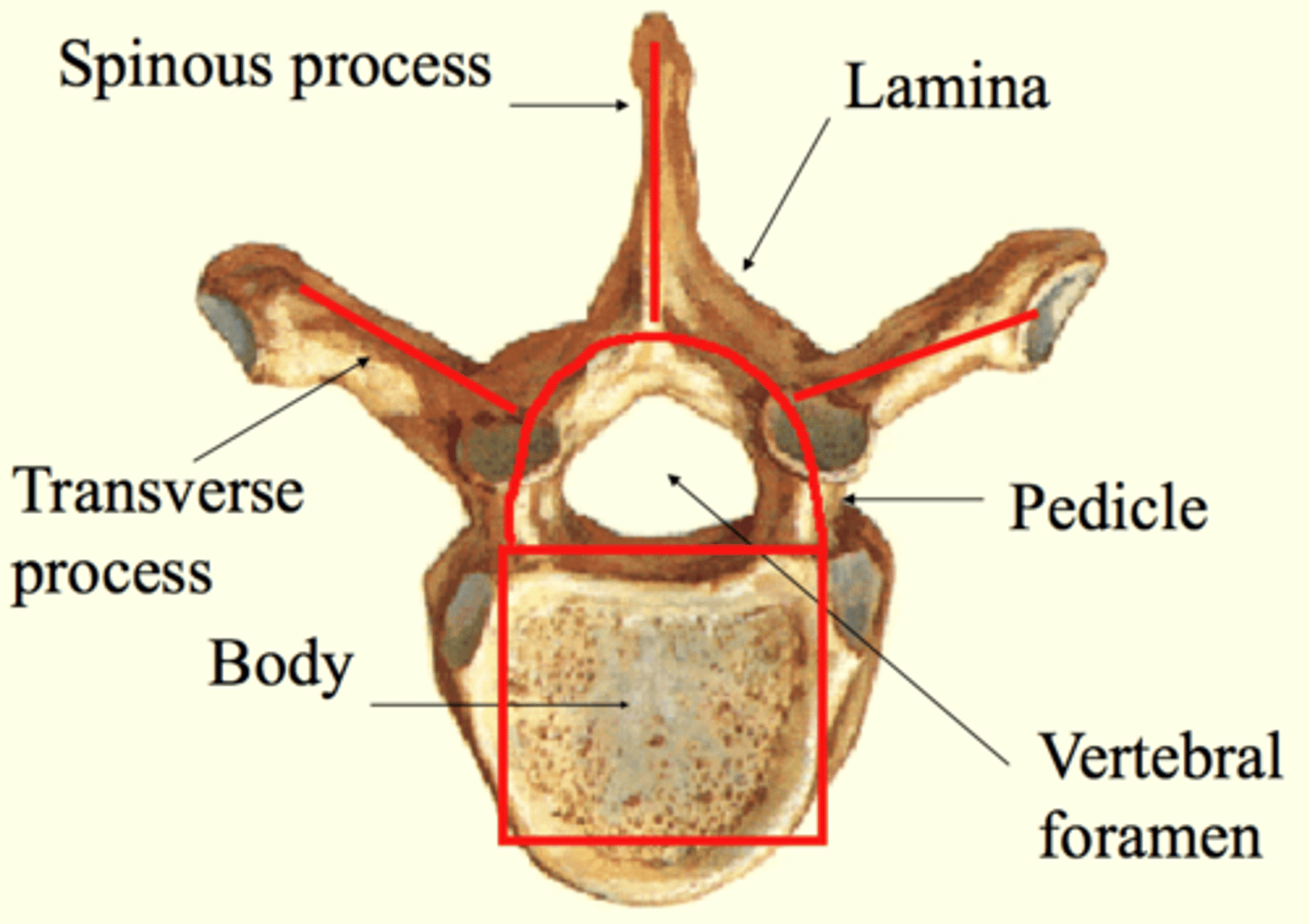
spinous process
extending posteriorly
transverse process
- paired, extending laterally
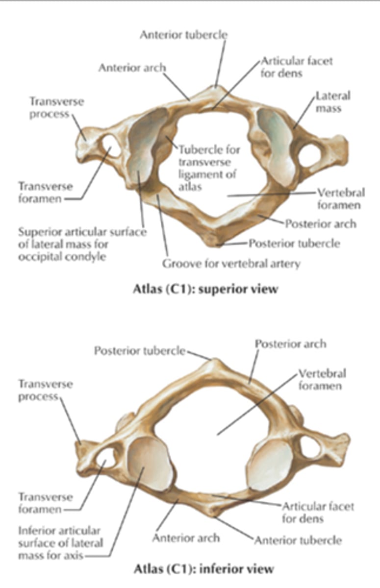
Vertebra that aren't the same (Atypical)
C1
C2
C1
- 1st cervical
2 features important for its articulation with other bones
ANTERIOR ARCH
- surface for articulation with the dens (C2)
LATERAL MASSES
- surface for articulation with the occipital condyles of the occipital bone
C2
- main feature:
DENS
- rests within the anterior arch of C1
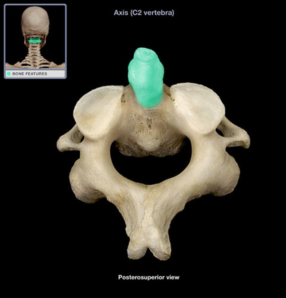
Movement of C1 and C2
- the occipital condyles rest on the lateral masses of C1, allowing for "yes" movement of head
- the dens of C2 articulates with the anterior arch of C1, allowing for the "no" movement of the head
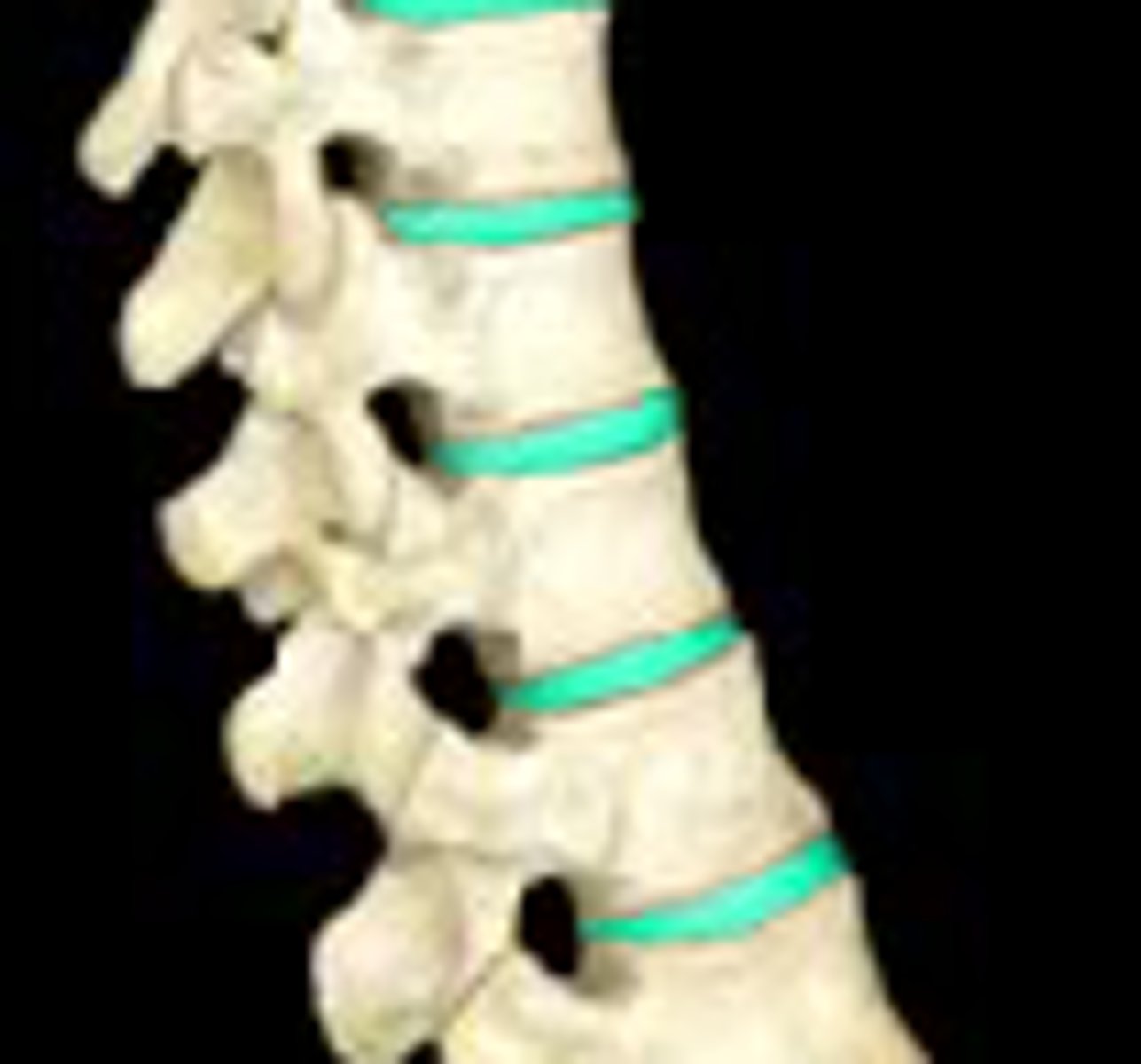
Vertebral articulations
INTRAVERTEBRAL DISC
- jelly between 2 vertebra
- acts as a shock absorber
- fibrocartilage: they resist compression of the bones
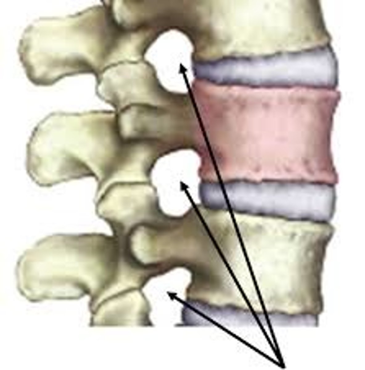
INTRAVERTABLE FORAMEN
- openings between vertebrae to allow for the passage of spinal nerves
intravertebral discs
intravertebral foramen
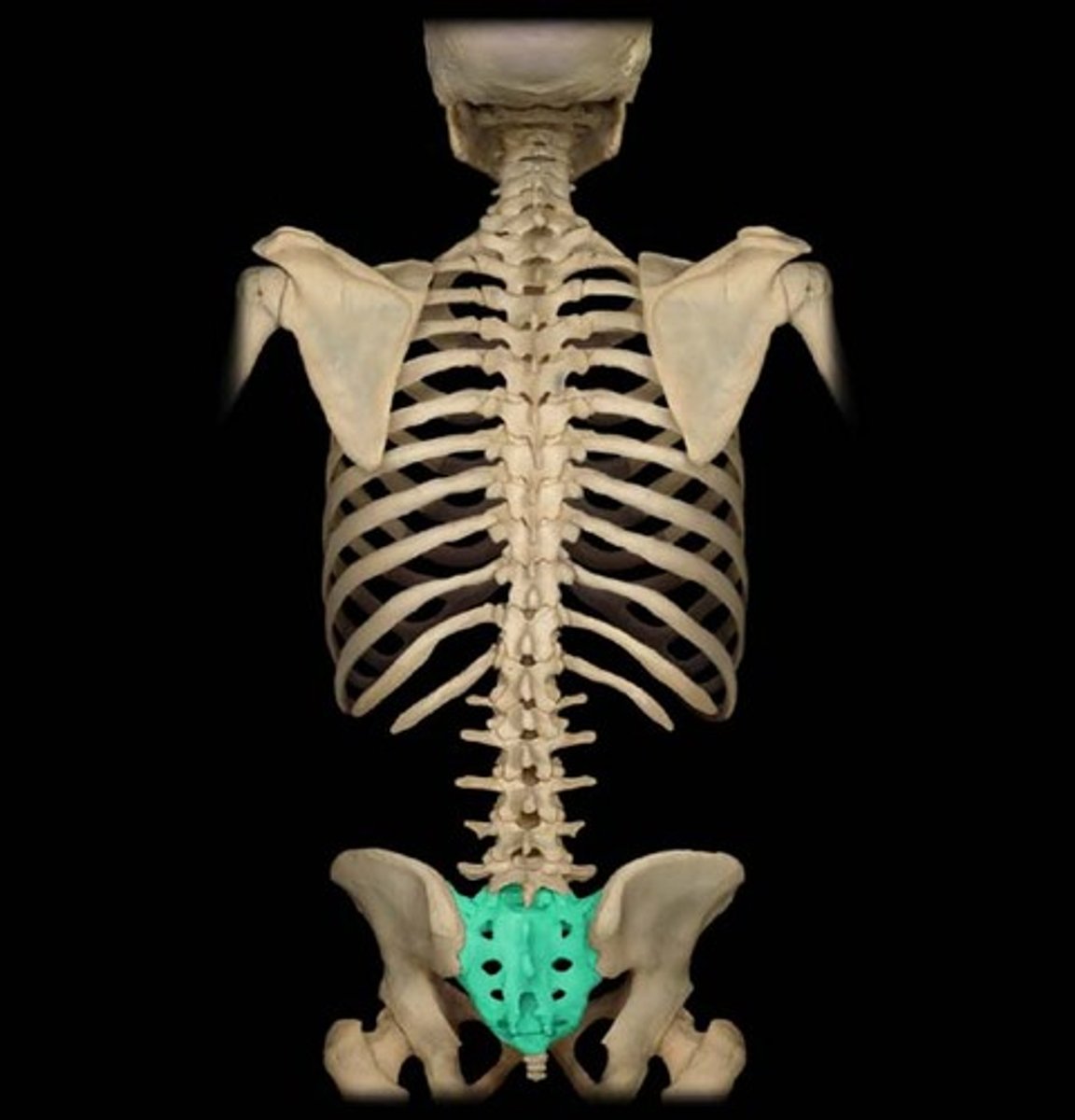
Sacrum
5 fused vertebrae
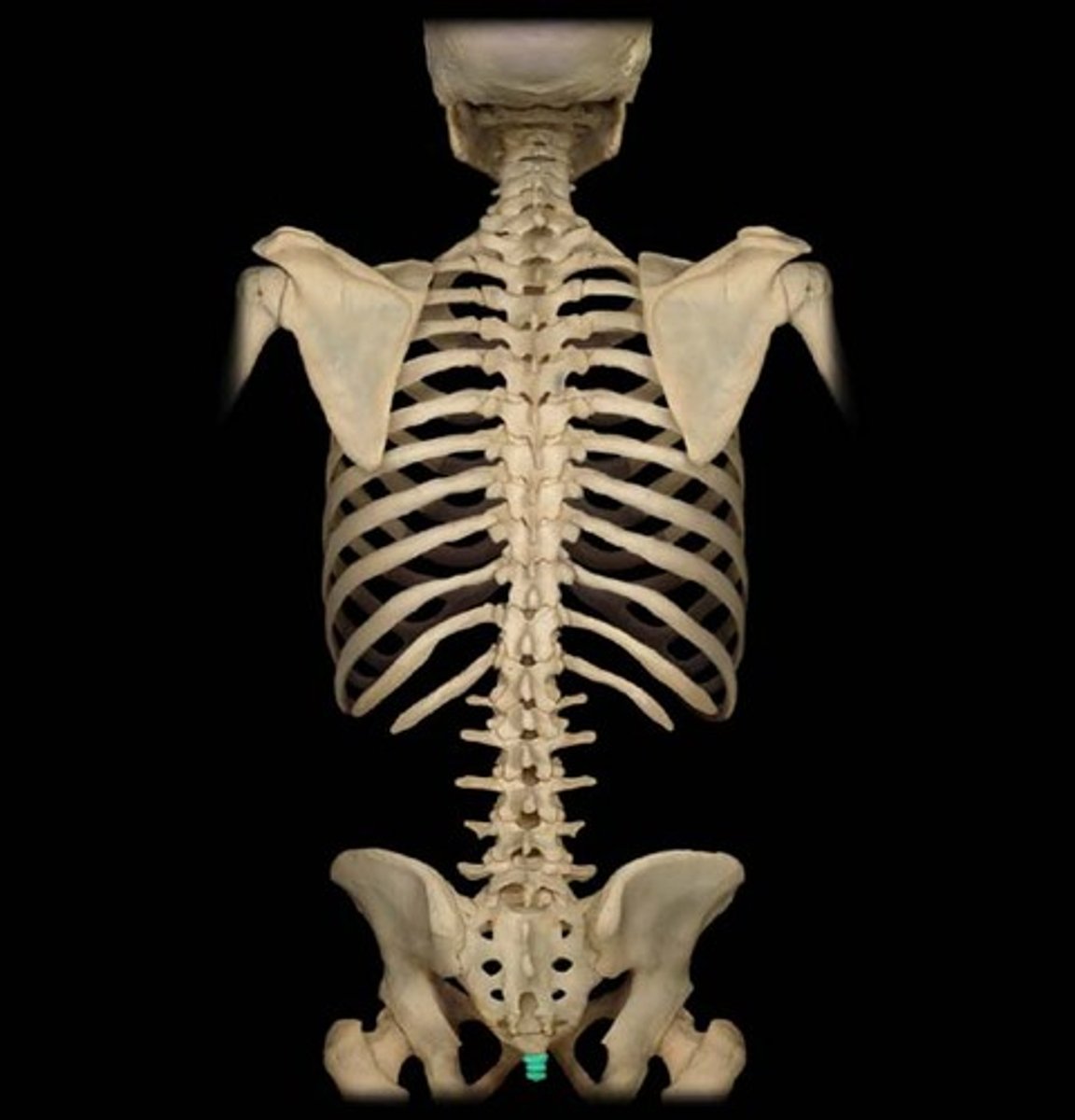
Coccyx
4 fused vertebrae
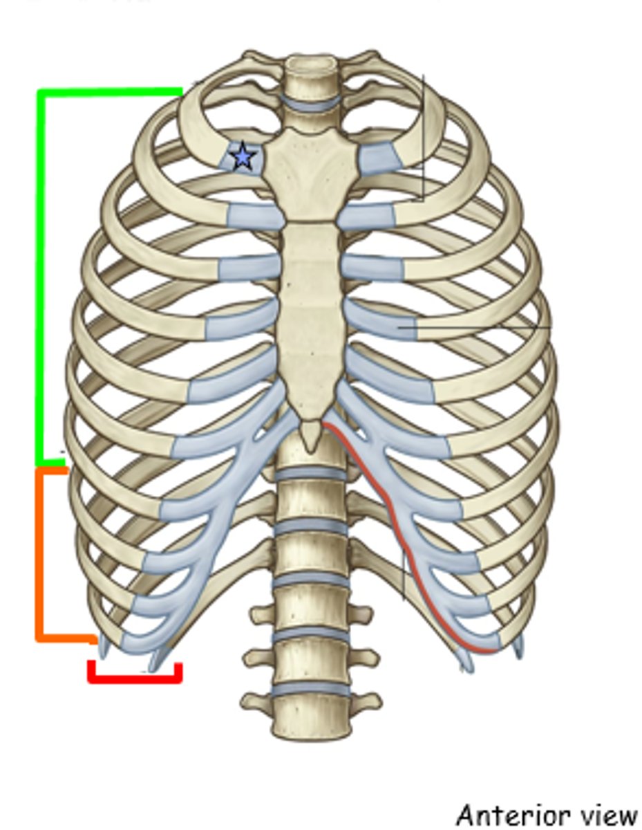
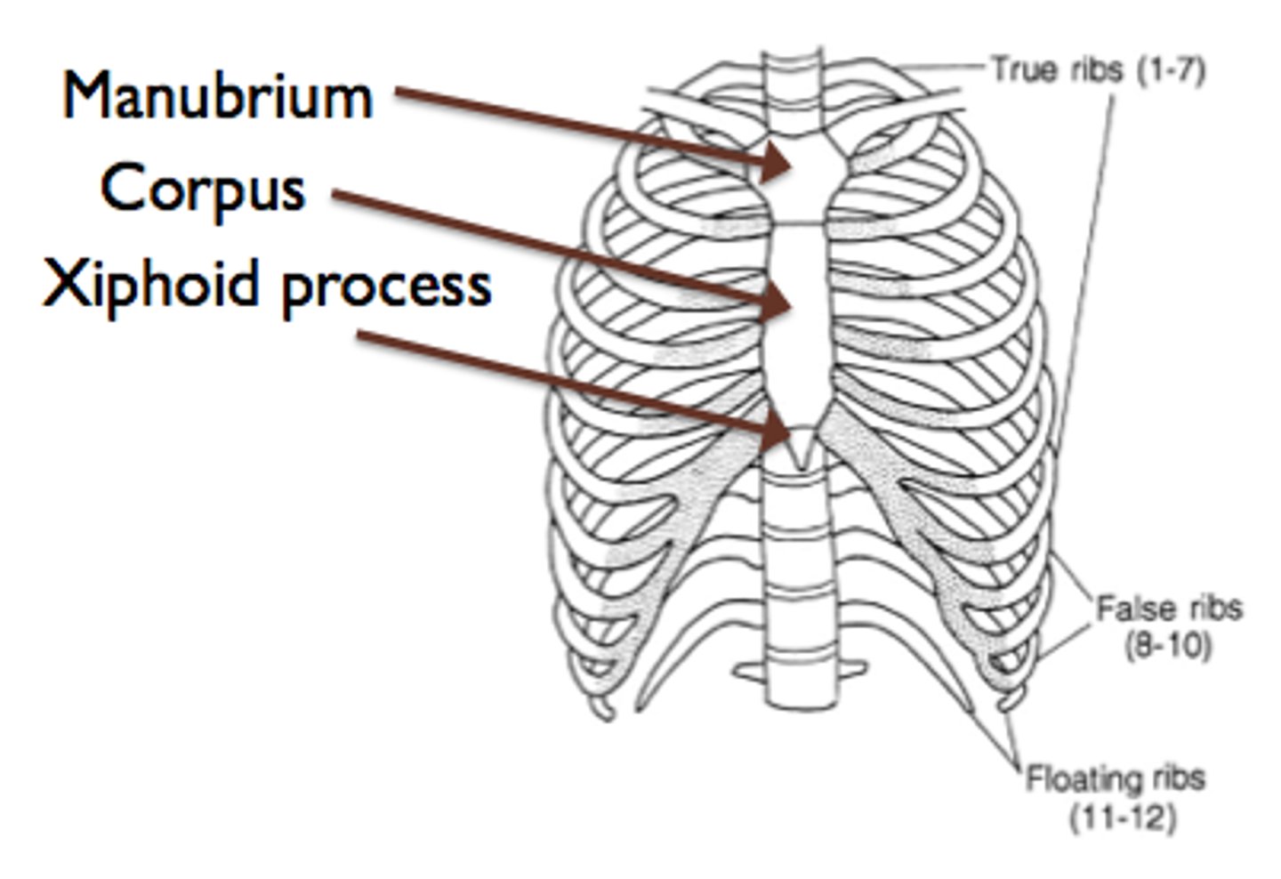
axial skeleton: ribs
We have 12 ribs
TRUE RIBS
- pairs 1-7
- attaches directly to the sternum
FALSE RIBS
- pairs 8-10
- they dont attach directly to the sternum, they attach to rib 7 and the sternum by cartilage
FLOATING RIBS
- pairs 11-12
- dont attach to the sternum
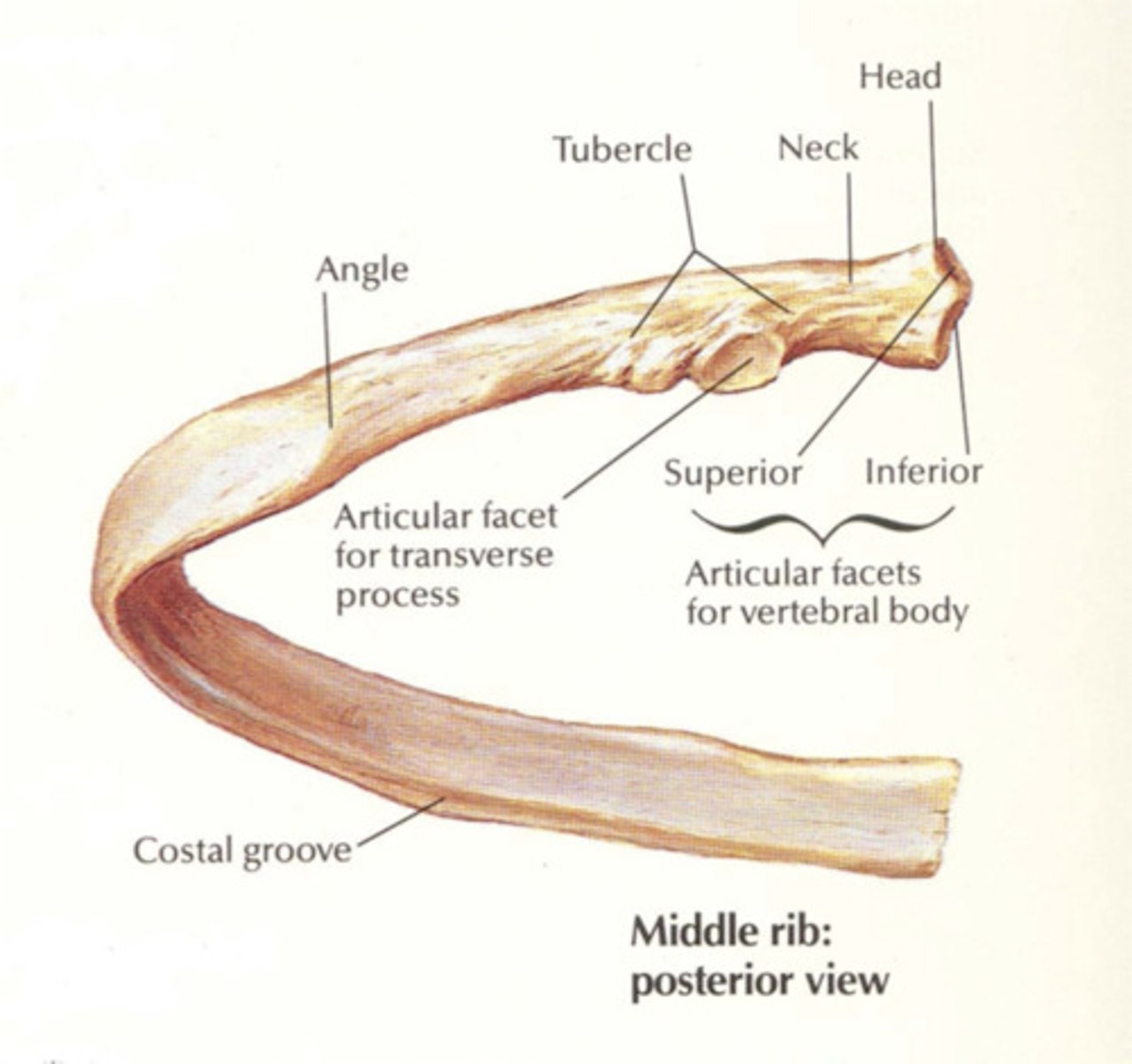
Structure of a rib
- head
- tubercle
- shaft (body)
- angle (where the shaft is bent)
- costal groove
Axial Skeleton: Sternum
- makes up the anterior portion of the thoracic cage
3 parts:
MANUBRIUM
- articulation with rib 1
BODY
- articulation with rib 2-7
THE XIPHOID PROCESS
- tip
Thoracic cage
- made up of thoracic vertebrae, ribs, and sternum
Appendicular Sketelon: Upper Limb
contains 30 bones
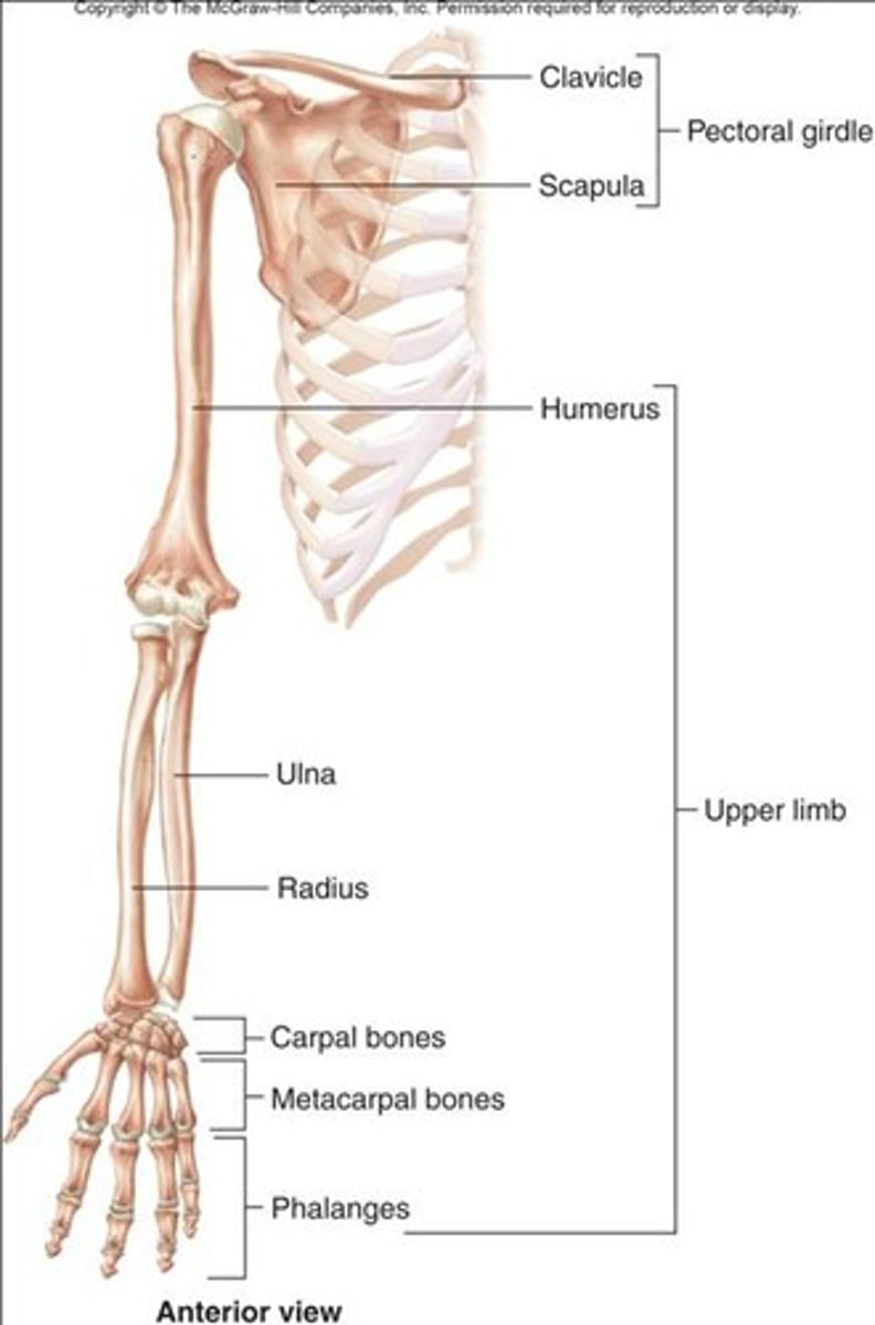
Upper Limb: Pectoral Girdle
- connects the upper limb to the axial skeleton
Formed by:
- clavicle
- scapula
pectoral girdle: Clavical
- s-shaped bone (collar bone)
- it joins with the manubrium of the sternum (proximally) and the scapula (distally)
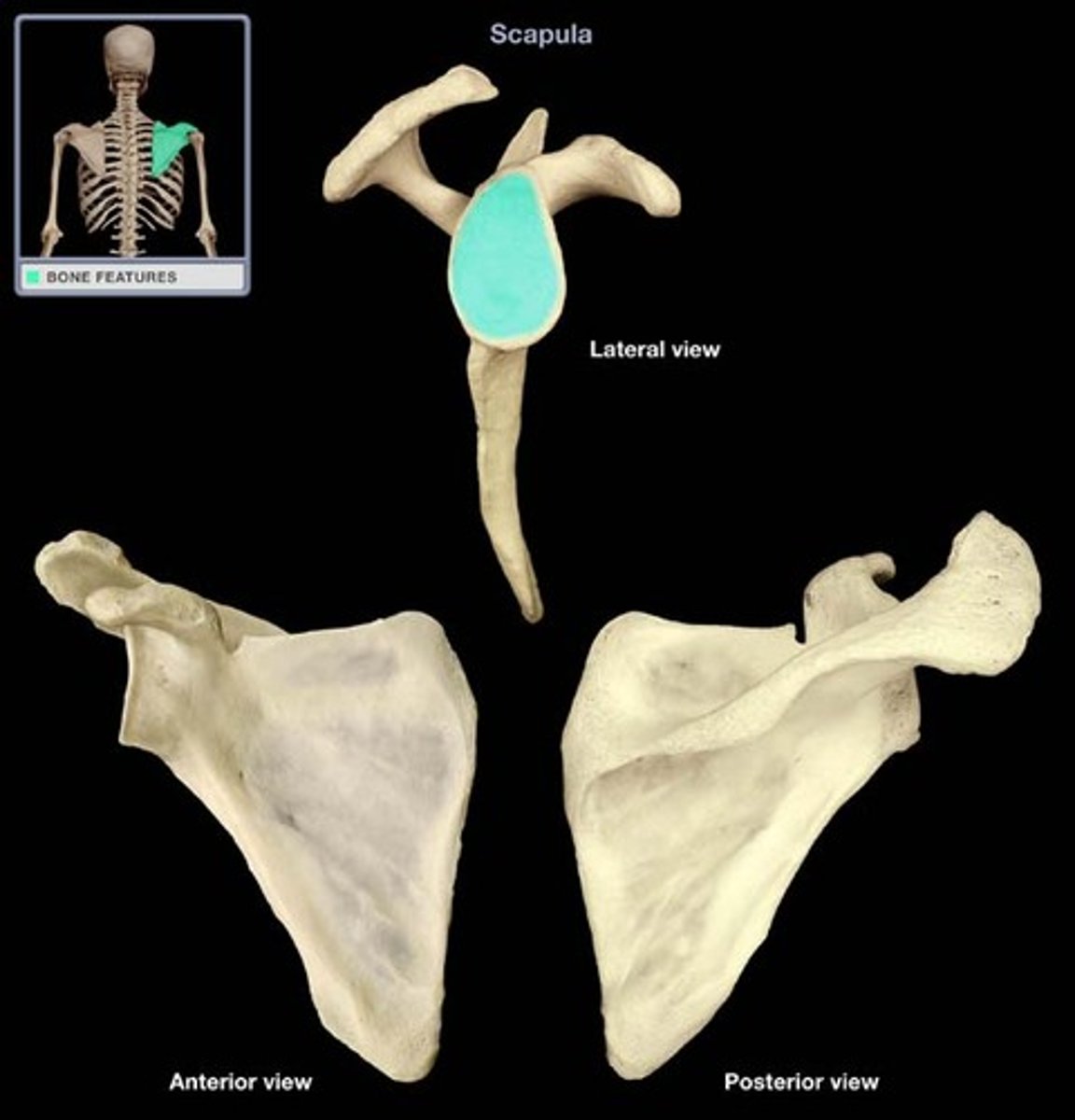
pectoral girdle: Scapula
- triangle shape
ANTERIOR SURFACE
- corticoid process
POSTERIOR SURFACE
- acromion
- spine
LATERAL SURFACE
-glenoid fossa
coracoid process
(with the acromion, the corticoid helps stabilize the shoulder joint)
LOOK AT PICTURE IN BOOK
acromion
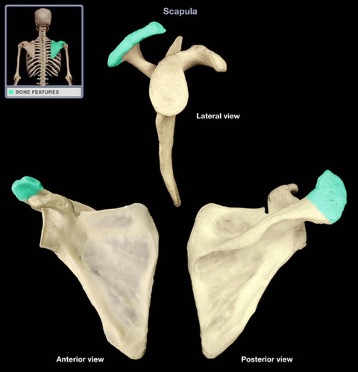
Spine (Pectoral Girdle)
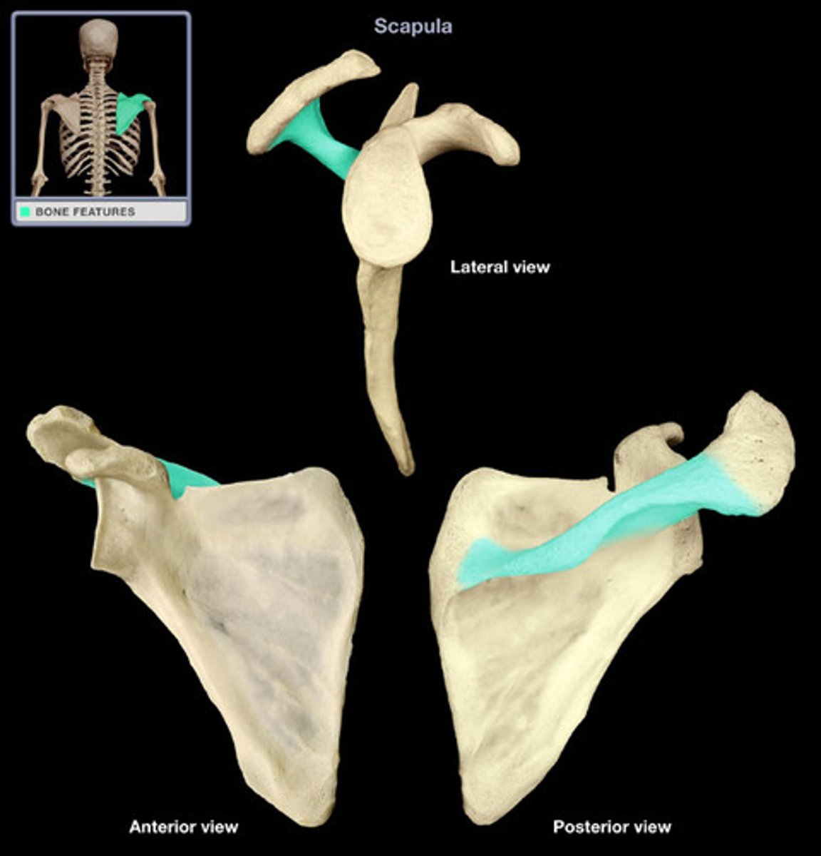
glenoid fossa
- articulates with the head of the humerus to contribute to the shoulder joint
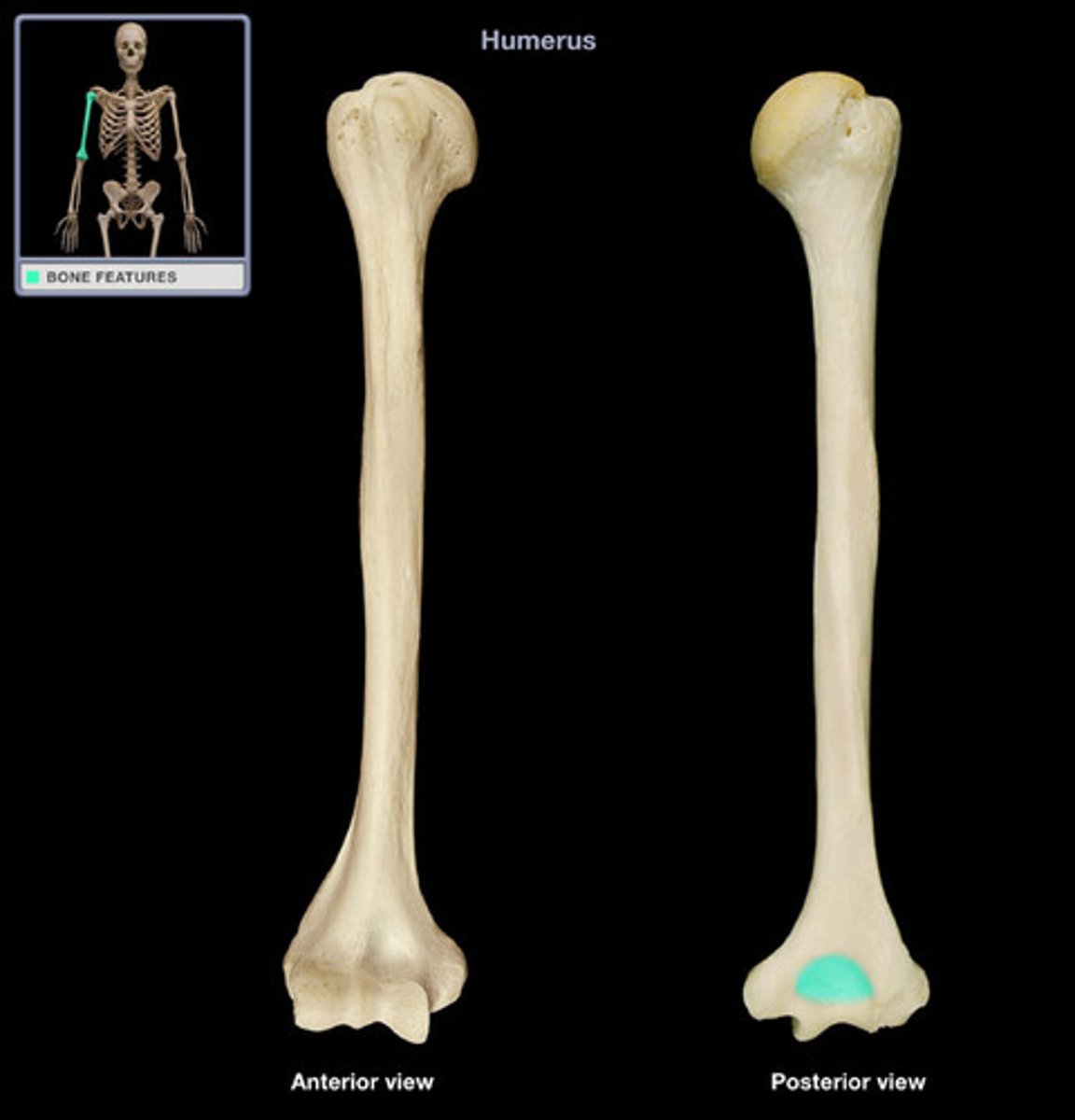
Arm- Humerus
- articulates with the glenoid fossa to form the shoulder joint, and distally with the radius and ulna to form the elbow joint

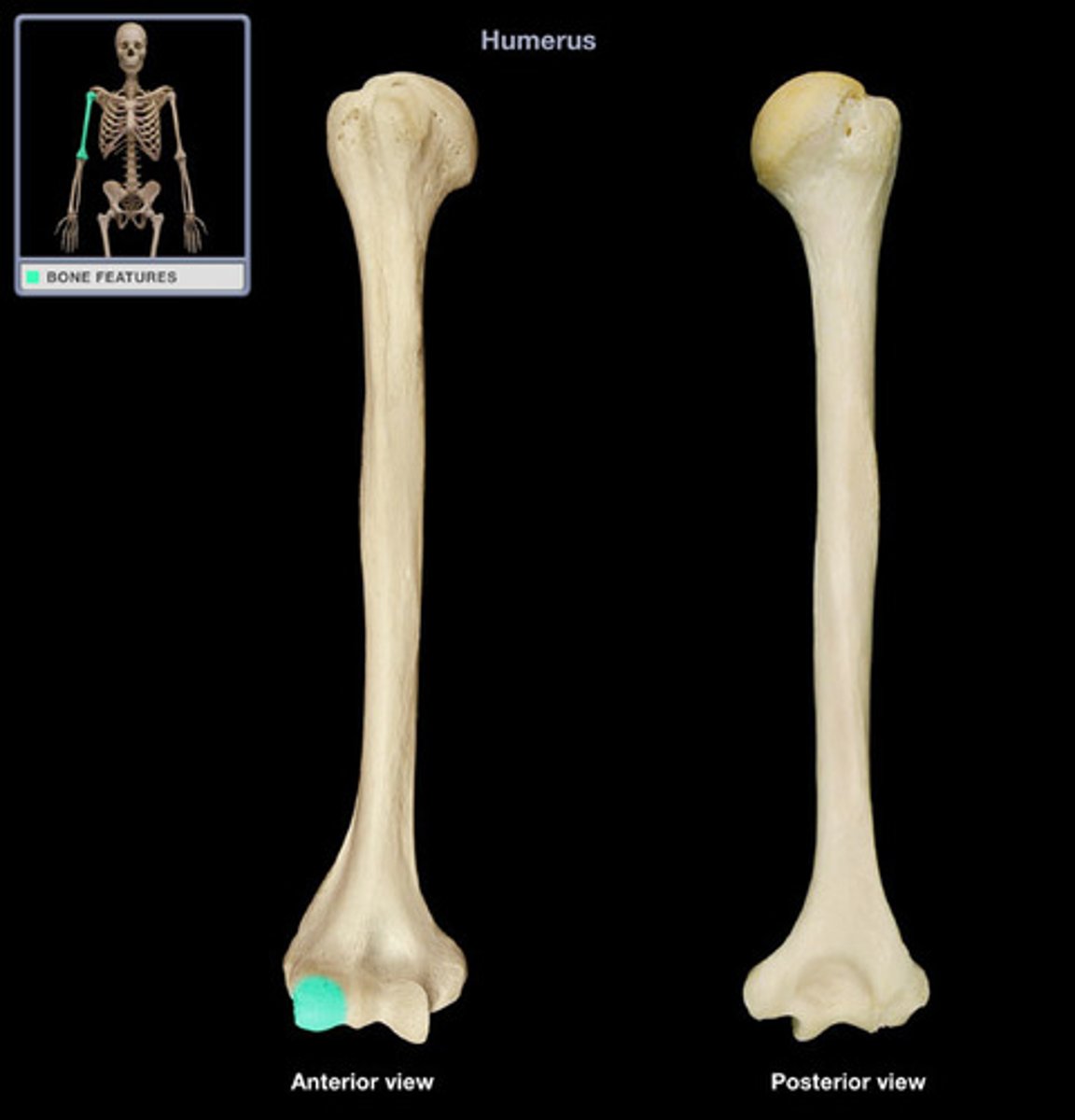
features of the humerus
- head, neck, shaft
ANTERIORLY
- lateral epicondyle
- medial epicondyle
- capitulum
- trochlea
POSTERIORLY
- olecranon fossa
lateral and medial epicondyles
(lateral away from body)
(medial towards body)
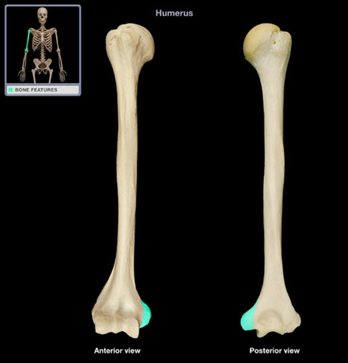
capitulum
trochlea
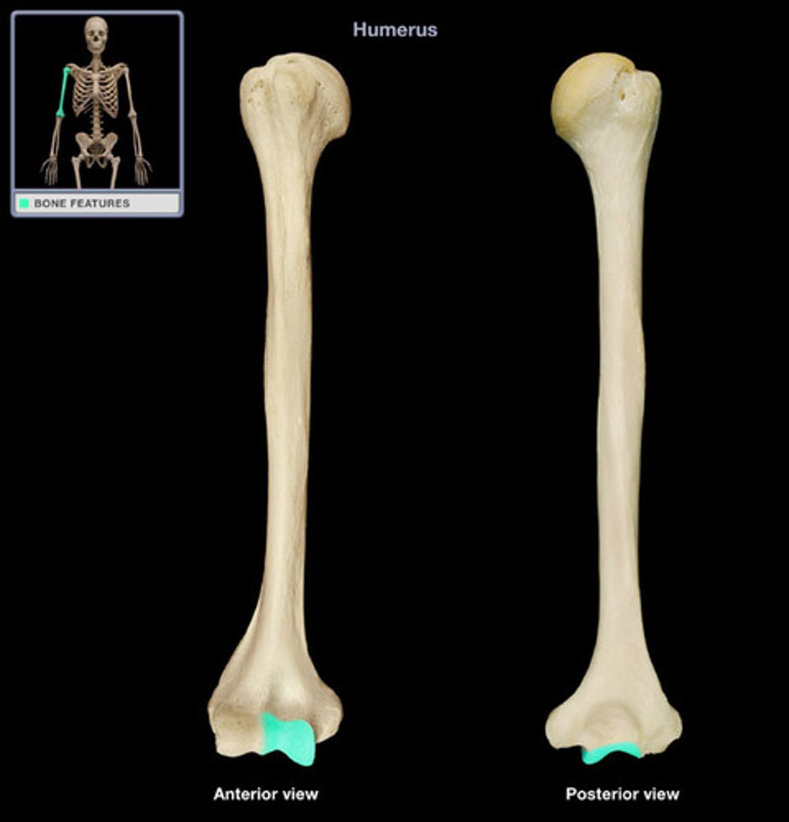
olecrannon fossa
Forearm
RADIUS
ULNA
forearm: Radius
- in anatomical position, the radius is on the thumb side
features:
HEAD (PROXIMAL END)
- round shape that articulates with the humerus and the ulna
SHAFT
NECK
DISTAL END
- articulates with the carpal (wrist) bones
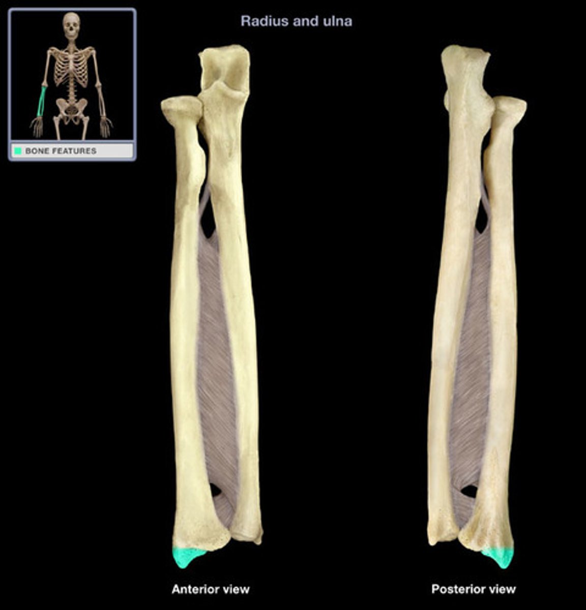
- styloid process
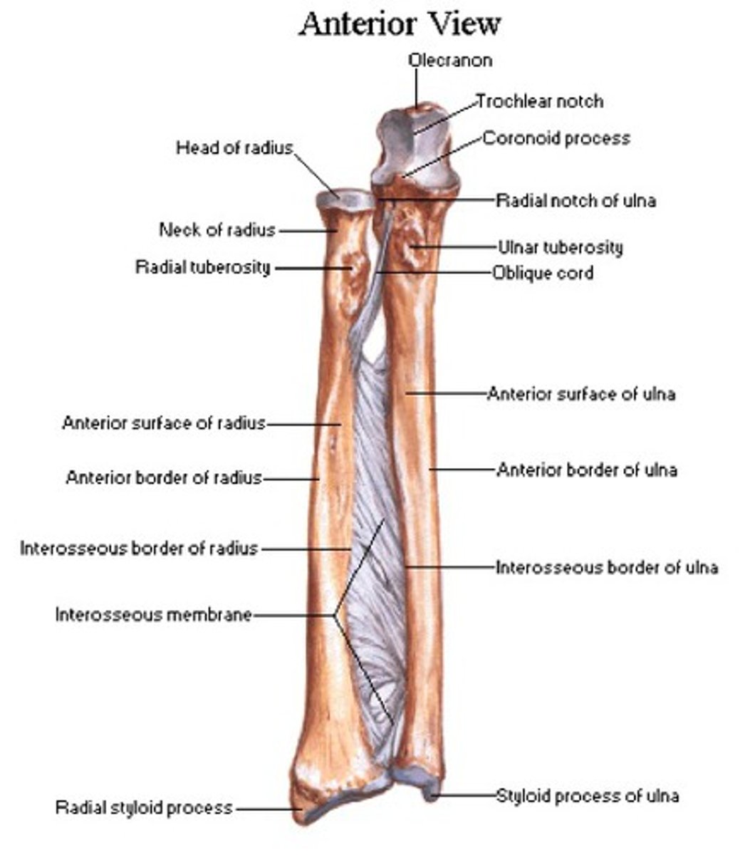
styloid process of radius
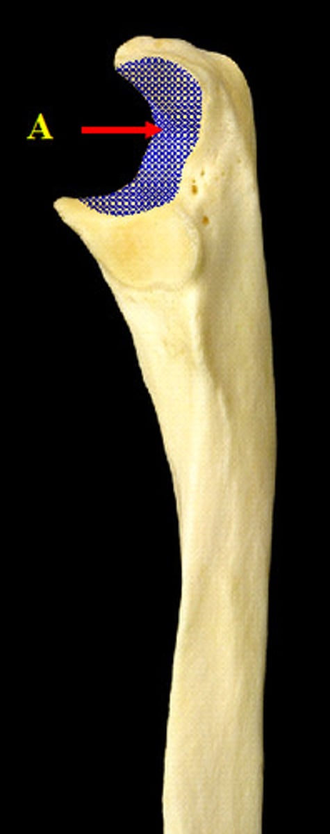
forearm: Ulna
- in anatomical position, it is on the pinky side
features:
PROXIMAL END
- articulates with the distal end of the humerus
- olecranon
- trochlear notch
SHAFT
NECK
HEAD (DISTAL END)
- styloid process
IMPORTANT TO NOTICE THAT UNLIKE THE RADIUS, THE HEAD IS DISTAL
olecranon
fits into the olecranon fossa of the humerus
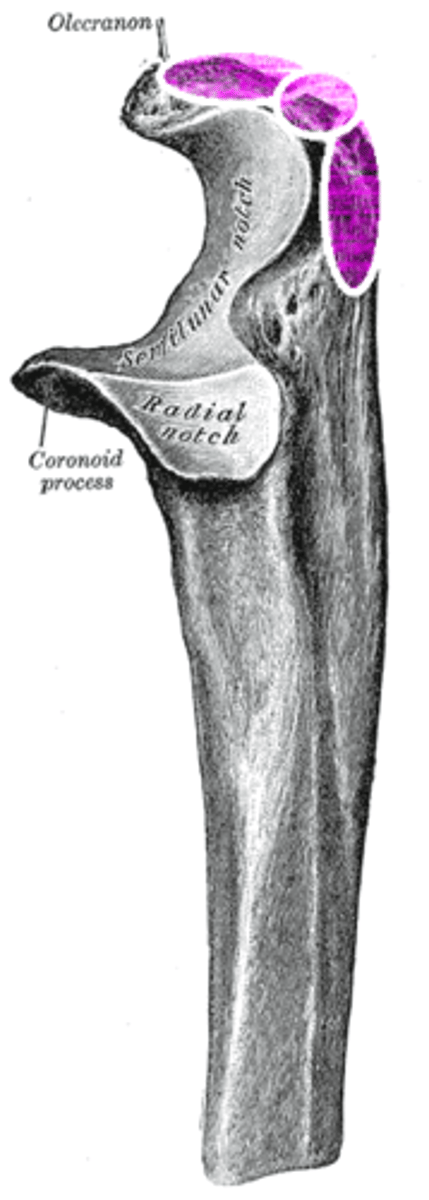
trochlear notch
C shaped interlocks with the trochlea of the humerus
styloid process of ulna
Tiny bulb at bottom of Ulna
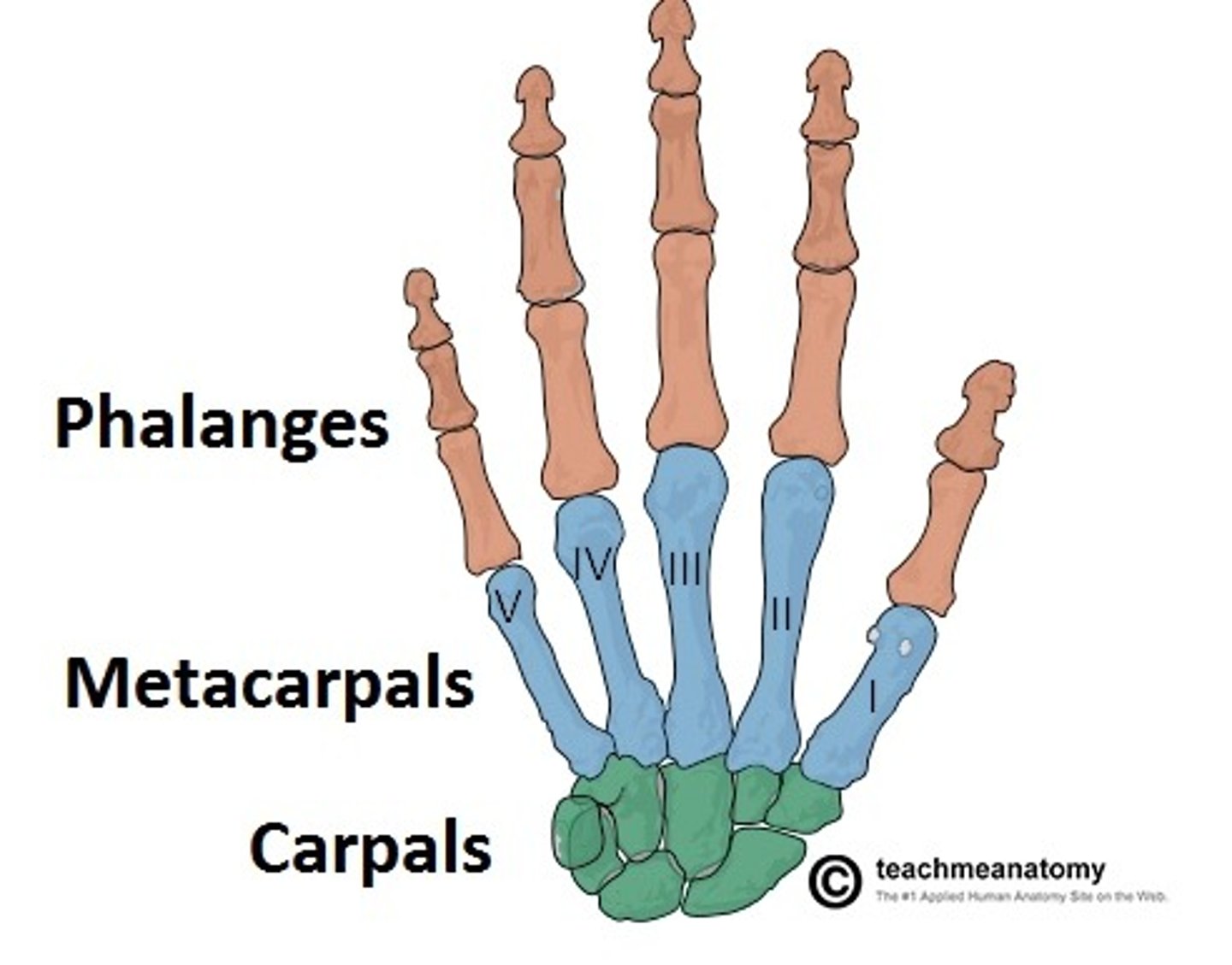
Wrist and Hand
Wrist
- 8 carpal bones (short bones)
Palm
- 5 metacarpal bones (long bones)
FIngers
- 14 phalanges (long bones) 3 each except the thumb has 2
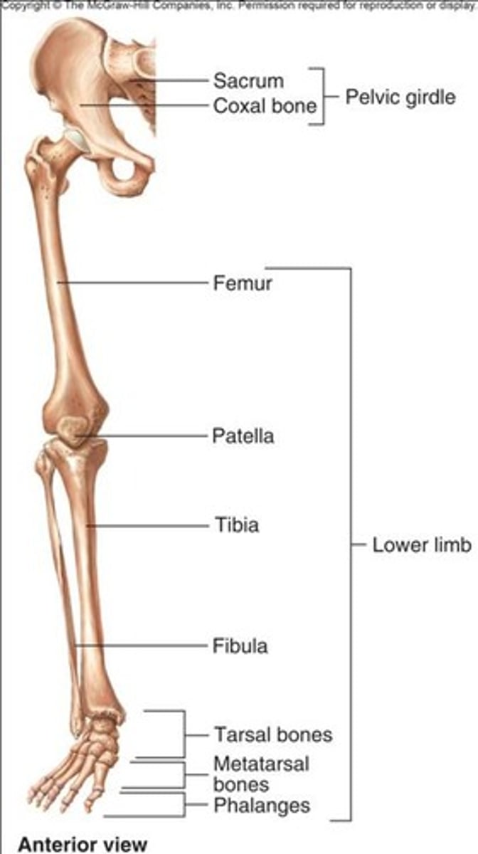
Appendicular Skeleton: Lower Limb
contains 30 bones +inanimate bone
Lower Limb: pelvic girdle
- attaches the lower limb to the axial skeleton
formed by:
ILIUM
ISCHIUM
PUBIS
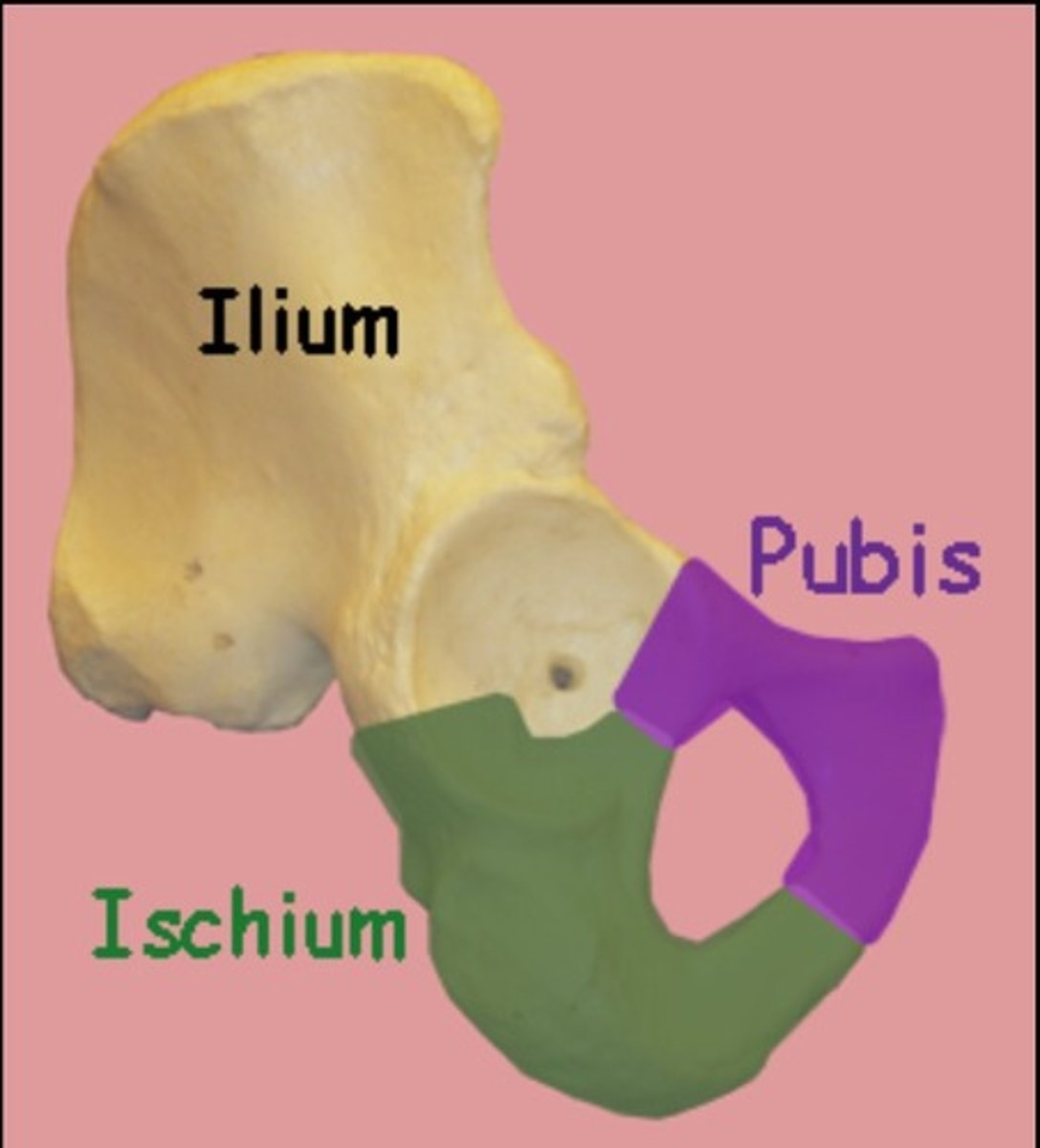
Pelvic Girdle: Ilium
- largest bone of the pelvic girdle
features:
- iliac crest
- anterior superior iliac spine
- anterior inferior iliac spine
- posterior superior iliac spine
- posterior inferior iliac spine
LOOK IN NOTE FOR A PICTURE
Pelvic Girdle: Pubis
- fuses with the ilium and the ischium
- fuses with the other pubic bone at the pubic symphysis (made of fibrocartilage)
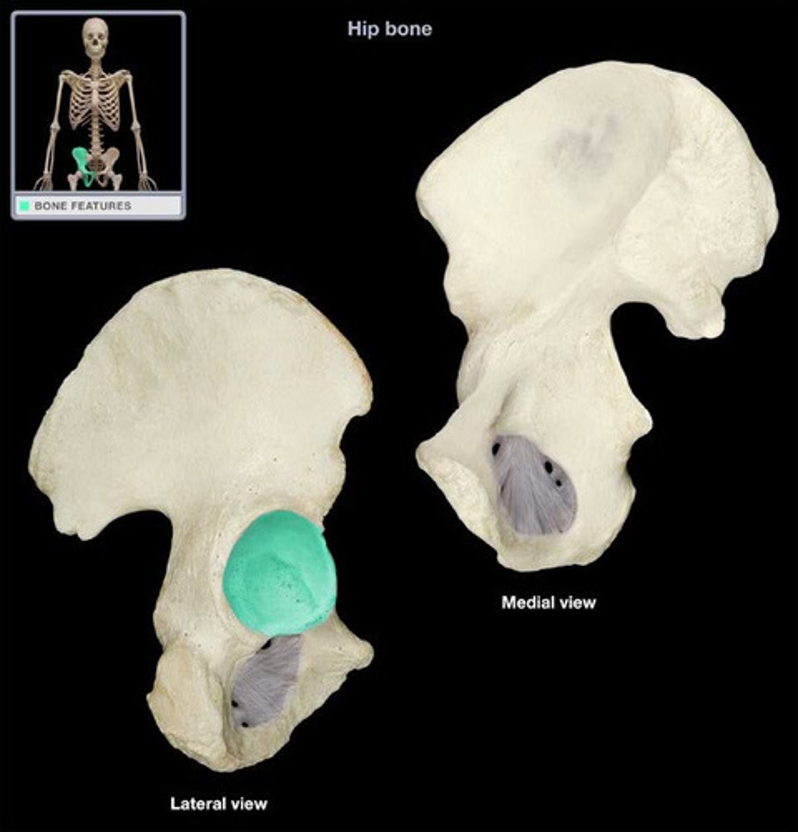
2 structures of the pelvic girdle
ACETABULUM
- deep depression for articulation with the head of the femur
- made up by the ilium, ischium, and pubis
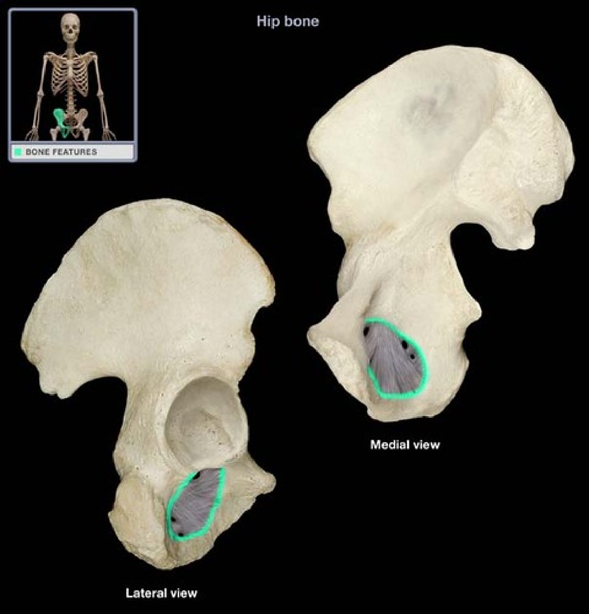
OBTURATOR FORAMEN
- large opening in the anterior of each pelvic girdle which is a passage for verse and blood vessels
- made up of the ischium and pubis
Acetabulum
obturator foramen
Also on the pelvis:
GREATER SCIATIC NOTCH
- located between the posterior inferior iliac spine
- allows the passage of major nerves and blood vessels from the pelvic cavity into the posterior region
LESSER SCIATIC NOTCH
- located between the posterior inferior iliac spine and the ischial tuberosity
- allows for the passage of structures from the pelvic cavity to the genital region
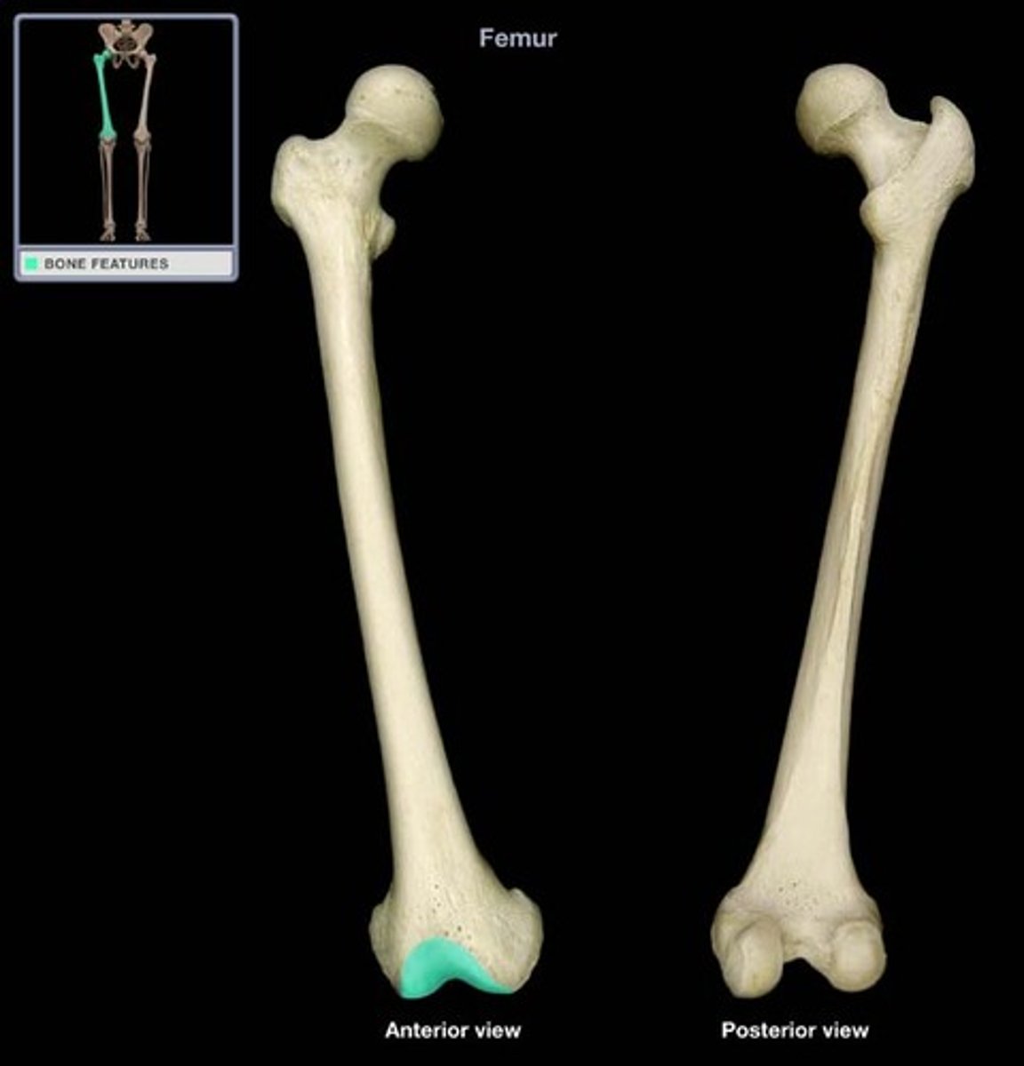
Thigh- Femur
- biggest bone in the body
features:
- head, neck, shaft
Proximal End of femur
- head articulates with the acetabulum of the pelvic girdle forming the hip joint
- bumps and ridges for muscle attachment
Distal End of the Femur
- articulates with the tibia and patella (knee cap)
- medial and lateral condyles
- patellar surface
medial and lateral condyles (on the femur)
- 2 bumps on the end
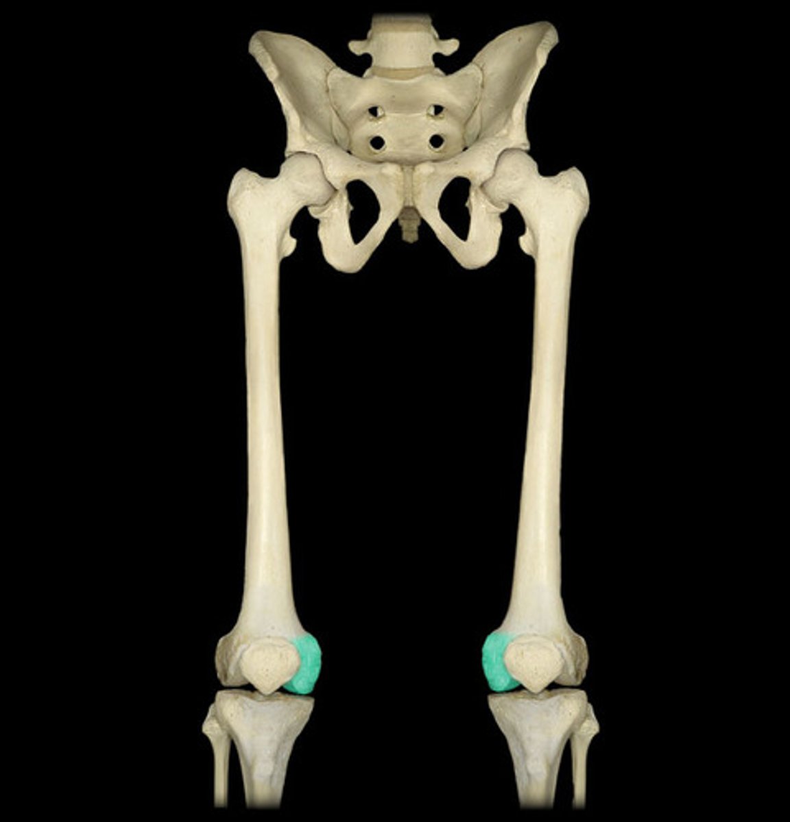
patellar surface
- surface which forms a groove for the patella
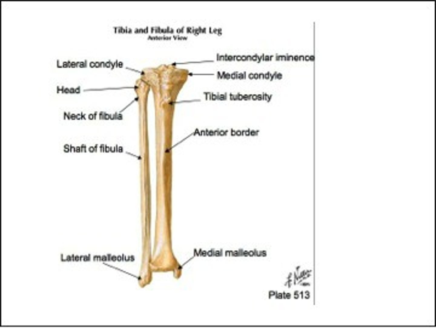
Leg
tibia and fibula
Leg: Tibia
- weight bearing bone of the leg
features
PROXIMAL END
- medial and lateral condyles (which articulate with the medial and lateral condyles of the femur)
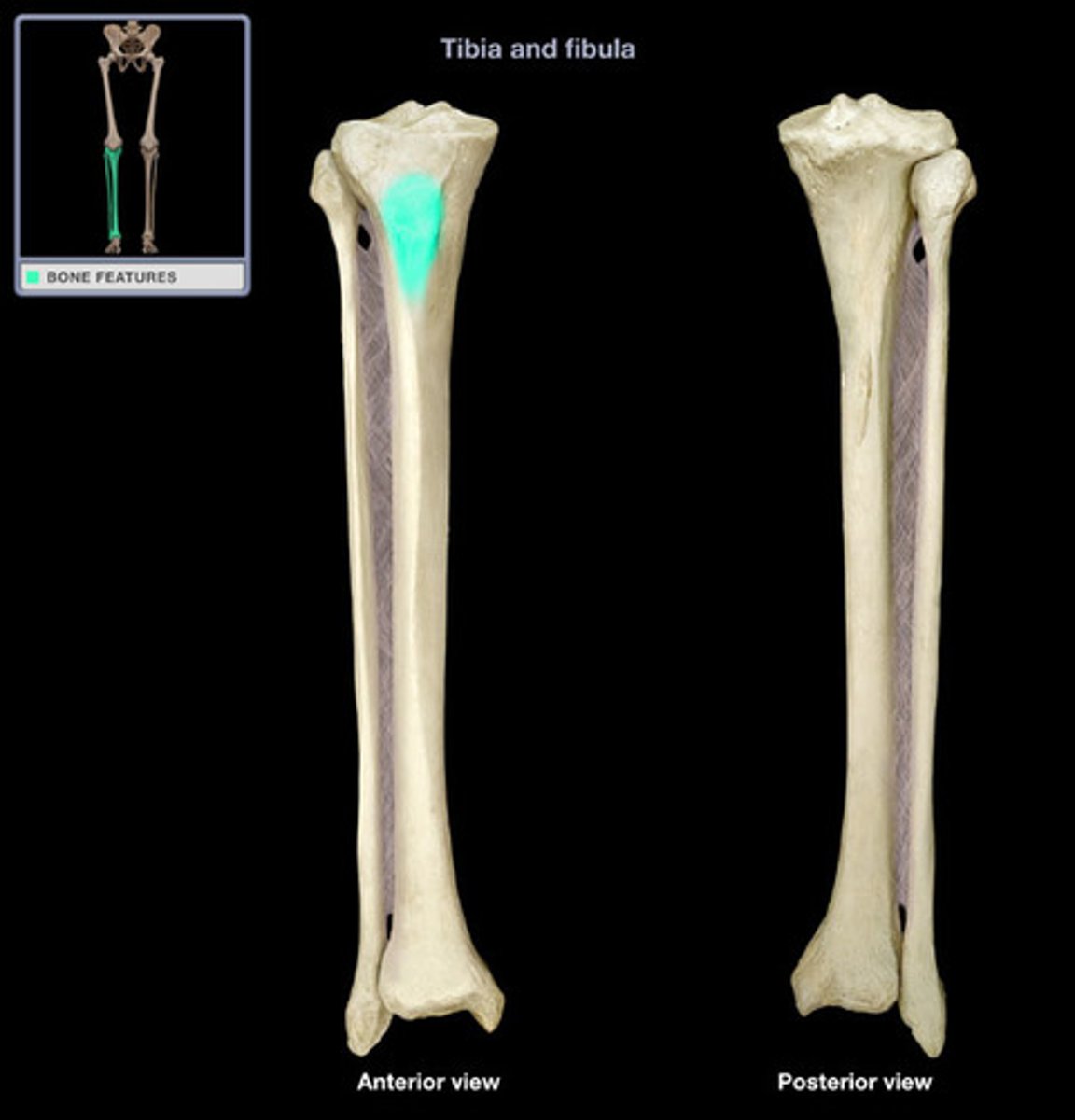
- tibial tuberosity
SHAFT
DISTAL END
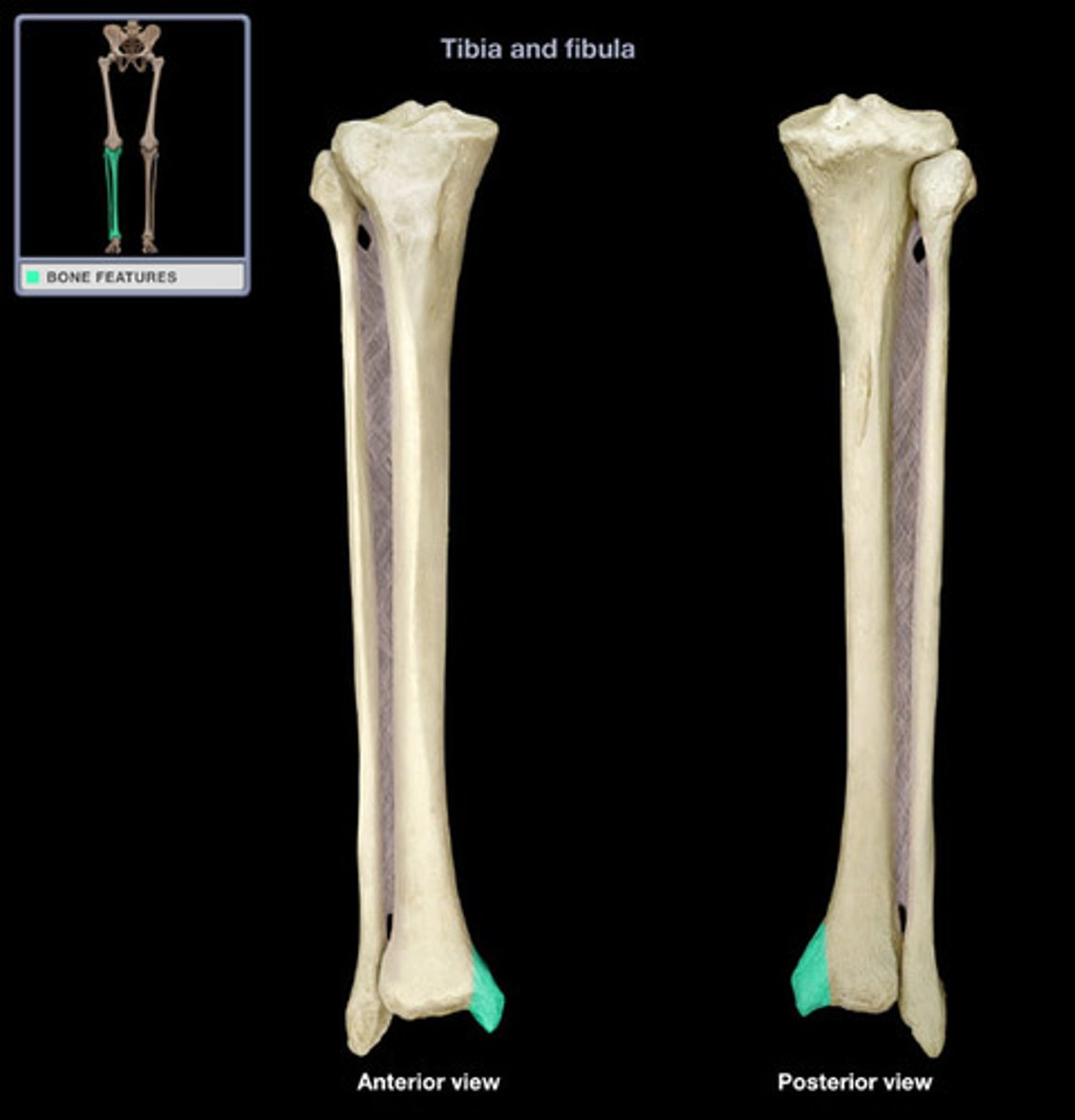
- medial malleolus
tibial tuberosity
medial malleolus
Fibula
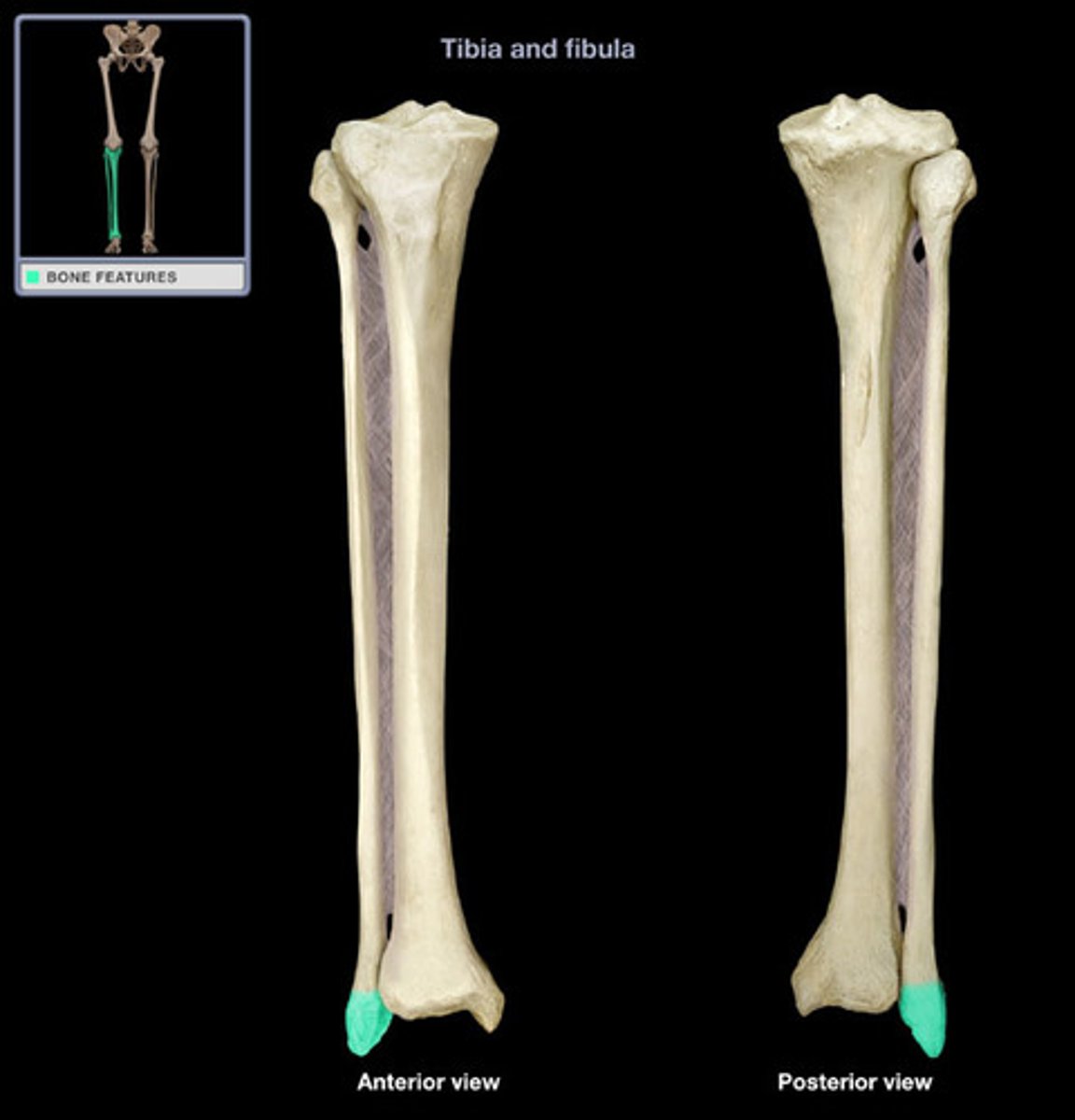
- the lateral malleolus (the distal tip of the fibula) provides stability to the ankle joint
features:
PROXIMAL END
- medial head which articulates with the tibia
NECK
SHAFT
DISTAL END
- lateral malleolus

lateral malleolus
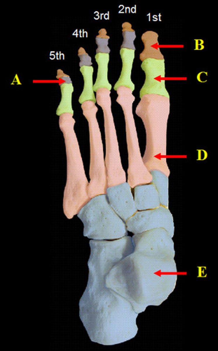
Ankle and foot
ankle
- 7 tarsal bones (short bones)
foot
- 5 metatarsal bones (long bones)
toes
- 14 phalanges (long bones)
articulation
- where bones meet other bone, cartilage, or teeth
the most mobile joints are unstable
the least mobile joints are stable
Classification of joints
Structural Classification
- based on the type of materials that unite the articulating bones
Functional Classification
- based on the extend of movement they permit
Synovial joints
- articulating bones are enclosed by a joint capsule with a fluid filled cavity
movements of synovial joints
- gliding
- angular
- rotation
- special movements
movements of synovial joints: Gliding
the motion of bones sliding on one another
movements of synovial joints: angular
Flexion
- decreasing angle between articulating bones (bending)
Extension
- increasing angle between articulating bones (extending)
Abduction
- movement away from the midline of the body
Adduction
- movement towards the midline of the body