MOD 9 - Skull Trauma and Facial Fractures – Study Flashcards
1/56
Earn XP
Description and Tags
Flashcards compiled from the lecture notes on skull trauma, skull fractures, head trauma, and facial fractures.
Name | Mastery | Learn | Test | Matching | Spaced | Call with Kai |
|---|
No analytics yet
Send a link to your students to track their progress
57 Terms
What are the two broad types of skull injuries?
Superficial injuries (bruising, soft tissue swelling) and internal injuries (brain swelling from increased intracranial pressure).
Why is imaging important in skull trauma?
Skull radiography and CT help guide treatment and distinguish sutures or artery grooves from fracture lines.
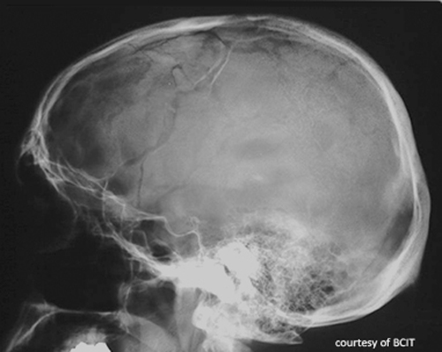
What two structures must be distinguished from fractures on imaging?
Sutures (jagged, interlocking edges) and middle meningeal artery grooves (normal grooves that can mimic fractures on lateral views).
What are the two main types of skull fractures?
Linear fractures and depressed fractures.
Describe a linear skull fracture.
Thin lines without splintering or depression; may be longitudinal or transverse; caused by low-impact blunt trauma.
Describe a depressed skull fracture.
Bone fragments pushed toward the brain, creating a depression; may be closed or open; caused by a direct blow to a small area.
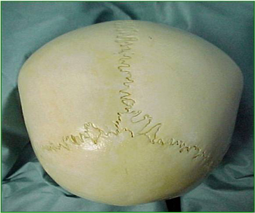
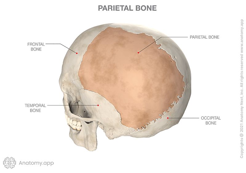
Where are skull fractures most common?
Temporal and parietal bones (temporal bone is the thinnest).
Why is CT often preferred over skull radiographs?
Skull radiographs are limited by bone thickness; CT provides better injury detail.
How are depressed open skull fractures treated?
Surgical reduction.
Name two possible complications of skull fractures.
Brain swelling and direct brain damage.
What is coup-contrecoup injury?
Brain damage at the site of impact and on the opposite side of the skull.
List five common signs of head trauma.
Nausea, vomiting, dizziness, altered consciousness, unequal pupils.
What is a concussion?
Temporary interruption of brain function from head jarring, with no structural changes. (Plain radiographs do not show the presence of a concussion, although, as mentioned above, patients are often sent for a CT to rule out any other pathologies which can have similar symptoms)
Is a concussion visible on plain X-rays?
No; CT is used to rule out bleeds or fractures.
What is the main treatment for a concussion?
Observation and rest for 24–48 hours.
What is a major risk of repeated concussions?
Long-term brain injury.
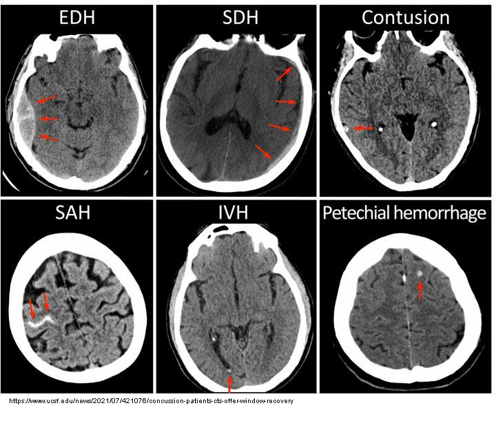
What is a cerebral contusion?
Micro-hemorrhages in brain tissue, often from coup-contrecoup injuries.
Where are cerebral contusions commonly located?
Frontal and temporal lobes.
How do fresh cerebral contusions appear on CT?
Hyperdense patches (due to fresh blood).
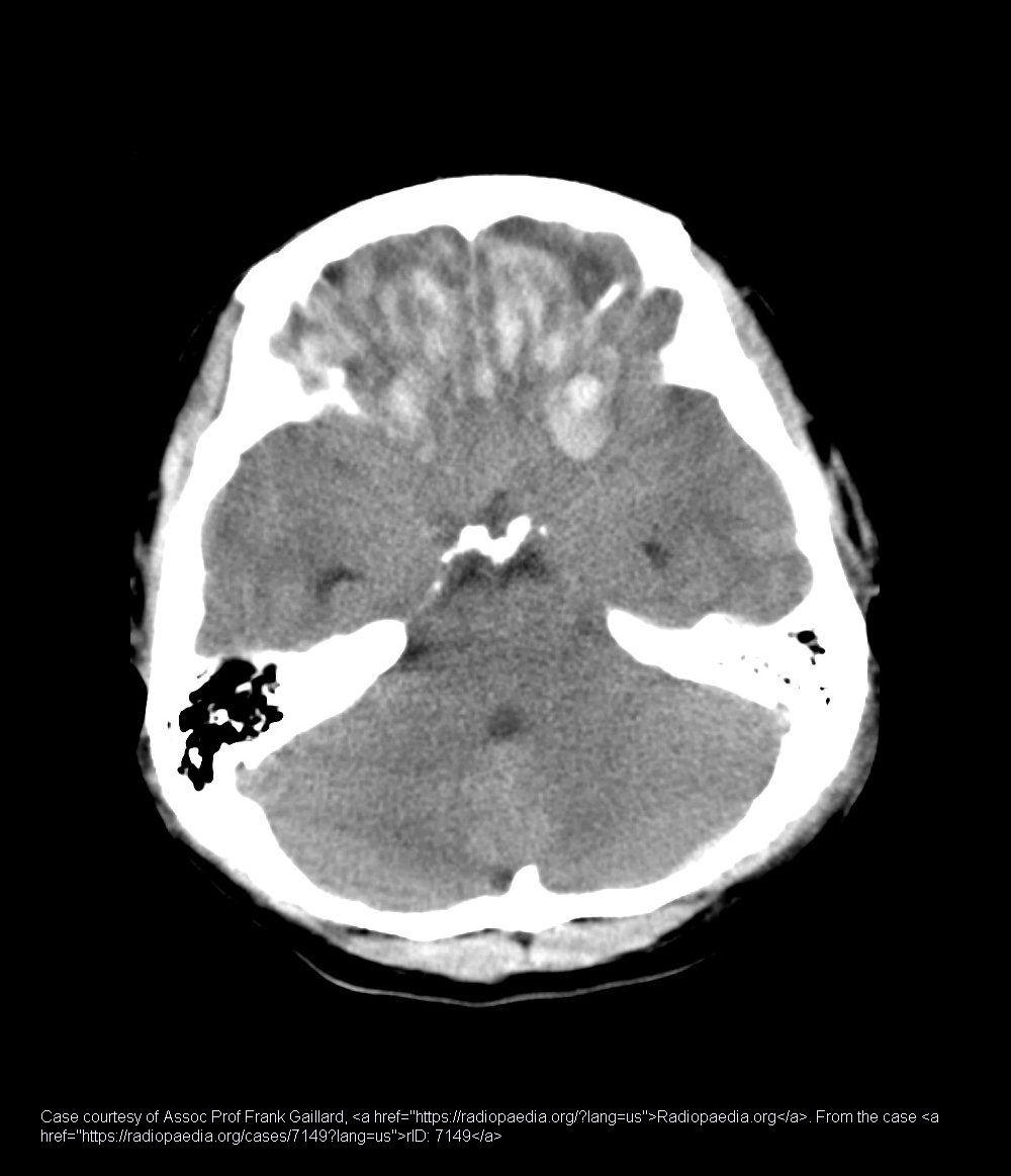
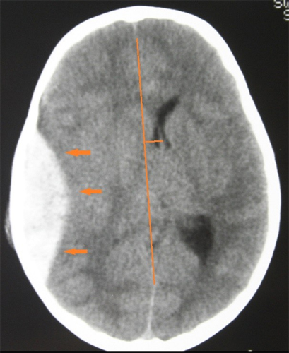
What is a cerebral hemorrhage?
Bleeding inside the brain; a medical emergency.
What causes cerebral hemorrhage in trauma?
Vessel damage from rapid acceleration/deceleration (e.g., MVA, shaken baby syndrome).
What complication can large bleeds cause on CT?
Midline shift of the Falx Cerebri.
Name two serious outcomes of untreated cerebral hemorrhage.
Paralysis and death.
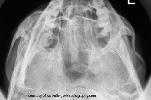
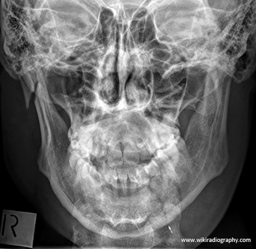
Which bones are most often fractured in facial trauma?
Mandible, maxilla, frontal bone, nasal bones, and zygoma.
What can major facial swelling require in emergency care?
Oral intubation.
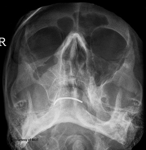
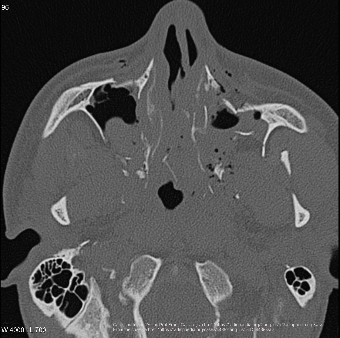
What is a common sign of an inferior orbital rim blow-out fracture?
Tear-drop opacity from trapped orbital muscles.
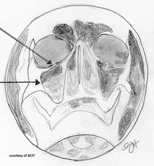
What is a possible complication of maxilla fracture affecting vision?
Restricted upward eye motion from optic nerve entrapment.
What is the typical cause of a zygomatic arch fracture?
Direct trauma.
Name one functional complication of a zygomatic arch fracture.
Lacrimal obstruction.
Name the three LeFort fracture types.
Type I (horizontal), Type II (pyramidal), Type III (transverse).
What is the treatment for LeFort fractures?
Surgical stabilization with a multidisciplinary team.
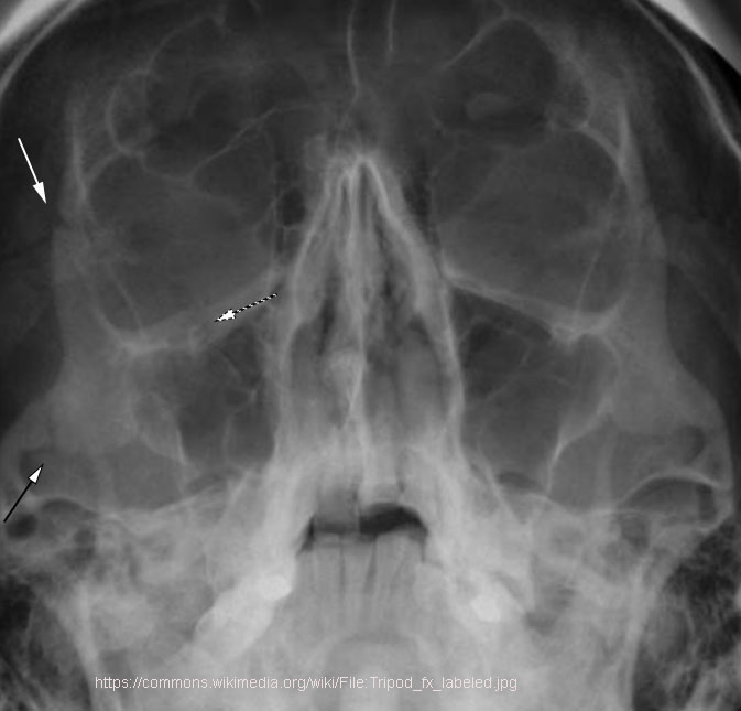
Which sites are involved in a tripod fracture?
Zygomatic arch, orbital floor/rim, and zygomatic-frontal suture.
What functional problem can a tripod fracture cause?
Chewing difficulty from temporalis muscle impingement.
Why are both sides of the mandible imaged if one side is fractured?
There is a high incidence of contrecoup fractures.
What is a major airway risk with unstable mandible fractures?
Tongue displacement obstructing the airway.
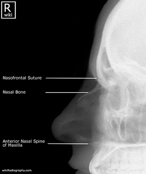
What is the most common facial fracture?
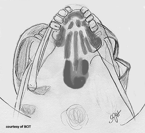
Nasal bone fracture.
What complication can a deviated septum cause?
Breathing difficulty.
How can sutures be differentiated from fracture lines on imaging?
Sutures appear jagged and interlocking; fracture lines are linear, sharp, and may cross edges unnaturally.
Why is the middle meningeal artery groove important in skull trauma imaging?
It is a normal groove for the artery and can mimic fracture lines on lateral views.
What type of trauma usually causes linear skull fractures?
Low-impact blunt trauma over a wide area (e.g., motor vehicle accidents, falls, sports injuries).
What differentiates open depressed skull fractures from closed ones?
Open fractures have a break in the skin exposing bone; closed fractures do not.
What does altered consciousness imply clinically in head trauma?
Possible brain injury affecting awareness or responsiveness, from confusion to coma.
Why is CSF or blood leakage from ears or nose concerning?
May indicate a basilar skull fracture with dural tear.
Why must concussion patients avoid physical activity for 24–48 hours?
To prevent worsening injury and allow brain recovery.
What should be done if concussion symptoms worsen?
Seek immediate medical evaluation to rule out complications like brain hemorrhage.
What symptoms might cerebral contusions cause besides imaging findings?
Headache, confusion, focal neurological deficits depending on injury location.
How can cerebral contusions progress?
They may cause swelling and increased intracranial pressure.
What is the difference between arterial and venous cerebral hemorrhages on CT?
Arterial bleeds are faster, larger, and cause more mass effect; venous bleeds may be slower and smaller.
What is angiographic embolization?
A procedure to block bleeding vessels to control hemorrhage.
What is malocclusion, and which facial fracture complication does it relate to?
Misalignment of teeth/jaw; common after maxillary or mandibular fractures.
How can infraorbital anesthesia occur after facial fractures?
Injury to the infraorbital nerve during fracture of the maxilla or orbital floor.
What does nonunion mean in fracture healing?
Failure of the bone fragments to heal together properly.
How can midface lengthening affect appearance after a zygomatic arch fracture?
Flattening or widening of the cheek area.
Which LeFort fracture type involves separation of the entire midface from the skull base?
Type III (transverse).
What causes nerve pain in tripod fractures?
Injury or impingement of the infraorbital nerve.
What muscle action can displace mandible fracture fragments?
Movement of muscles used for chewing and talking.
Why is the nasal bone prone to fractures?
It is thin and prominent on the face.