Bio 202 - Cell Cycle
1/14
There's no tags or description
Looks like no tags are added yet.
Name | Mastery | Learn | Test | Matching | Spaced | Call with Kai |
|---|
No analytics yet
Send a link to your students to track their progress
15 Terms
DNA monomers and their structure
DNA monomers are nucleotides with the following structure:
1) a phosphate group
2) a deoxyribose sugar
3) nucleotide base(s) in ring shape
A polymer consists of the sugar molecule of one binding to the phosphate group of another.
Chromatin
eukaryotic chromosomes that associate with proteins to be packaged in the nucleus
1) euchromatin: chromatin with DNA that is more loosely packed and accessible for transcription
2) heterochromatin: chromatin with DNA that is densely packed and not really transcribed
Interphase
the first stage of the cell cycle consisting of three subphases:
g1: the cell grows in mass
S-phase: DNA replicates
g2: the cell grows until a point where it is committed to enter mitosis.
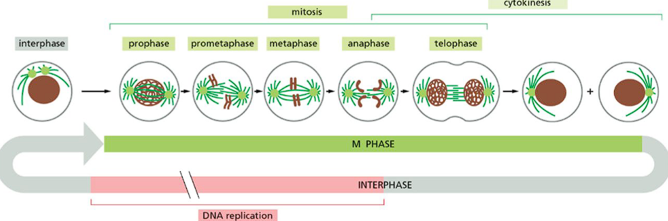
Mitotic phase
Mitosis occurs, the replicated chromosomes are separated and the nucleus splits in two. Then, cytokenesis occurs where cytoplasmic division leads to 2 new cells. There are 5 subphases: prophase, prometaphase, metaphase, anaphase, and telophase.
What is happening to the chromosomes during interphase?
In g1, each chromosome in the nucleus is one long strand of DNA.
In the S-phase, the DNA replicates to create two sister chromatids held together by cohesin proteins.
In g2, the chromatids condense and pack together through condensin proteins.
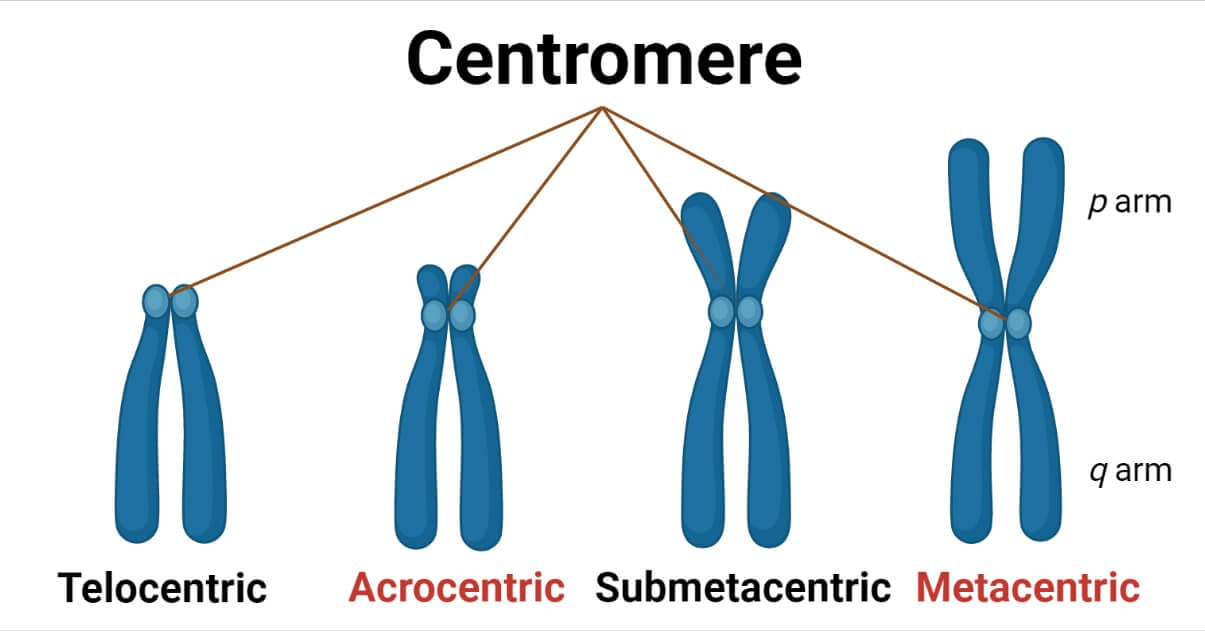
What are the chromatid centromeres and arms?
Each sister chromatid has a centromere, a region made up of repetitive sequences in the chromosomal DNA where sister chromatids are closely attached.
The portion of a chromatid on either side of the centromere is a called an arm. Each chromatid has two.
What is happening to chromosomes during mitosis?
The condensed sister chromatids are split by the mitotic spindle to double the number of chromosomes. An equal portion travels to the polar ends of the cell. Chromosomes decondense and once the cell enters cytokinesis, each daughter cell has identical DNA.
How is the mitotic spindle made?
After the S-Phase and in G2, the cell has two centrosomes (organelles which organize microtubules) adjacent to the nuclear membrane. They begin to nucleate microtubules. As the microtubules grow in prophase, the centrosomes are pushed apart. By metaphase, each centrosome is at opposite ends of the cell and the spindle is set up. The spindle provides the mechanical force needed toa lign chromosomes during metaphase.
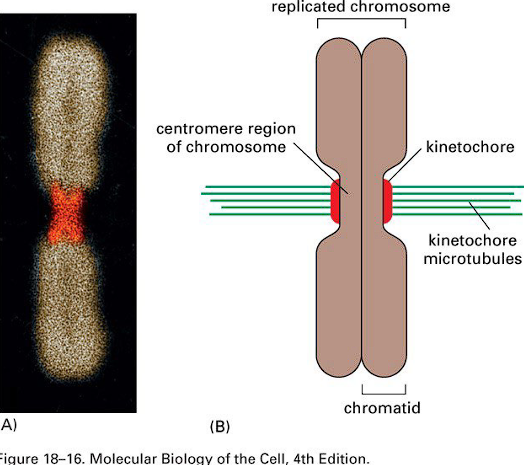
Kinetochores
Microtubule “links” on chromosomes that assembles on centromeres. Each sister chromatid assembles one kinetochore. Microtubules from the spindle attach to chromosome by binding the kinetochores.
Prophase
Chromosomes condense as condensin II binds DNA and extrudes loops.
• The mitotic spindle begins to form.
• Centrosomes are pushed toward opposite cell poles by lengthening microtubules between them.
Prometaphase
The nuclear membrane breaks down and fragments.
• Microtubules from the spindle begin to attach to the chromosomes via the kinetochores.
• Each sister chromatid has a kinetochore.
Condensin I forms new loops on the existing loops generated by condensin II, leading to further chromosome condensing.
Metaphase
Microtubules have captured all the kinetochores from opposite poles. Tug of war between pulling and pushing of kinetochore microtubules aligns chromosomes at the metaphase plate, an imaginary plane where all chromosomes are aligned.
Anaphase
Each sister chromatid is separated from each other and pulled towards the pole it is attached to by spindle fibers. After separation, each sister chromatid is considered a chromosome with equivalent genetic content. Each pole now has an equivalent number of chromosomes.
Telophase
Duplicated groups of chromosomes have arrived at opposite ends of the elongated cell. The nuclear membrane begins to
reassemble, and chromatin becomes less tightly coiled. A cleavage furrow forms at the midplane during cytokinesis, which
will separate daughter cells.
Image of all cell phases