The Human Eye
1/14
There's no tags or description
Looks like no tags are added yet.
Name | Mastery | Learn | Test | Matching | Spaced | Call with Kai |
|---|
No analytics yet
Send a link to your students to track their progress
15 Terms
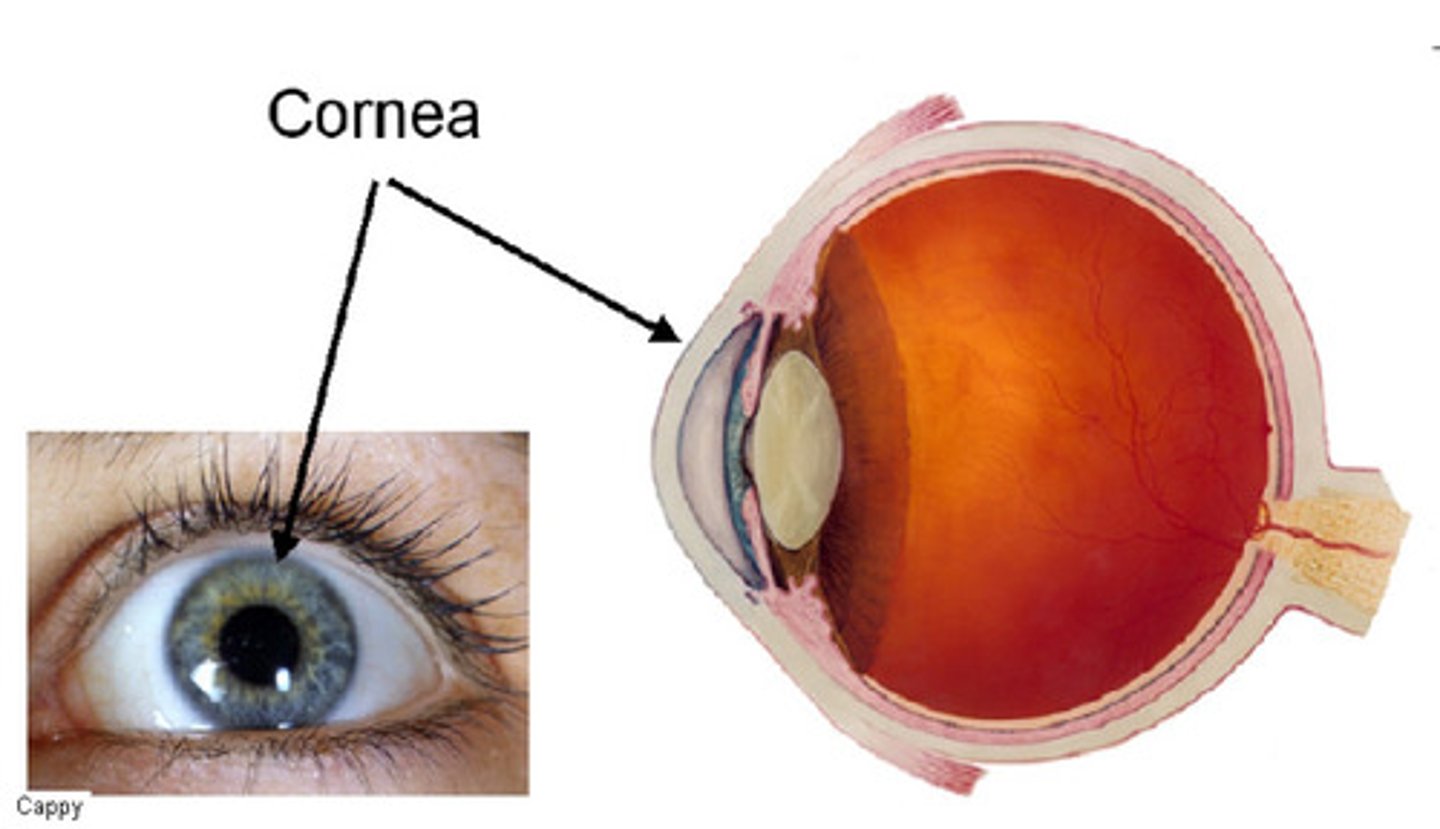
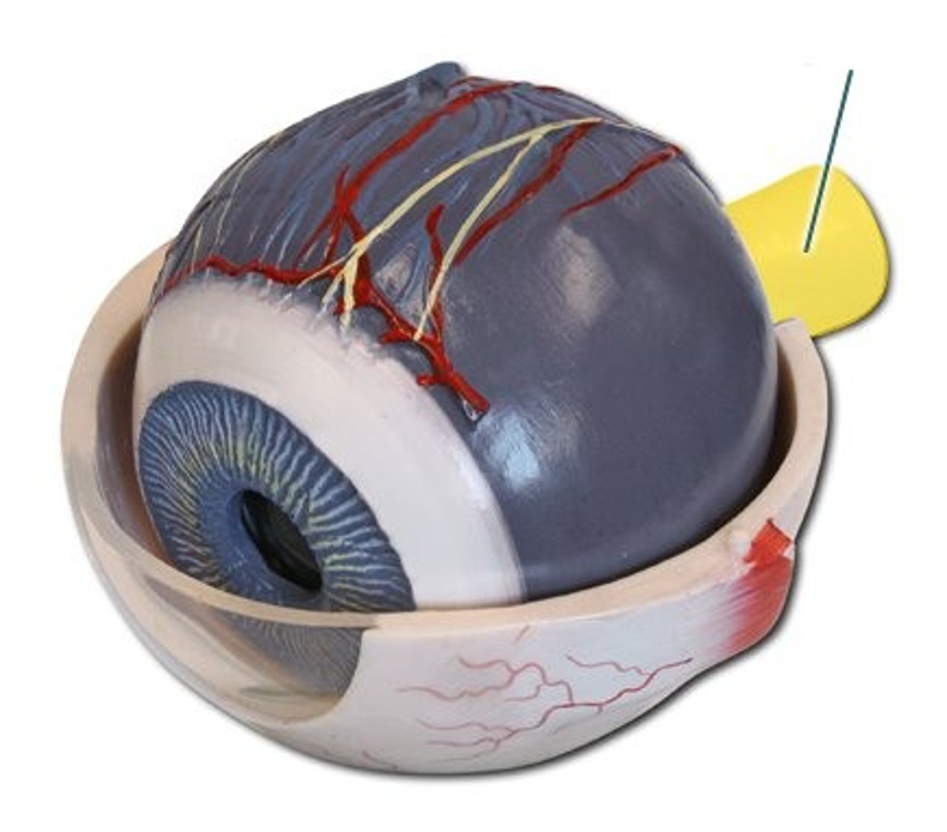
cornea
clear protective layer in the front of the eye
pupil
a circular opening that controls how much light enters the eye
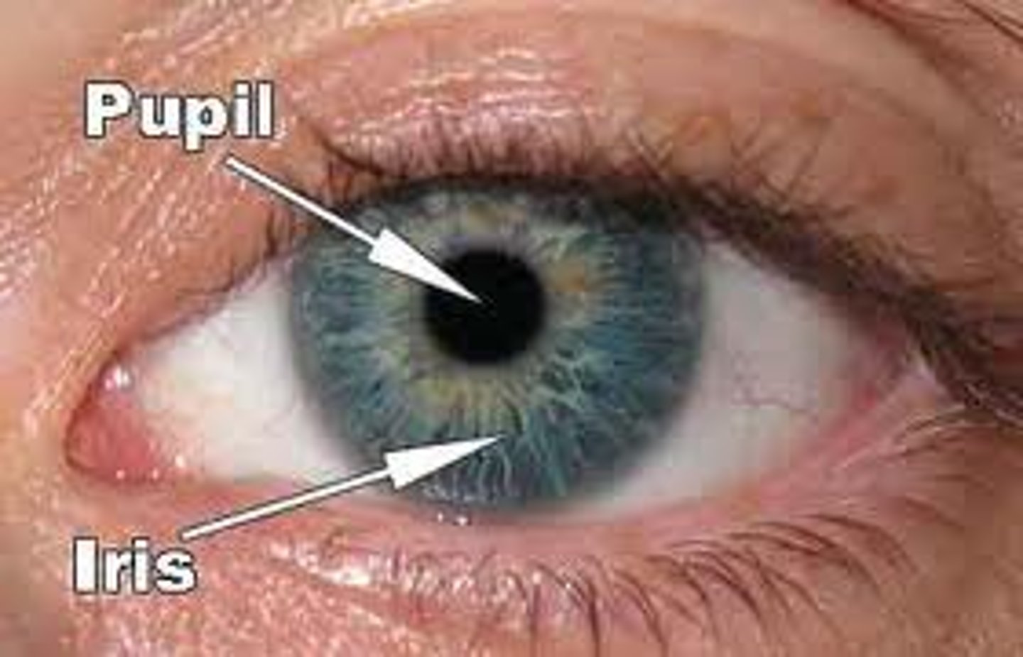
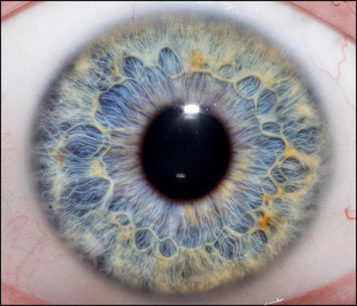
iris
the colored part of your eye, adjusts the size of the pupil
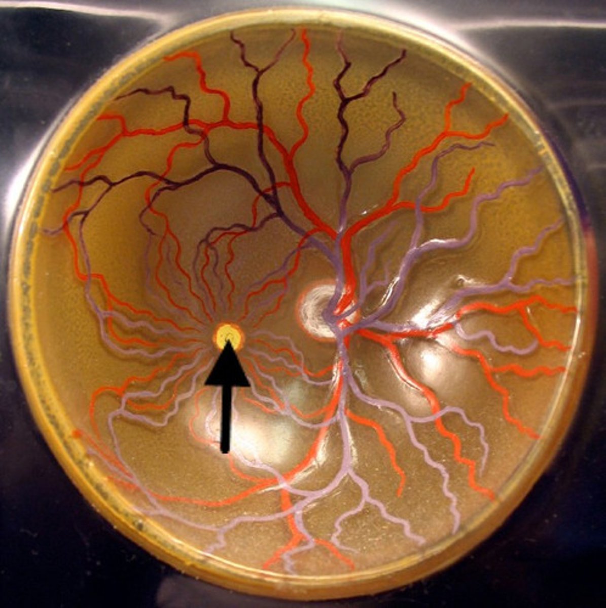
retina
the back of the the inside of the eyeball, contains light-sensitive rods and cones
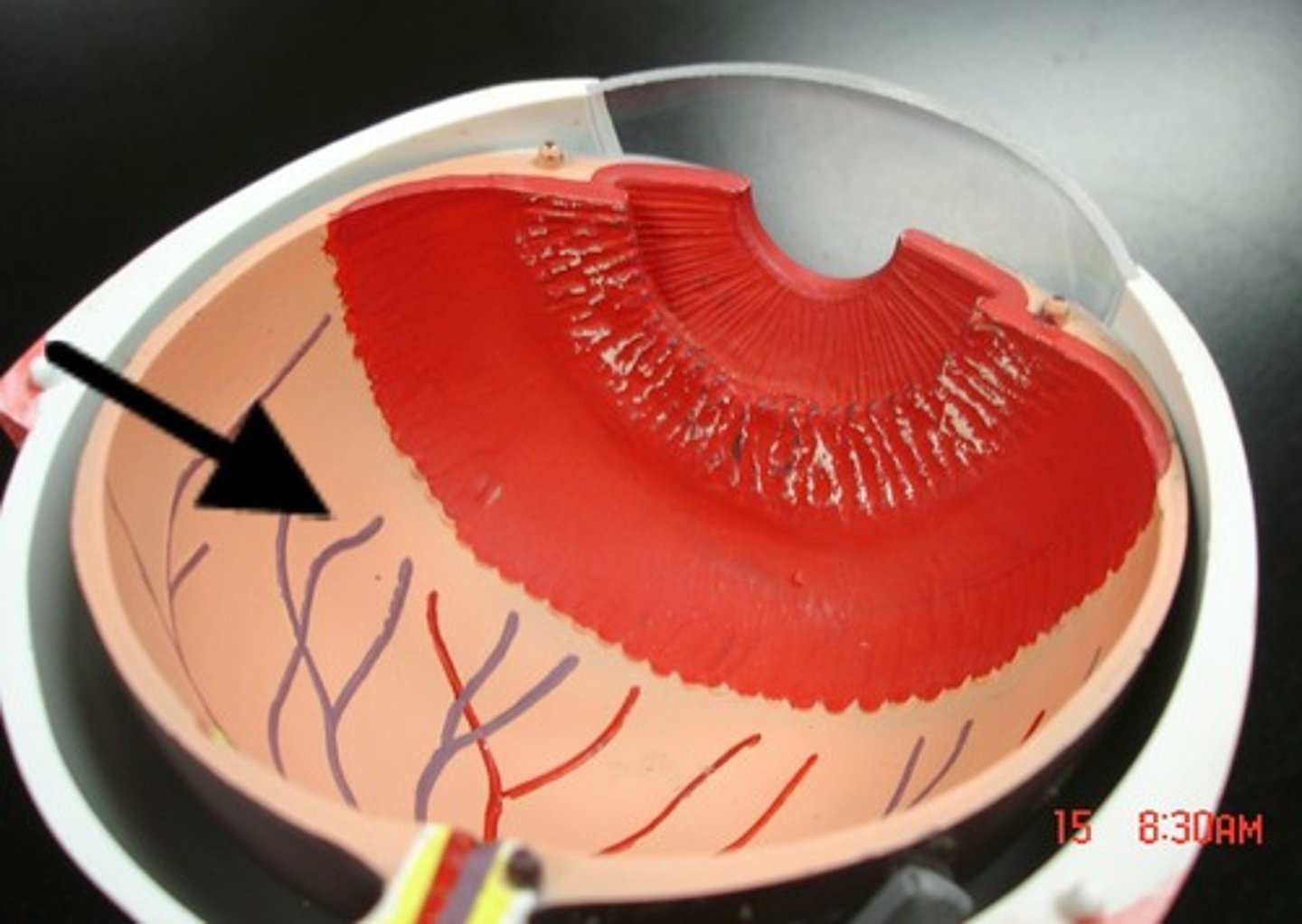
optic nerve
receives messages from the retina and then transmits messages to the brain
rod cells
cells located in the retina that respond to and recognize only black and white
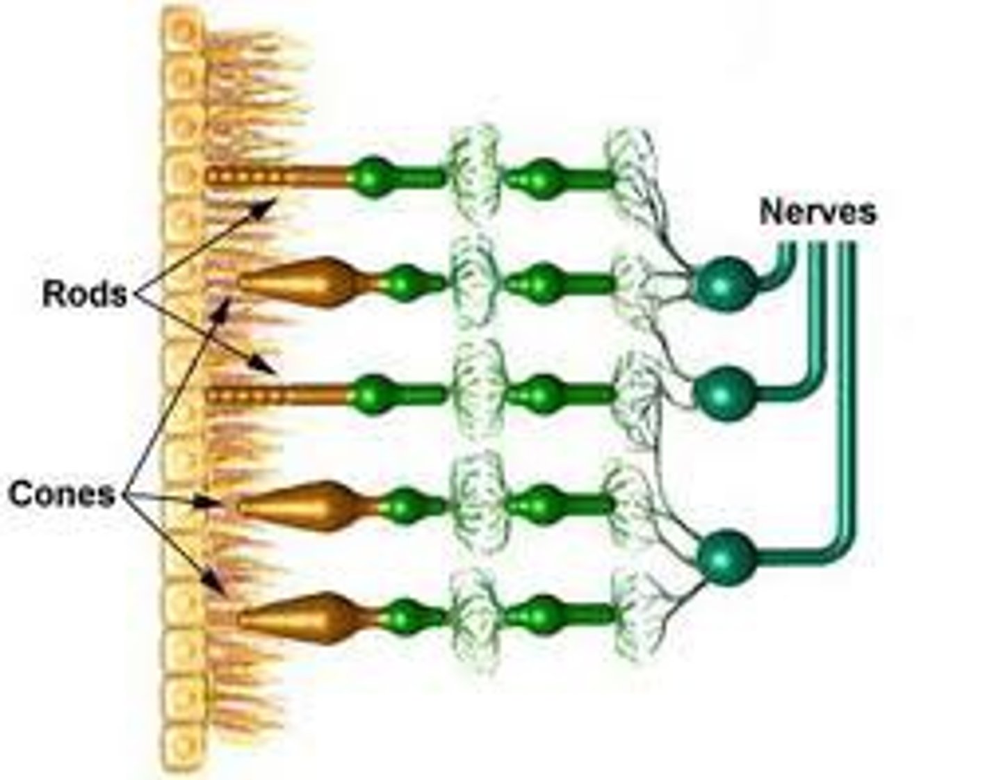
cone cells
cells located in the retina that respond to and recognize color and make shapes

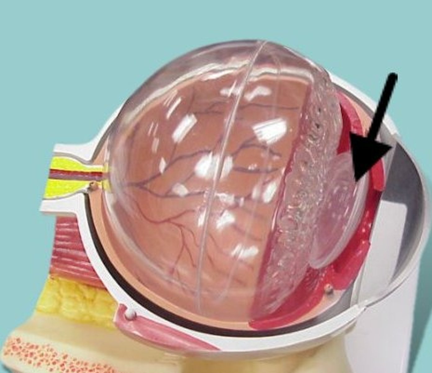
vitreous humor
the transparent, jelly-like tissue filling the eyeball behind the lens.
sclera
the white outer layer of the eye, provides support for the eyeball and allows attachment of muscles to the eyeball
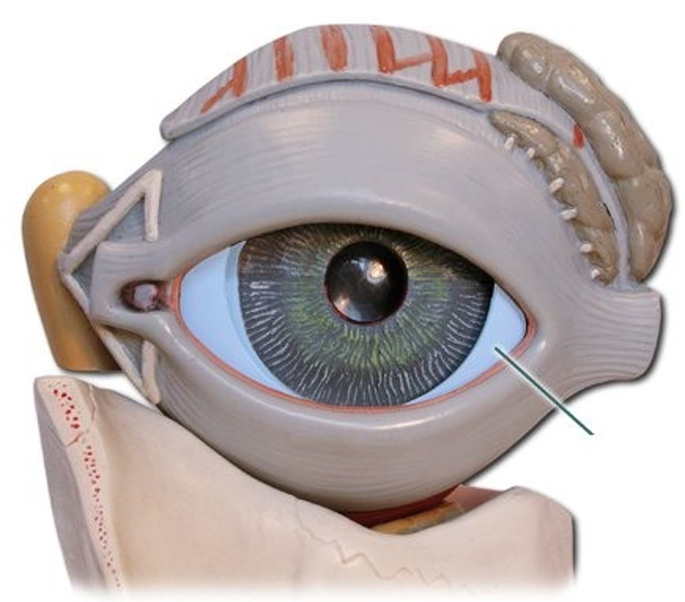
aqueous humor
watery liquid found between the cornea and lens
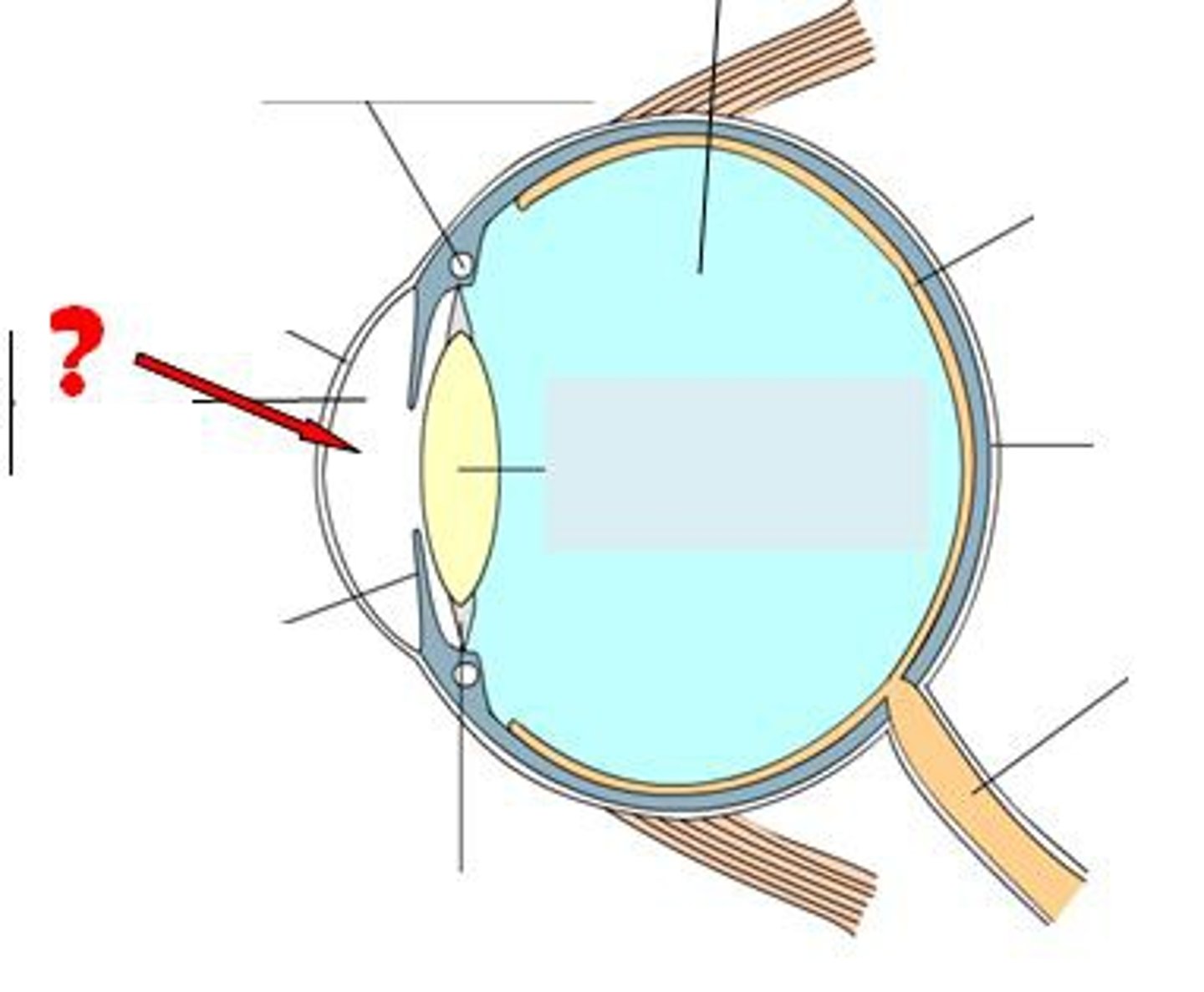
fovea
the central focal point in the retina, around which the eye's cones cluster
ciliary body
ring of tissue behind the iris that holds the lens in place and changes the shape of the lens
lens
the transparent structure behind the pupil that changes shape to help focus images on the retina
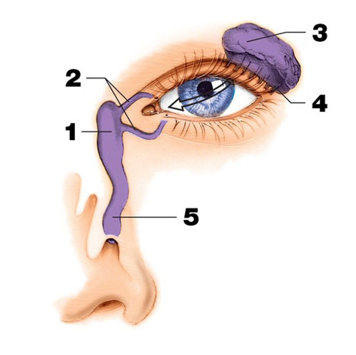
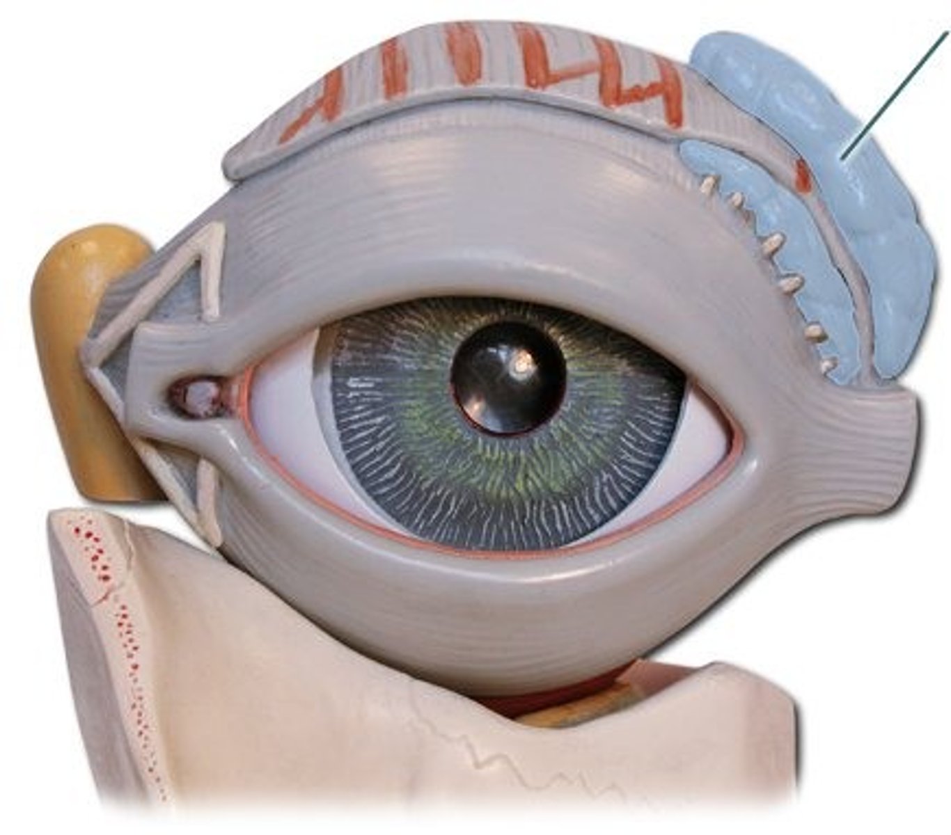
lacrimal gland
gland located in the upper outer region above the eyeball that secretes tears
lacrimal duct
the passageway that drains excess tears into the nose (#5 in the image)