Lab 6: Pectoral Girdle, Limb Bones, and their Muscles
1/76
There's no tags or description
Looks like no tags are added yet.
Name | Mastery | Learn | Test | Matching | Spaced | Call with Kai |
|---|
No analytics yet
Send a link to your students to track their progress
77 Terms
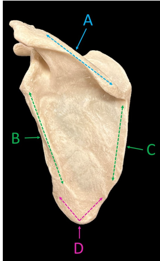
Label A-D
A: scapular spine
B: lateral border
C: medial border
D: inferior angle
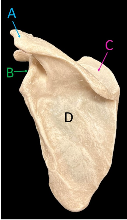
Label A-D.
A: acromion
B: glenoid cavity
C: supraspinous fossa
D: infraspinous fossa
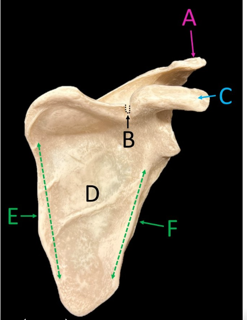
Label A-F.
A: acromion
B: suprascapular notch
C: coracoid process
D: subscapular fossa
E: medial border
F: lateral border
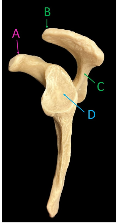
Label A-D.
A: coracoid process
B: acromion
C: scapular spine
D: glenoid cavity
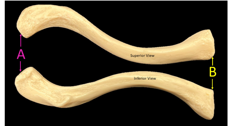
Label A-B.
A: acromial end
B: sternal end
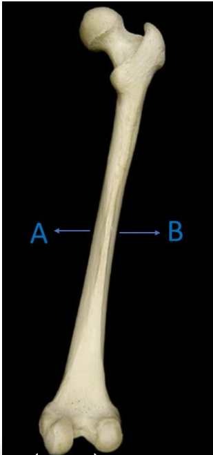
The image is of the _____ (posterior/anterior) view of the humerus.
The distal end of the humerus is represented by the letter (A/B)?
The proximal end of the humerus is represented by the letter (A/B)?
posterior
B represents the distal end of the humerus
A represents the proximal end of the humerus
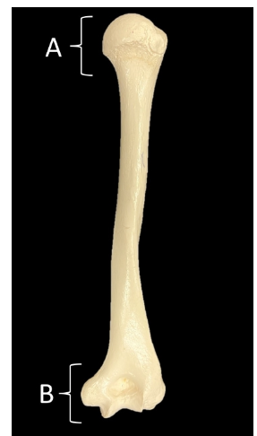
Label A-E.
A: head of humerus
B: olecranon fossa
C: medial epicondyle
D: trochlea
E: lateral epicondyle
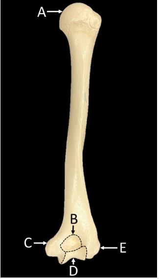
Label A-G.
A: head of humerus
B: radial fossa
C: coronoid fossa
D: lateral epicondyle
E: capitulum
F: trochlea
G: medial epicondyle
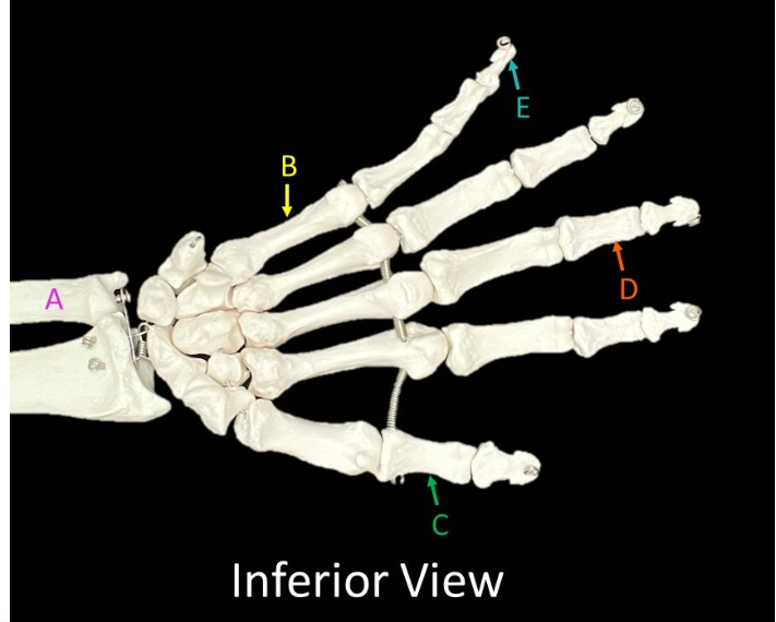
Label A-E.
A: ulna
B: metacarpal
C: proximal phalanx
D: middle phalanx
E: distal phalanx
Does the image show an anterior or posterior view?
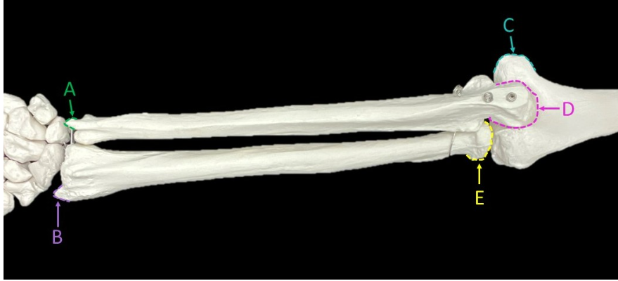
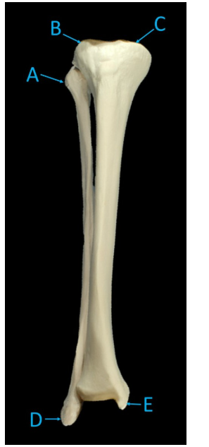
Label A-E.
POSTERIOR VIEW
A: styloid process of the ulna
B: styloid process of the radius
C: medial epicondyle
D: olecranon
E: head of radius
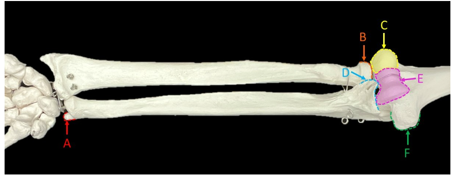
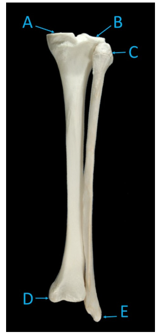
Label A-F.
A: styloid process of the ulna
B: head of radius
C: capitulum
D: coronoid process
E: trochlea
F: medial epicondyle
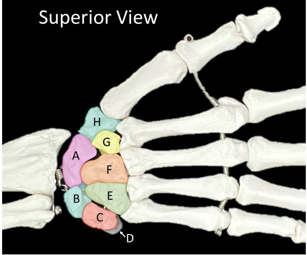
Label A-H
A: scaphoid
B: lunate
C: triquetrum
D: pisiform
E: hamate
F: capitate
G: trapezoid
H: trapezium
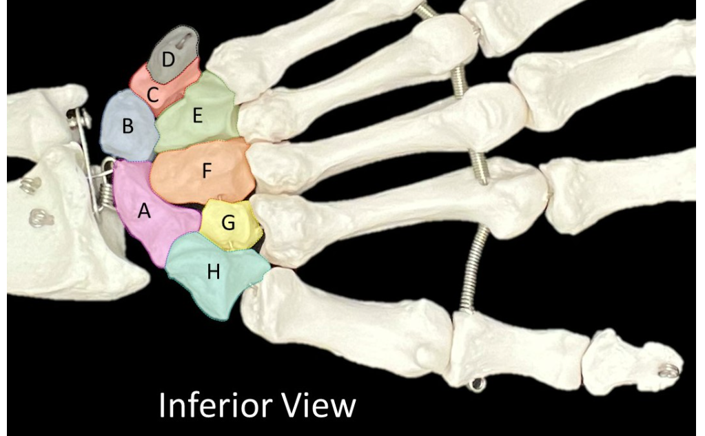
Label A-H.
A: scaphoid
B: lunate
C: triquetrum
D: pisiform
E: hamate
F: capitate
G: trapezoid
H: trapezium
Which letter on the image indicates the medial side and which one indicates the lateral side of the femur?
MEDIAL: a
LATERAL: b
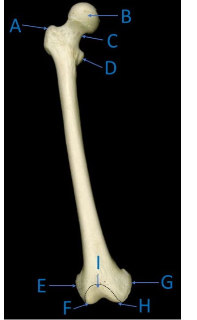
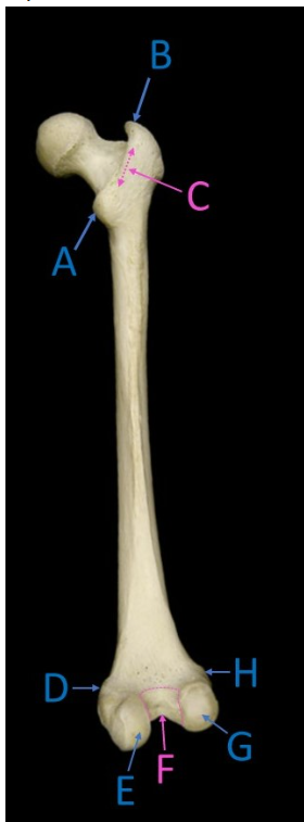
Label A-I.
A: greater trochanter
B: head
C: neck
D: lesser trochanter
E: lateral epicondyle
F: lateral condyle
G: medial epicondyle
H: medial condyle
I: patellar surface
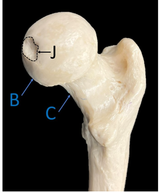
Label B, C, & J.
B: head
C: neck
J: fovea
Label A-H.
A: lesser trochanter
B: greater trochanter
C: intertrochanter crest
D: medial epicondyle
E: medial condyle
F: intercondylar fossa
G: lateral condyle
H: lateral epicondyle
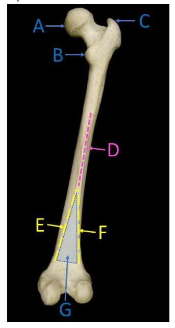
Label A-G.
A: head
B: lesser trochanter
C: greater trochanter
D: linae aspera
E: medial supracondylar line
F: lateral supracondylar line
G: popliteal surface
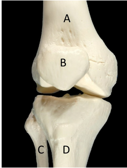
Label A-D.
A: femur
B: patella
C: fibula
D: tibia
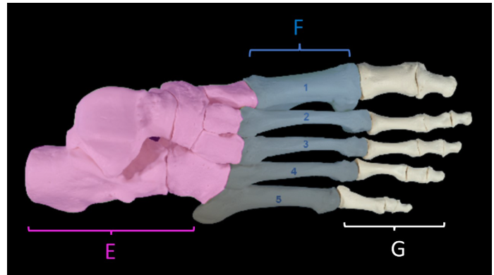
Label E-G.
E: tarsals
F: metatarsals
G: phalanges
Label A-E.
A: head of fibula
B: lateral condyle
C: medial condyle
D: lateral malleolus
E: medial malleolus
Label A-E.
A: medial condyle
B: lateral condyle
C: head of fibula
D: medial malleolus
E: lateral malleolus
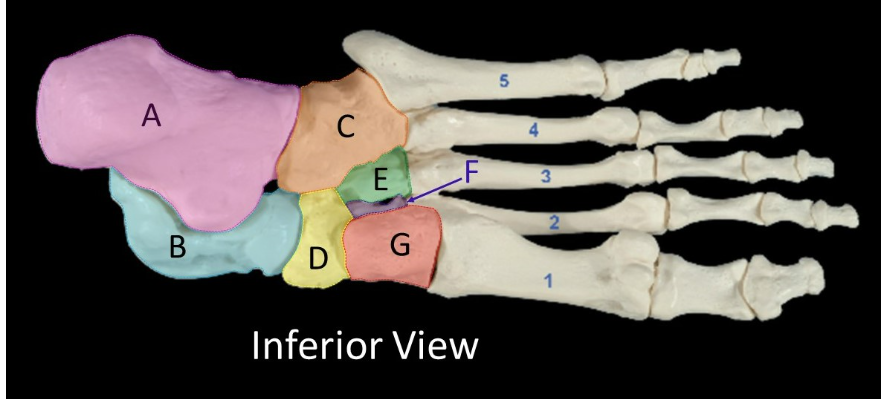
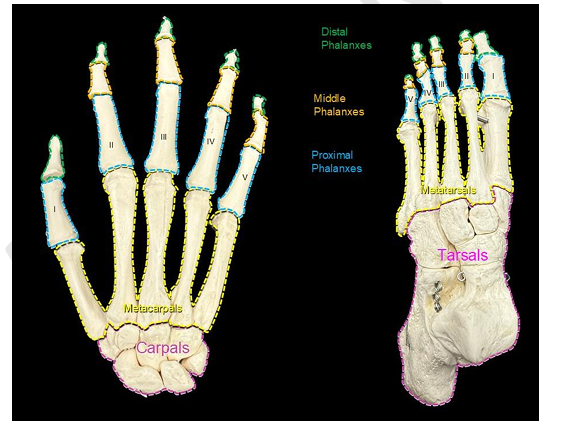
Label A-G.
A: calcaneus
B: talus
C: cuboid
D: navicular
E: lateral cuneiform
F: intermediate cuneiform
G: medial cuneiform
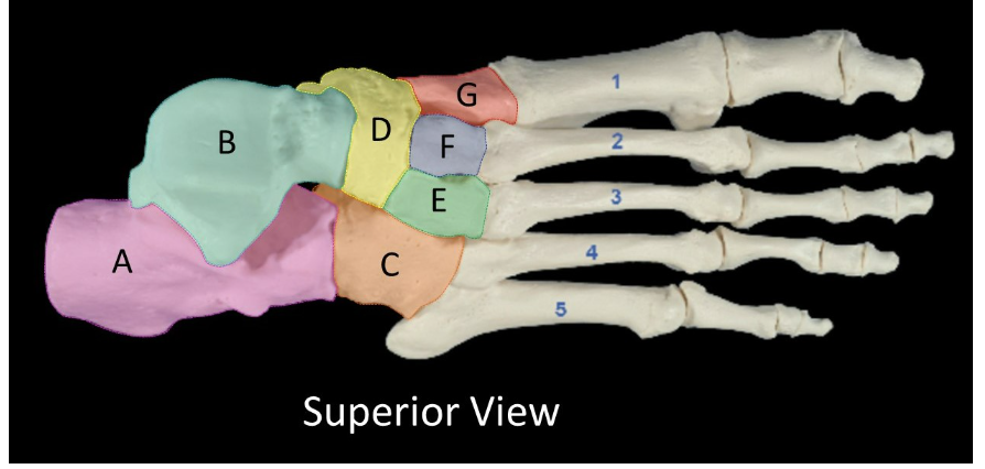
Label A-G.
A: calcaneus
B: talus
C: cuboid
D: navicular
E: lateral cuneiform
F: intermediate cuneiform
G: medial cuneiform
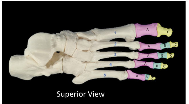
Label A-C.
A: proximal phalanges
B: middle phalanges
C: distal phalanges
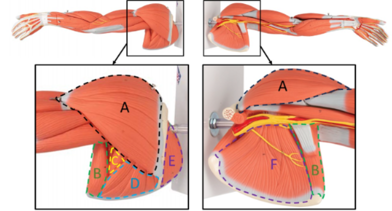
Label A-F.
A: deltoid
B: teres major
C: teres minor
D: infraspinatus
E: supraspinatus
F: subscapularis
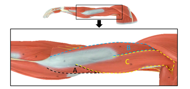
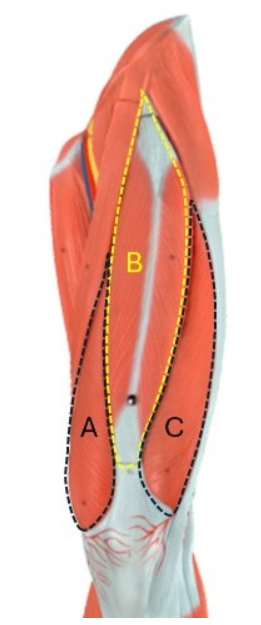
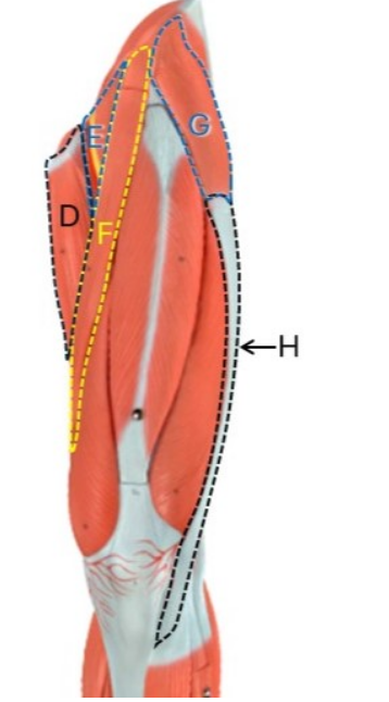
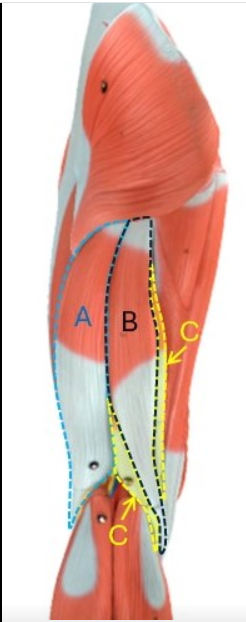
Label A-C.
A: triceps brachii - medial head
B: triceps brachii - lateral head
C: triceps brachii - long head
Label D-E.
D: biceps brachii - long head
E: biceps brachii - short head
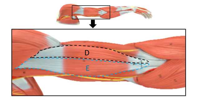
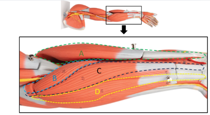
Label A-E.
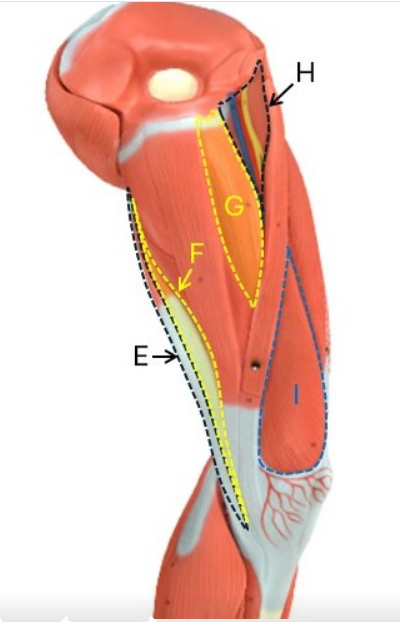
A: coracobrachialis
B: biceps brachii- short head
C: biceps brachii- long head
D: triceps brachii- medial head
E: brachialis
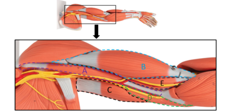
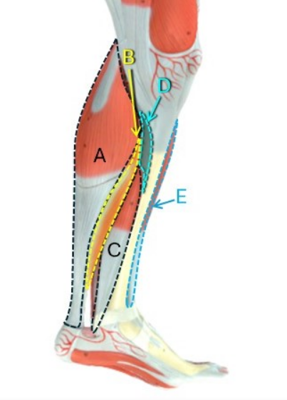
Label A-D.
A: brachioradialis
B: pronator teres
C: flexor carpi radialis
D: palmaris longus
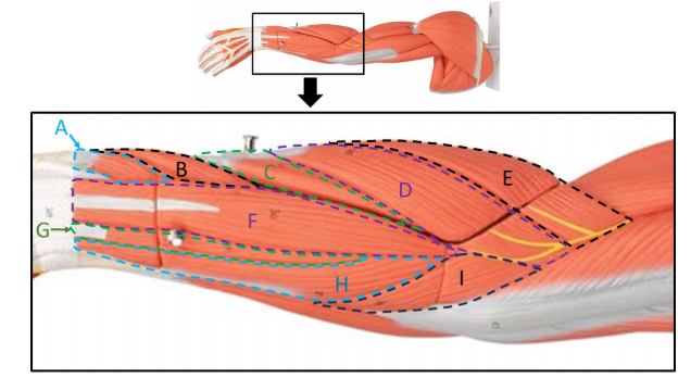
Label A-I.
A: extensor pollicis brevis
B: abductor pollicis longus
C: extensor carpi radialis brevis
D: extensor carpi radialis longus
E: brachioradialis
F: extensor digitorum
G: extensor digiti minimi
H: extensor carpi ulnaris
I: anconeus
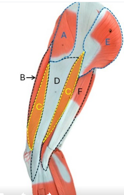
Label A-C.
A: vastus medialis
B: rectus femoris
C: vastus lateralis
Label D-H.
D: adductor longus
E: iliopsoas
F: sartorius
G: tensor fasciae latae
H: iliotibial tract
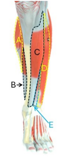
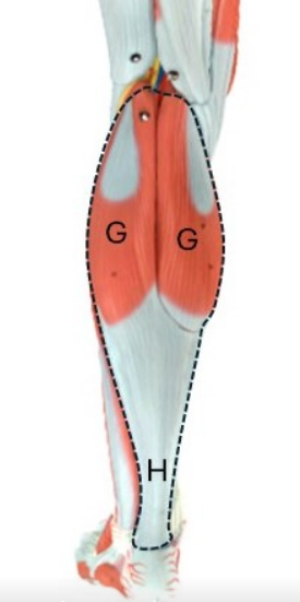
Label A-E.
A: gastrocnemius
B: flexor digitorum longus
C: tibialis anterior
D: extensor digitorum longus
E: extensor hallucis longus
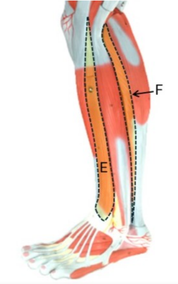
Label A-F.
A: tensor fasciae latae
B: rectus femoris
C: vastus lateralis
D: iliotibial tract
E: gluteus maximus
F: biceps femoris
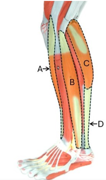
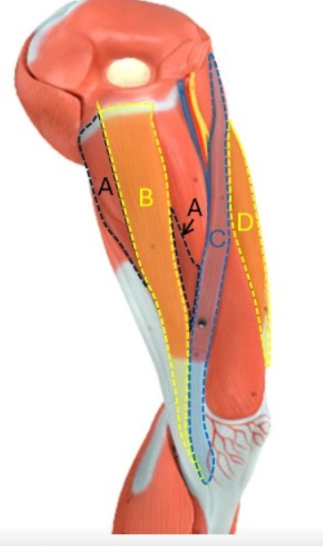
Label A-D.
A: tibialis anterior
B: fibularis longus
C: gastrocnemius
D: calcaneal tendon
Label E-F.
E: extensor digitorum longus
F: soleus
Label A-F.
A: biceps femoris
B: semitendinosus
C: semimembranosus
D: gluteus maximus
E: adductor magnus
F: gracilis
Label G-H.
G: gastrocnemius
H: calcaneal tendon
Label A-D.
A: adductor magnus
B: gracilius
C: sartorius
D: rectus femoris
Label E-I.
E: semitendinosus
F: semimembranosus
G: adductor longus
H: iliopsoas
I: vastus medialis
Label A-E.
A: gastrocnemius
B: soleus
C: flexor digitorum longus
D: popliteus
E: tibialis anterior
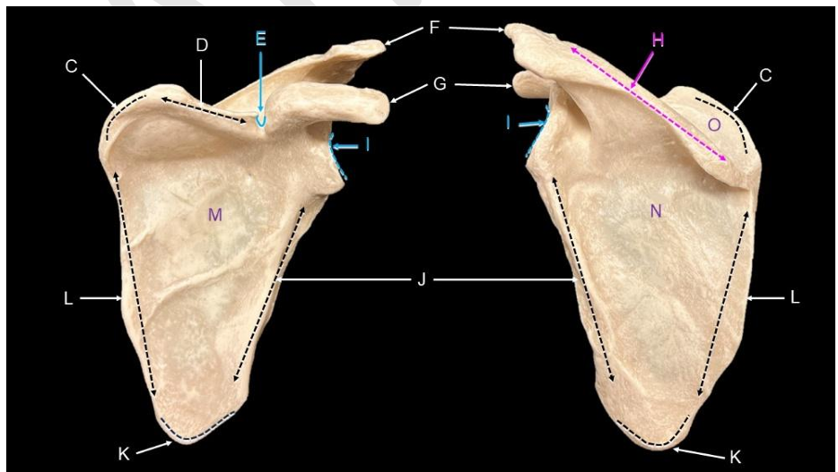
Label C-O.
C: superior angle
D: superior border
E: suprascapular notch
F: acromion
G: coracoid process
H: spine of the scapula
I: glenoid cavity
J: lateral border
K: inferior angle
L: medial border
M: subscapular fossa
N: infraspinous fossa
O: supraspinous fossa
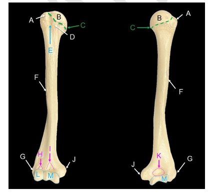
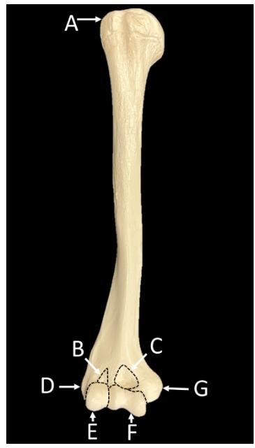
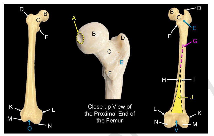
Label A-M.
A: greater tubercle
B: head of humerus
C: anatomical neck of humerus
D: lesser tubercle
E: intertubercular sulcus
F: deltoid tuberosity
G: lateral epicondyle
H: radial fossa
I: coronoid fossa
J: medial epicondyle
K: olecranon fossa
L: capitulum
M: trochlea
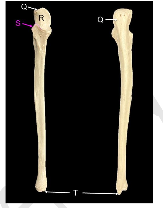
Label Q-T.
Q: olecranon process
R: trochlear notch
S: coronoid process
T: styloid process of ulna
a. What part of the humerus articulates with the scapula – the proximal or distal end?
The head of the humerus (proximal end) articulates with the glenoid cavity region of the scapula
b. What bone articulates the humerus at the olecranon fossa?
The ulna
c. Structurally, how can you differentiate between the medial and lateral epicondyle?
The medial epicondyle is much more prominent and is next to the trochlea while the lateral epicondyle is much less prominent and next to the capitulum.
d. Which bone is more medial – the radius or the ulna?
ulna
e. How do the proximal shapes of the radius and ulna compare?
The radius has a flatter; more level head compared to the “U-shaped” hook of the ulna.
f. What particular bone features occur where the distal end of the humerus articulates with the proximal end of the radius?
The head of the radius articulates with the capitulum of the humerus.
g. What particular bone features occur where the distal end of the humerus articulates with the proximal end of the ulna?
The trochlear notch of the ulna articulates with the trochlea of the humerus.
h. What part of the humerus articulates with the scapula – the proximal or distal end?
The head of the humerus (proximal end) articulates with the glenoid cavity region of the scapula.
i. What bone articulates the humerus at the olecranon fossa?
The ulna
j. Structurally, how can you differentiate between the medial and lateral epicondyle?
The medial epicondyle is much more prominent and is next to the trochlea while the lateral epicondyle is much less prominent and next to the capitulum.
Label A-V.
A: fovea
B: head of femur
C: neck of femur
D: greater trochanter
E: intertrochanter crest
F: lesser trochanter
G: linea aspera
H: medial supracondylar line
I: lateral supracondylar line
J: popliteal surface
K: lateral epicondyle
L: medial epicondyle
M: lateral condyle of femur
N: medial condyle of femur
O: patellar surface
V: intercondylar fossa
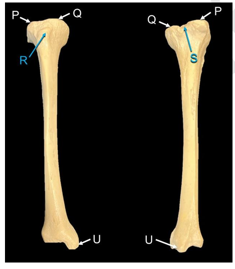
Label P, Q, R, S, U.
P: lateral condyle of tibia
Q: medial condyle of tibia
R: tibial tuberosity
S: intercondylar eminence
U: medial malleolus
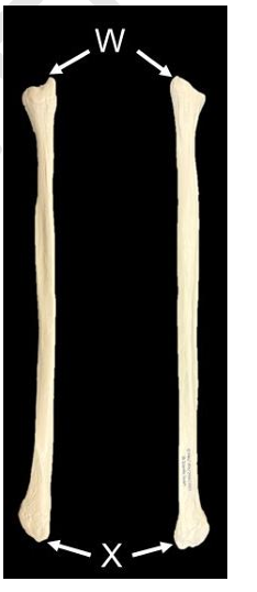
Label W, X.
W: head of fibula
X: lateral malleolus
a. Does the head of the femur point medial or lateral?
Medial
b. What features did you use to determine between the anterior and posterior sides of the tibia?
The anterior side as the tibia is where the tibial tuberosity is located and the posterior side has the intercondylar eminence.
c. What bones and bone features make up the bony protrusions of the ankle that you can palpate (both medial and lateral sides)?
The medial malleolus of the tibia (medial ankle protrusion) and the lateral malleolus of the fibula (lateral ankle protrusion).

STUDY THIS!
DO IT!!
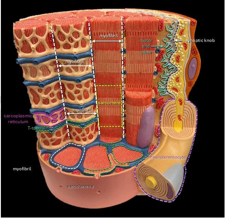
Label A-H.
A: myofibril
B: t-tubule
C: sarcoplasmic reticulum
D: sarcomere
E: motor end plate
F: synaptic knob
G: neurolemmocyte
H: sarcolemma
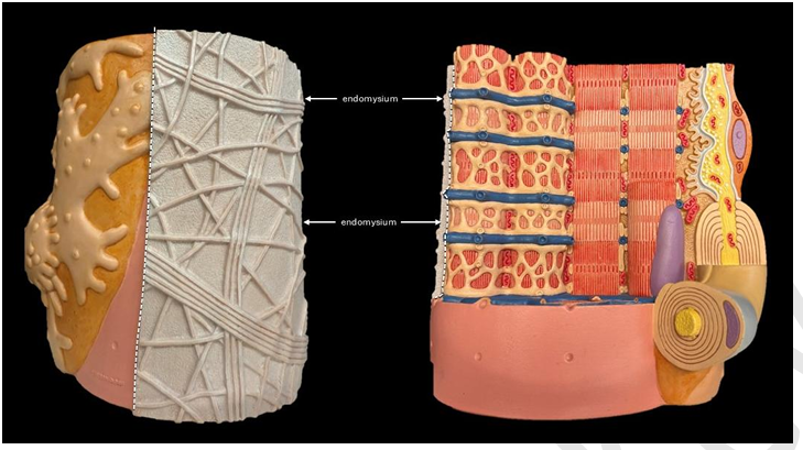
Label M.
M: endomysium
Label A-B.
A: sarcoplasmic reticulum
B: t-tubule
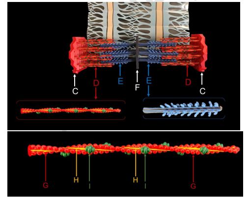
Label C-I.
C: z- disc
D: thin filament
E: thick filament
F: m-line
G: actin
H: tropomyosin
I: troponin
a. This model is of a sarcomere. These are lined up end to end on what specific structure?
Myofibril
b. This model is movable, allowing you to either increase or decrease the overlap of the thin and thick filaments. What area do you move, the M-line or the Z-discs to make this happen?
Z-discs
c. There is an orientation to the myosin molecules. What structure do the tails of the myosin attach to, the M-line or Z-disc?
M-line
d. What structure is the thin filament directly attached to, the M-line or Z-disc?
Z-disc
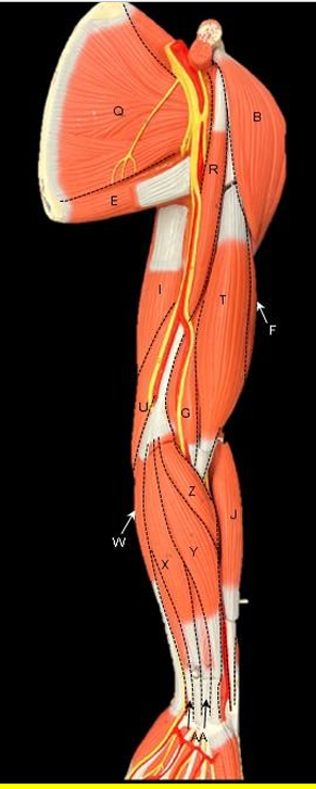
Label A-Z. (not all letters alphabetically included)
AA: flexor digitorum superficialis
B: deltoid
E: teres major
F: biceps brachii- long head
G: brachialis
I: triceps brachii - long head
J: brachioradialis
Q: subscapularis
R: coracobrachialis
T: biceps brachii - short head
U: triceps brachii - medial head
W: flexor carpi ulnaris
X: palmaris longus
Y: flexor carpi radialis
Z: pronator teres
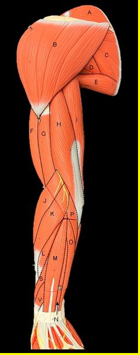
Label A-V. (not all letters included alphabetically)
A: supraspinatus
B: deltoid
C: infraspinatus
D: teres minor
E: teres major
F: biceps brachii - long head
G: brachialis
H: triceps brachii - lateral head
I: triceps brachii - long head
J: brachioradialis
K: extensor carpi radialis longus
L: extensor carpi radialis brevis
M: extensor digitorum
N: extensor digiti minimi
O: extensor carpi ulnaris
P: anconeus
S: abductor pollicis longus
V: extensor pollicis brevis
Label A-N.
A: adductor longus
B: iliopsoas
C: sartorius
D: tensor fasciae latae
E: vastus medialis
F: rectus femoris
G: vastus lateralis
H: iliotibial tract
I: gastrocnemius - medial head
J: flexor digitorum longus
K: tibialis anterior
L: extensor hallucis longus
M: extensor digitorum longus
N: fibularis longus
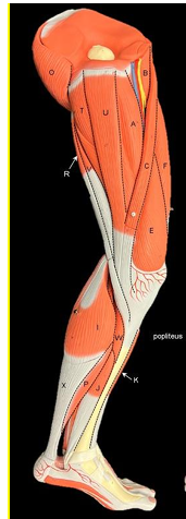
Label I-X. (not all letters included alphabetically)
I: gastrocnemius - medial head
O: gluteus maximus
Q: biceps femoris
S: gastrocnemius - lateral head
T: adductor magnus
U: gracilis
V: semimembranosus
X: calcaneal tendon
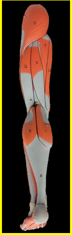
Label A-X. (not all letters included alphabetically)
A: adductor longus
B: iliopsoas
C: sartorius
E: vastus medialis
F: rectus femoris
I: gastrocnemius - medial head
J: flexor digitorum longus
K: tibialis anterior
O: gluteus maximus
P: soleus
R: semitendinosus
T: adductor magnus
U: gracilis
V: semimembranosus
W: popliteus
X: calcaneal tendon
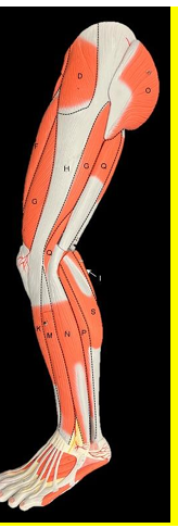
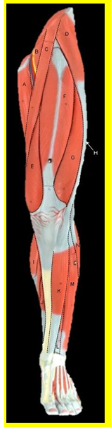
Label D-S. (not all letters included alphabetically)
D: tensor fasciae latae
F: rectus femoris
G: vastus lateralis
H: iliotibial tract
I: gastrocnemius - medial head
K: tibialis anterior
M: extensor digitorum longus
N: fibularis longus
O: gluteus maximus
P: soleus
Q: biceps femoris
S: gastrocnemius - lateral head
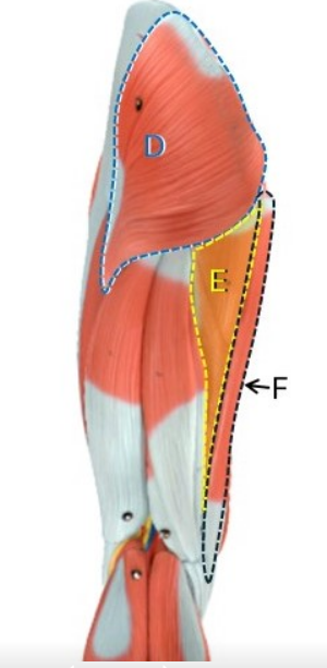
Label D-F.
D: gluteus maximus
E: adductor magnus
F: gracilis