Anatomy 17 | Foregut
1/50
Earn XP
Description and Tags
https://youtu.be/aBN15euQuXQ
Name | Mastery | Learn | Test | Matching | Spaced |
|---|
No study sessions yet.
51 Terms
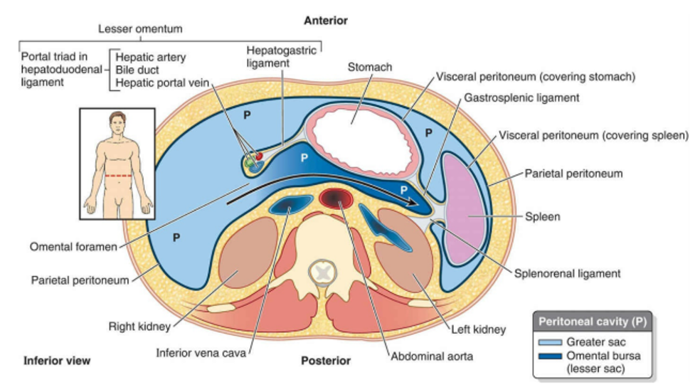
What is the peritoneal cavity?
Space between parietal and visceral layers of peritoneum
What is the mesentery?
A double layer of peritoneum that connects organs to the abdominal wall and allows blood vessels, nerves, and lymphatics to pass through.
Video
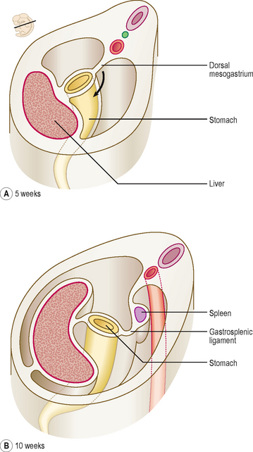
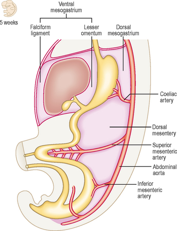
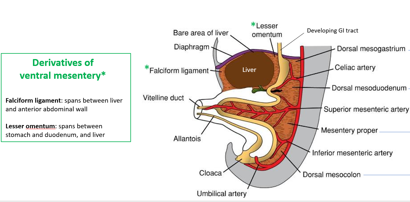
During development, the mesentery is split into two parts. What are they?
Ventral and dorsal parts
During development, what does the dorsal mesentery develop into?
Different structures that support and connect digestive organs:
Greater omentum: Supports the stomach.
Dorsal mesoduodenum: Supports the duodenum.
Mesentery proper: Supports the jejunum and ileum (small intestine).
Dorsal mesocolon: Supports the large intestine.
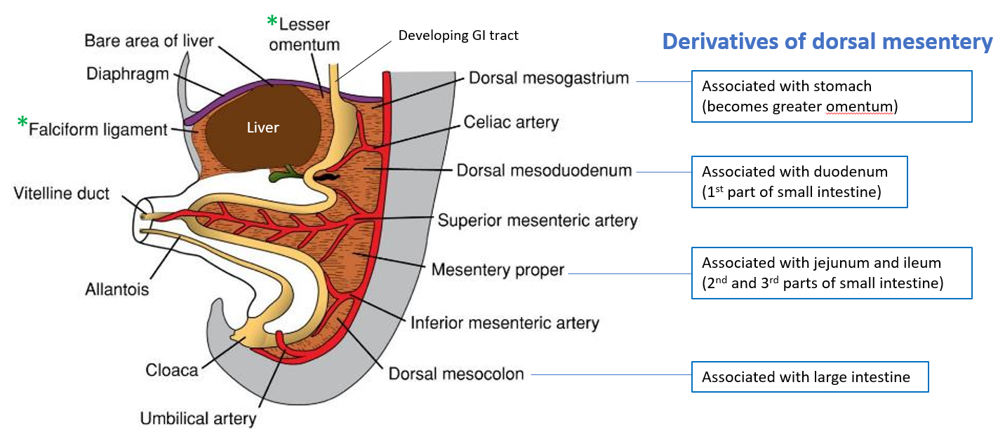
During development, what does the ventral mesentery develop into?
Falciform ligament: Connects the liver to the anterior abdominal wall.
Lesser omentum: Connects the stomach and duodenum to the liver.
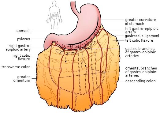
What is the greater omentum?
a large, apron-like fold of peritoneum that hangs from the greater curvature of the stomach and covers the intestines.
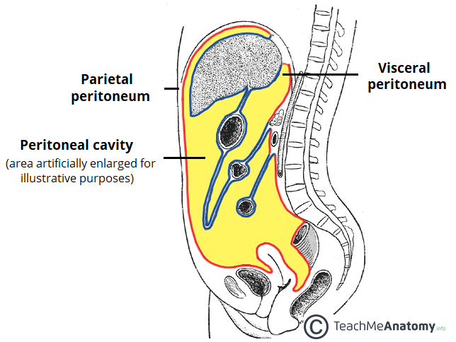
What is the peritoneal cavity?
The space between the parietal and visceral peritoneum, filled with a thin fluid that allows smooth movement of abdominal organs.
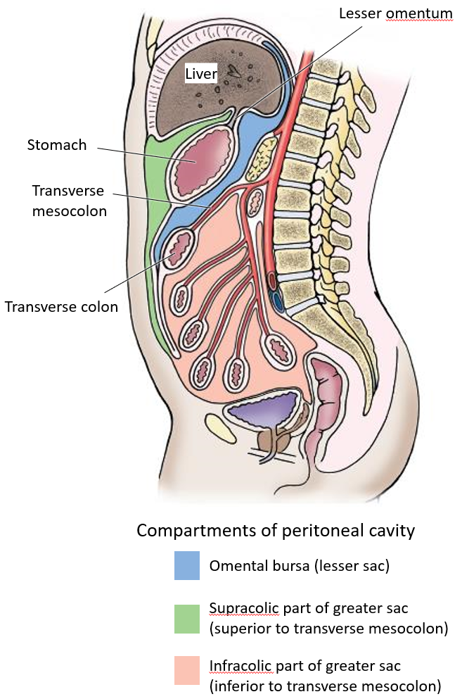
The peritoneal cavity is divided into two sacs. What are they called?
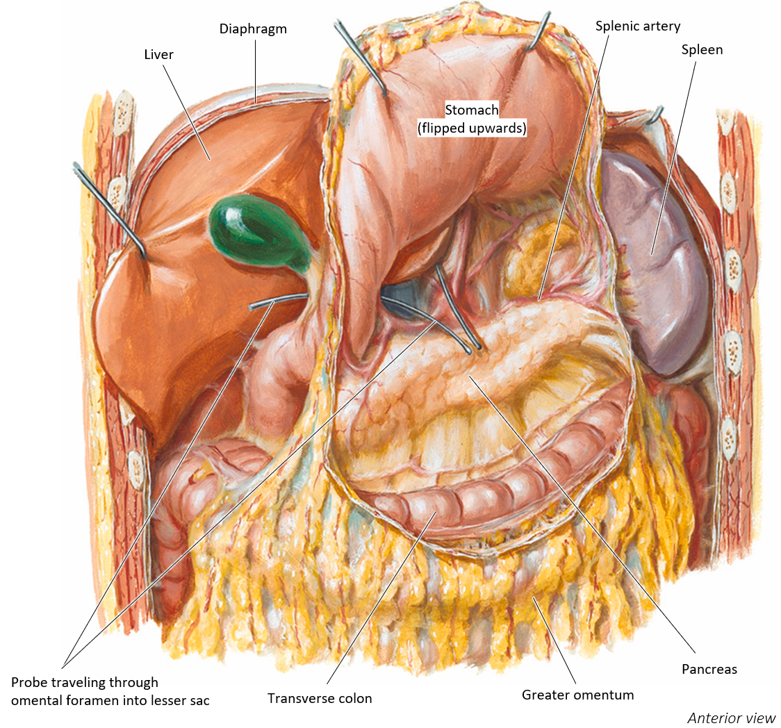
Lesser sac (omental bursa): A small extension behind the stomach, accessed through the omental foramen.
Greater sac: The main part of the peritoneal cavity.

The peritoneal cavity is divided into two sacs (lesser and greater sac). The greater sac is further divided into which compartments?
Supracolic compartment (above the transverse mesocolon).
Infracolic compartment (below the transverse mesocolon).
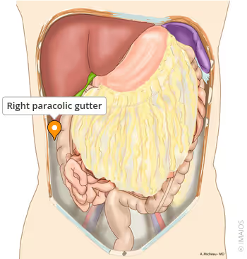
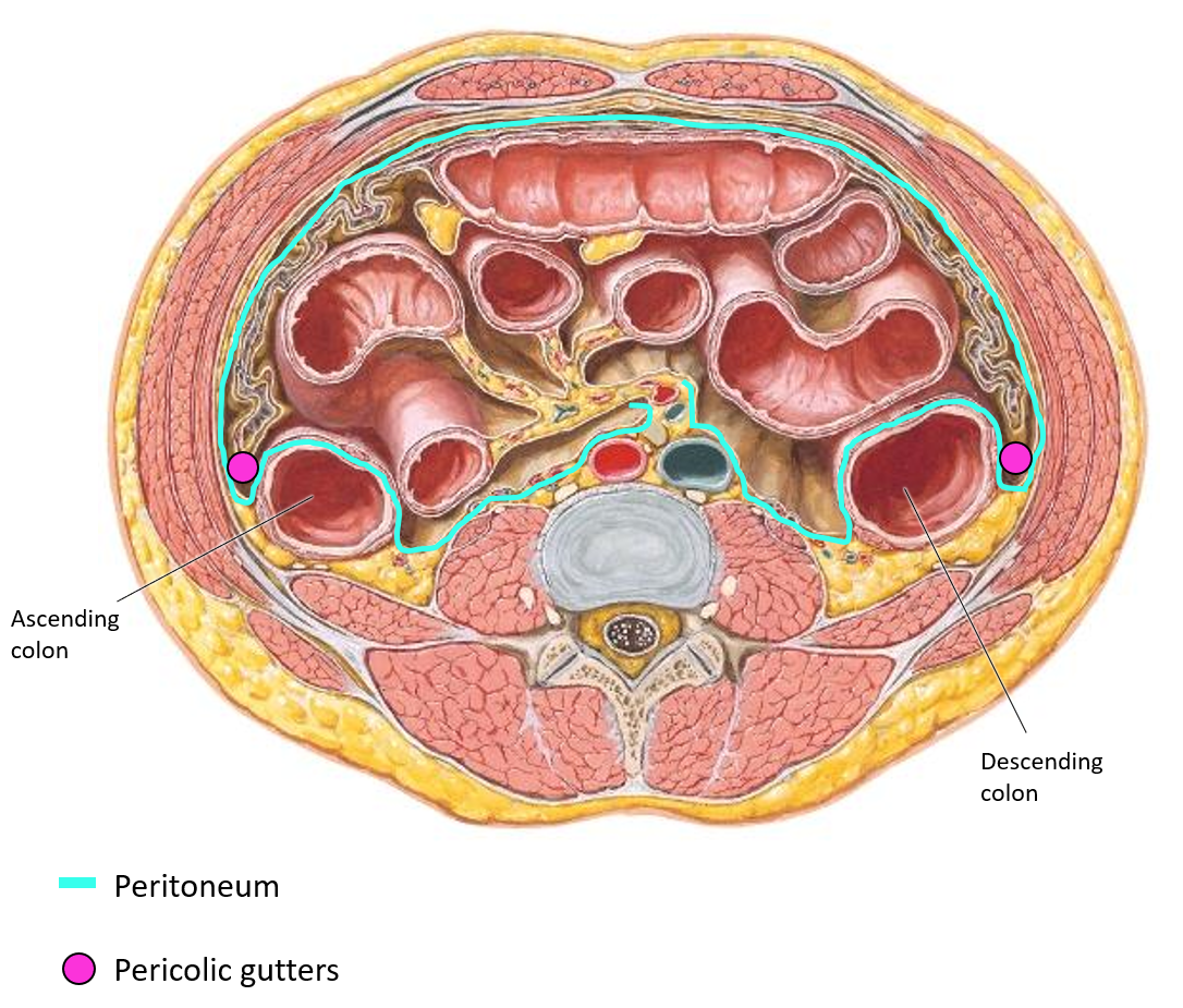
What are paracolic gutters?
Trough-like spaces between the colon and the abdominal wall, formed by the visceral peritoneum.
They allow fluid movement within the abdominal cavity.
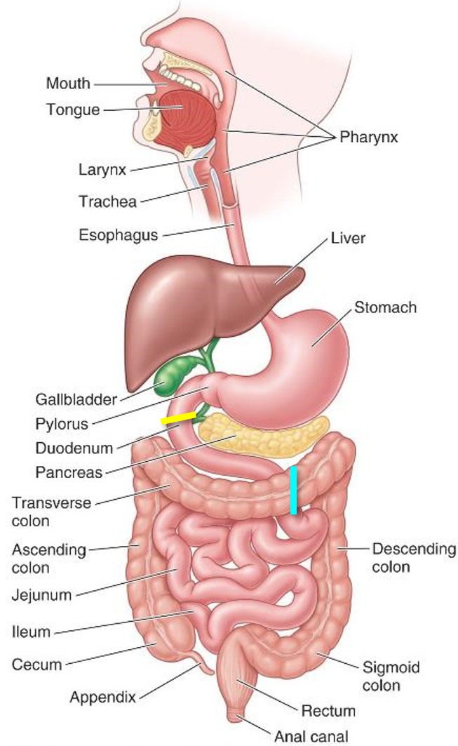
What is the GI tract/alimentary canal?
A hollow muscular tube responsible for:
Breaking down food (mechanically and chemically).
Absorbing nutrients.
Excreting waste.
The GI tract/alimentary canal consists of which structures?
Esophagus, stomach, small intestine (duodenum, jejunum, ileum).
Large intestine (cecum, ascending, transverse, descending colon, rectum, anal canal).
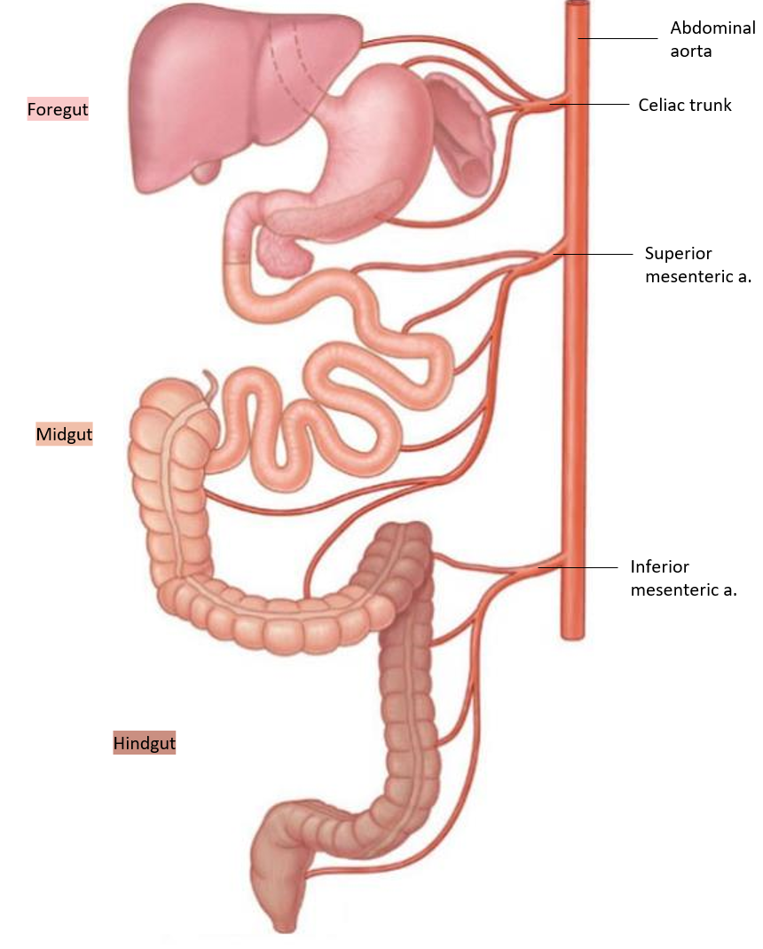
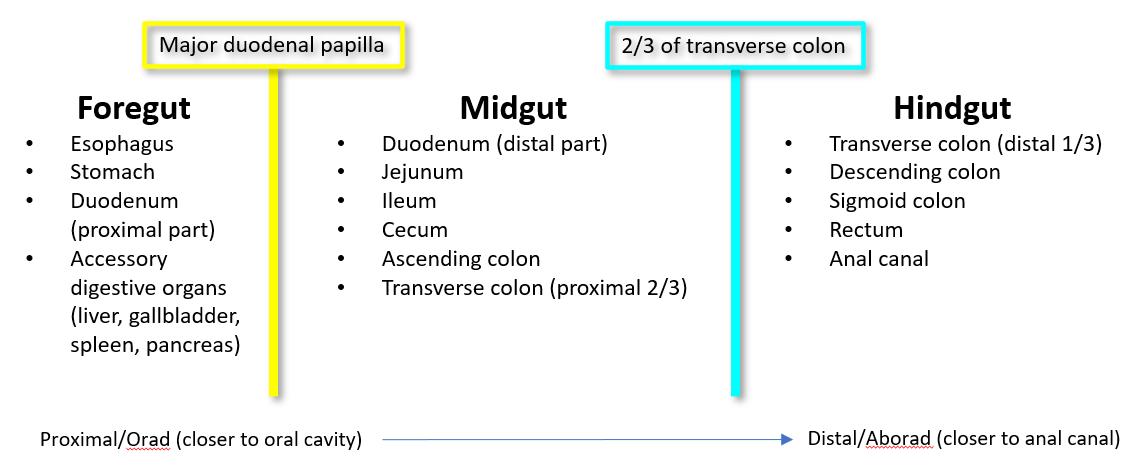
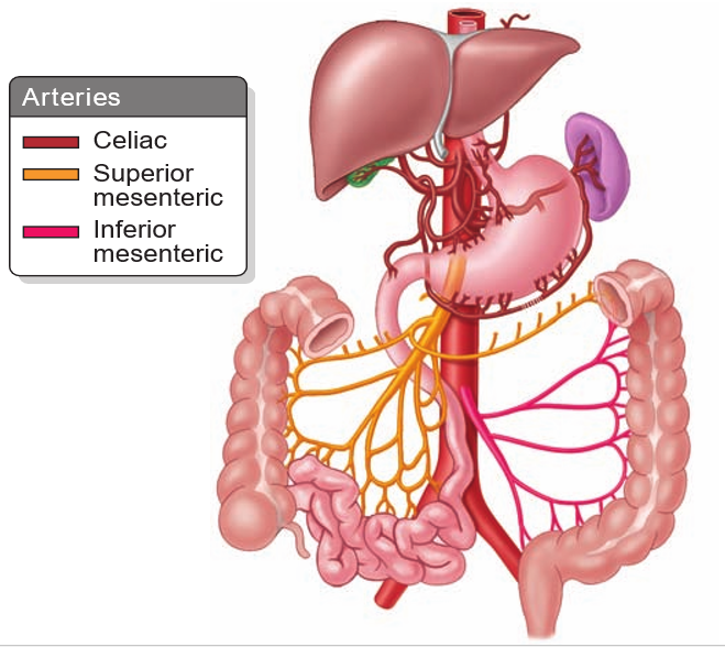
How is the GI tract divided?
It is divided into foregut, midgut, and hindgut based on blood supply.
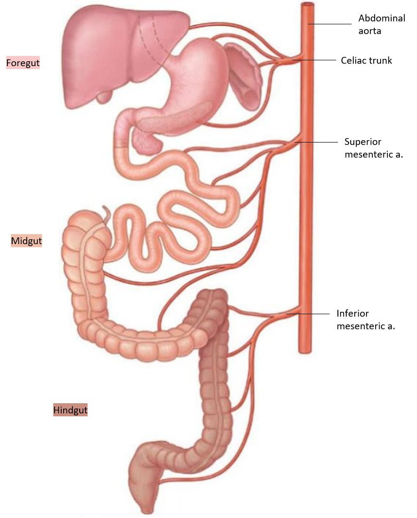
How does the GI tract get its blood supply?
From three major unpaired branches of the abdominal aorta
Celiac trunk: Supplies foregut structures
Superior mesenteric artery: Supplies midgut structures
Inferior mesenteric artery: Supplies hindgut structures
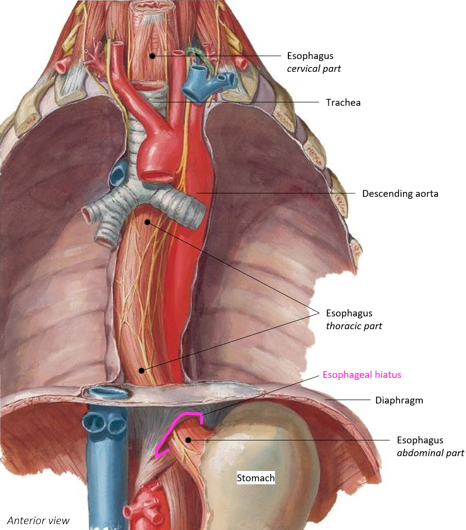
Through what opening does the esophagus enter the abdominal cavity?
It passes through the esophageal hiatus of the diaphragm to enter the abdominal cavity
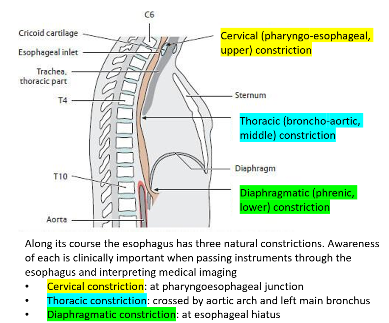
Where are the three natural constrictions of the esophagus located?
Cervical constriction:at the pharyngoesophageal junction.
Thoracic constriction: where the esophagus is crossed by the aortic arch and left main bronchus.
Diaphragmatic constriction: at the esophageal hiatus of the diaphragm.
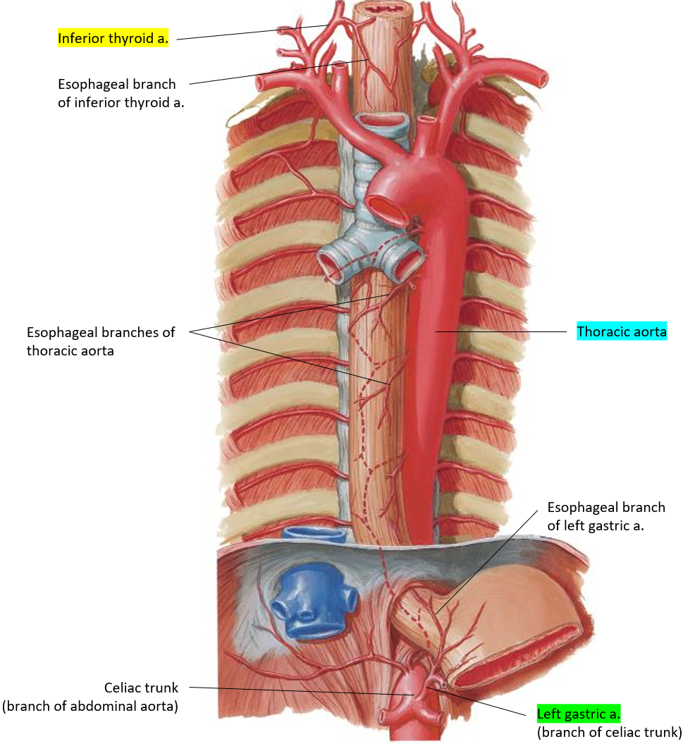
What arteries supply blood to the esophagus?
Cervical part: esophageal branches of the inferior thyroid artery.
Thoracic part: esophageal branches of the thoracic aorta.
Abdominal part: esophageal branch of the left gastric artery.
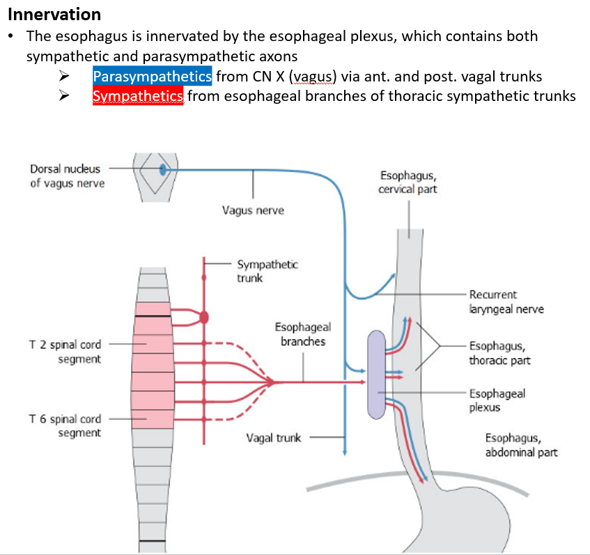
How is the esophagus innervated?
Parasympathetic innervation from the vagus nerve (CN X).
Sympathetic innervation from the esophageal branches of thoracic sympathetic trunks.
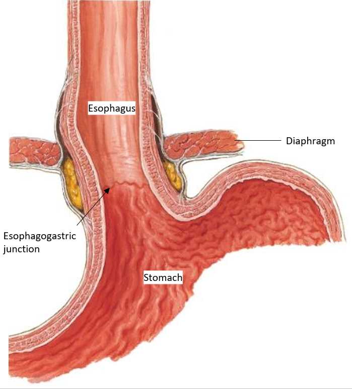
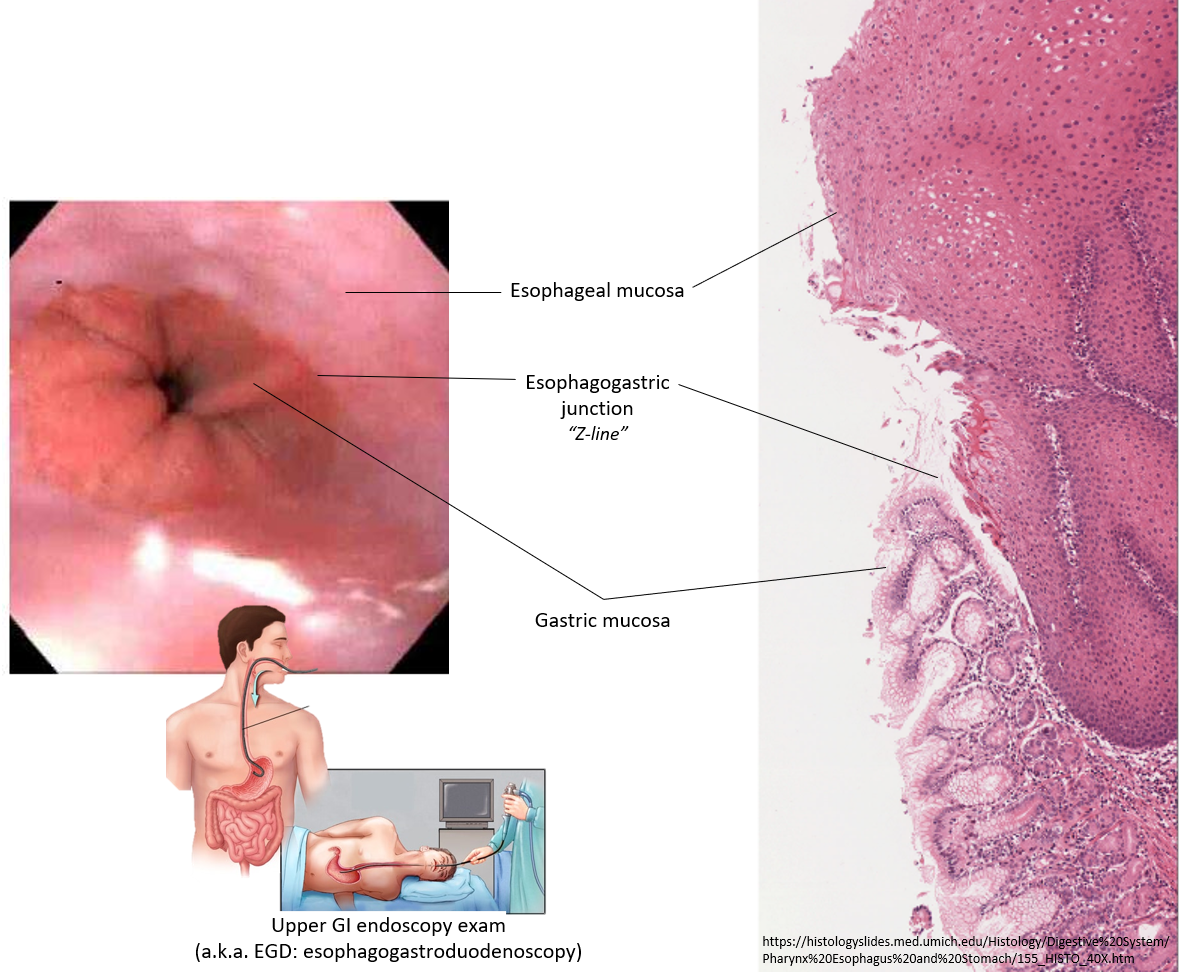
What is the esophagogastric junction?
Where the esophagus connects to the stomach.
The Z-line marks the spot where the lining of the esophagus changes to the stomach lining.
What part of the stomach is the entry point where the esophagus meets the stomach.
Cardia

What part of the stomach is the upper part that stores extra food, liquid, or gas.
Fundus
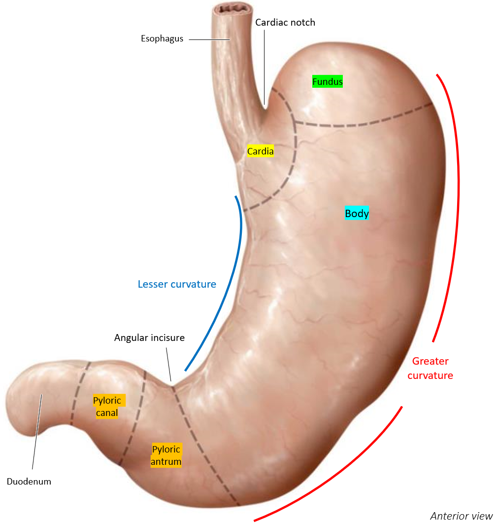
What part of the stomach is the largest section where digestion mainly happens.
Body

What part of the stomach is the funnel-shaped end that leads to the small intestine.
Pylorus

What are gastric folds (rugae)?
Ridges in the mucosal lining of the stomach that give it a wrinkled appearance when empty and flatten out as the stomach fills.
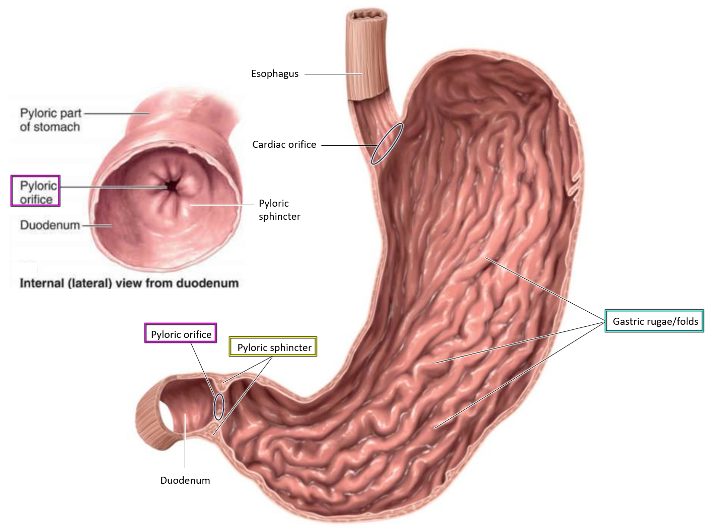
What is the function of the pyloric sphincter?
Controls the passage of food from the stomach to the duodenum by staying contracted and only relaxing during emptying.

Where does food pass after the pyloric orifice?
into the duodenum, the first part of the small intestine.

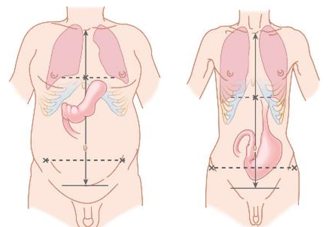
Describe the stomach’s position in the body
posterior to the anterior abdominal wall and the left lobe of the liver.
anterior to the lesser sac, pancreas, splenic artery, and spleen.
inferior to the diaphragm.
superior to the transverse colon and greater omentum.
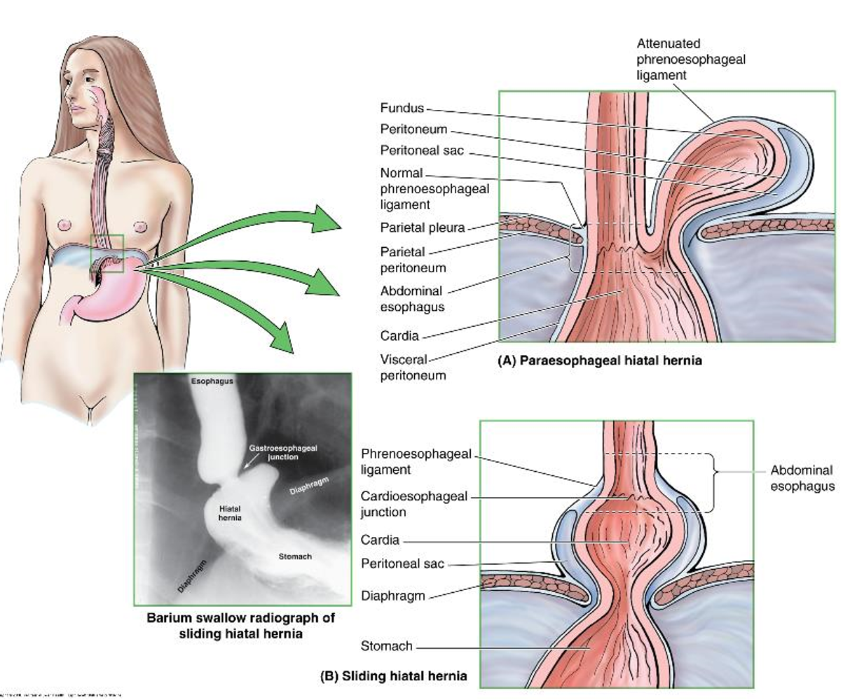
What is a hiatal hernia?
Occurs when part of the stomach moves through the esophageal hiatus of the diaphragm into the chest.
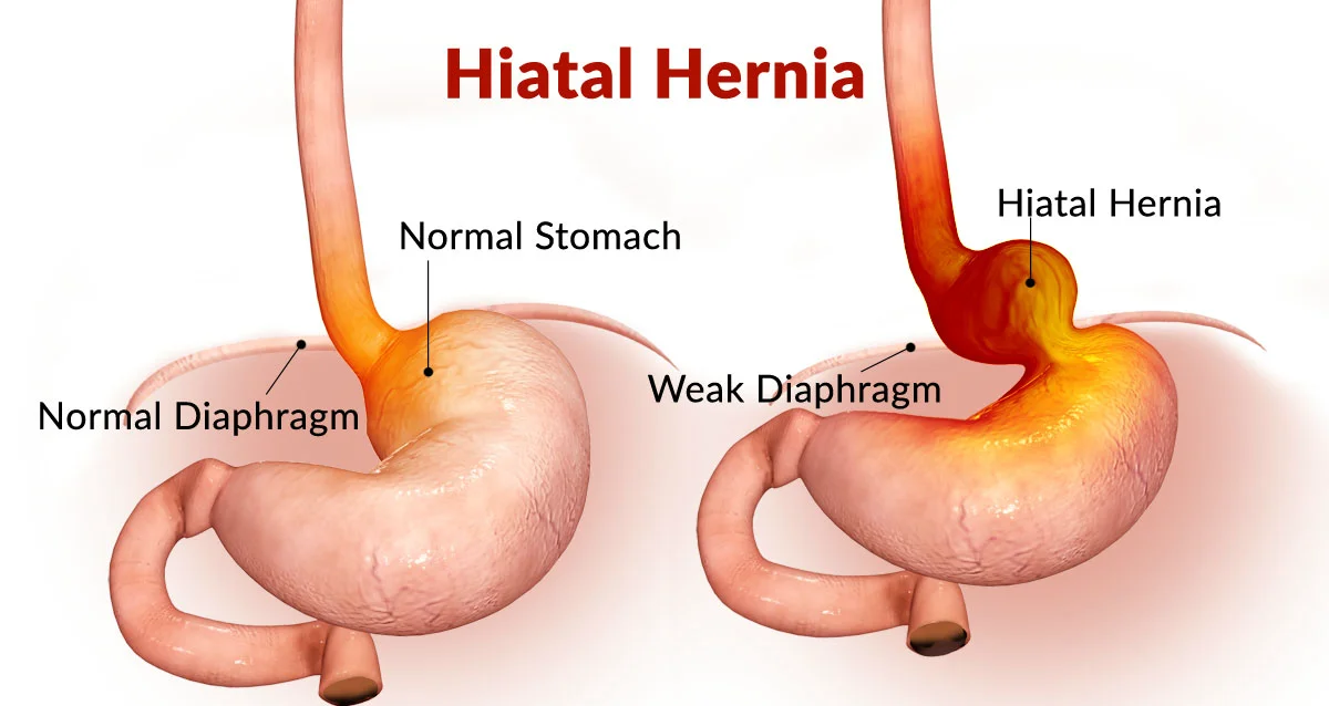
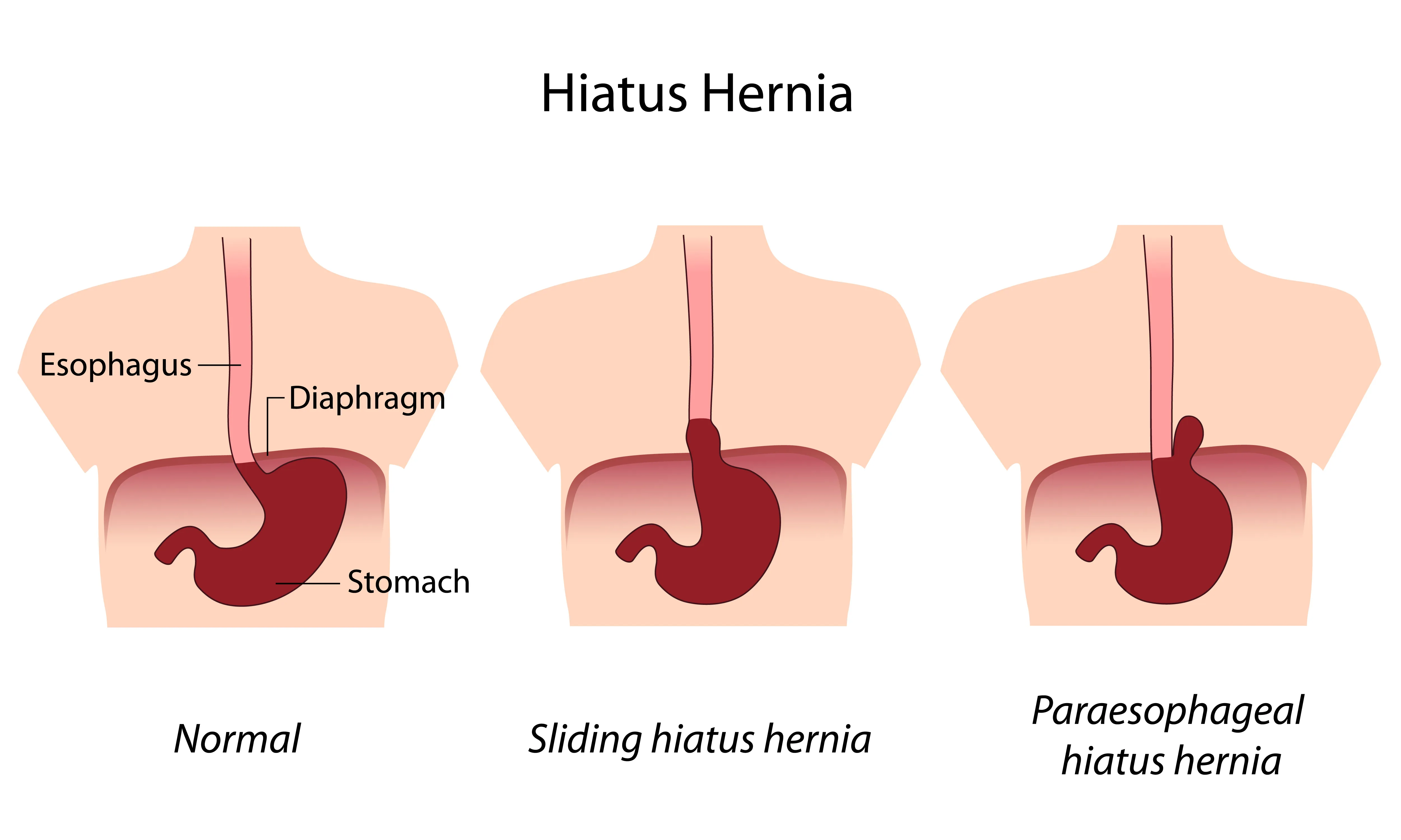
What is the difference between a paraesophageal and a sliding hiatal hernia?
A paraesophageal hiatal hernia involves the stomach's fundus moving up while the cardia stays in place and usually does not cause reflux.
A sliding hiatal hernia involves both the cardia and part of the esophagus moving up, often leading to acid reflux.

What is the duodenum?
the first part of the small intestine, located just beyond the pyloric sphincter, and is C-shaped as it wraps around the head of the pancreas.

What are the four parts of the duodenum?
Superior (1st) part
Descending (2nd) part
Horizontal (3rd) part
Ascending (4th) part
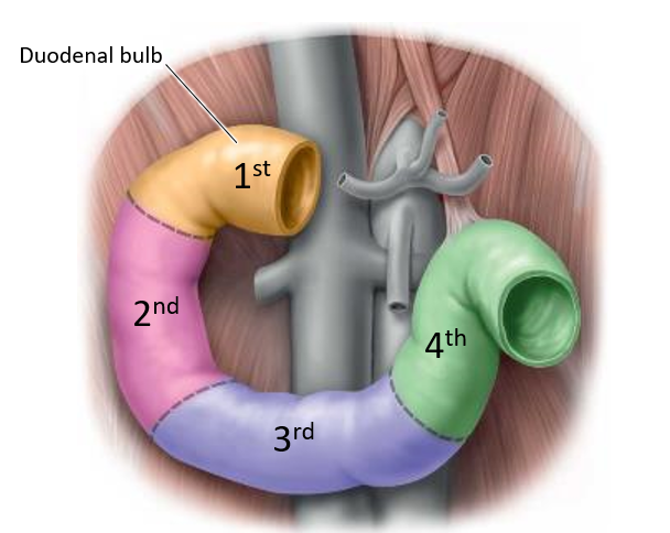
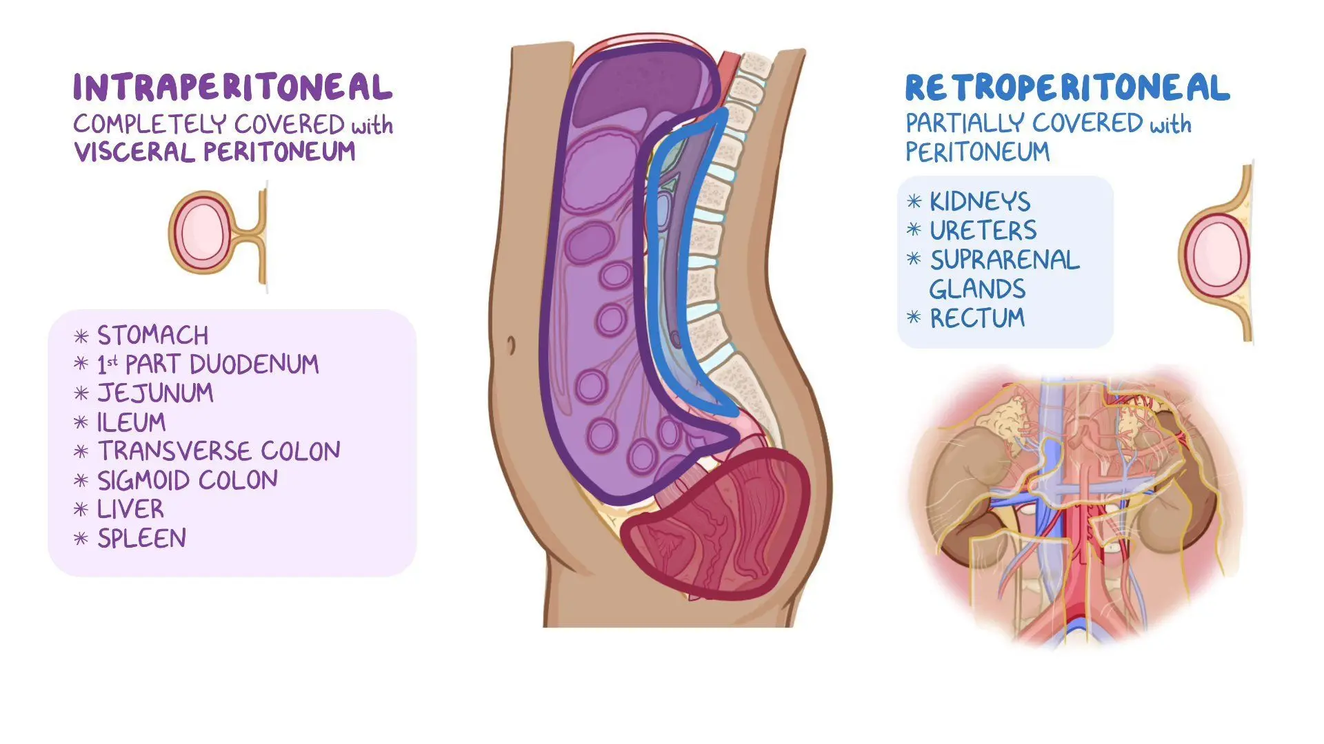
Which part of the duodenum is intraperitoneal?
Only the duodenal bulb (proximal portion of the 1st part) is intraperitoneal*; the rest is secondarily retroperitoneal.
*a structure is completely surrounded by the visceral peritoneum and is suspended within the peritoneal cavity by a mesentery.

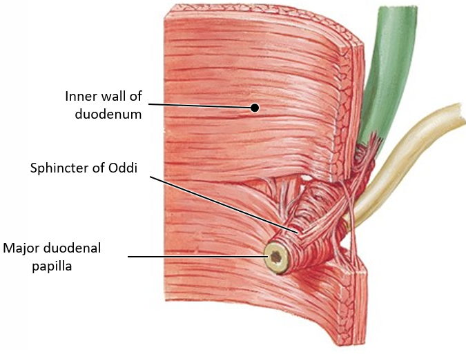
What is the major duodenal papilla?
An opening in the 2nd part of the duodenum where bile and pancreatic enzymes enter the small intestine for digestion.
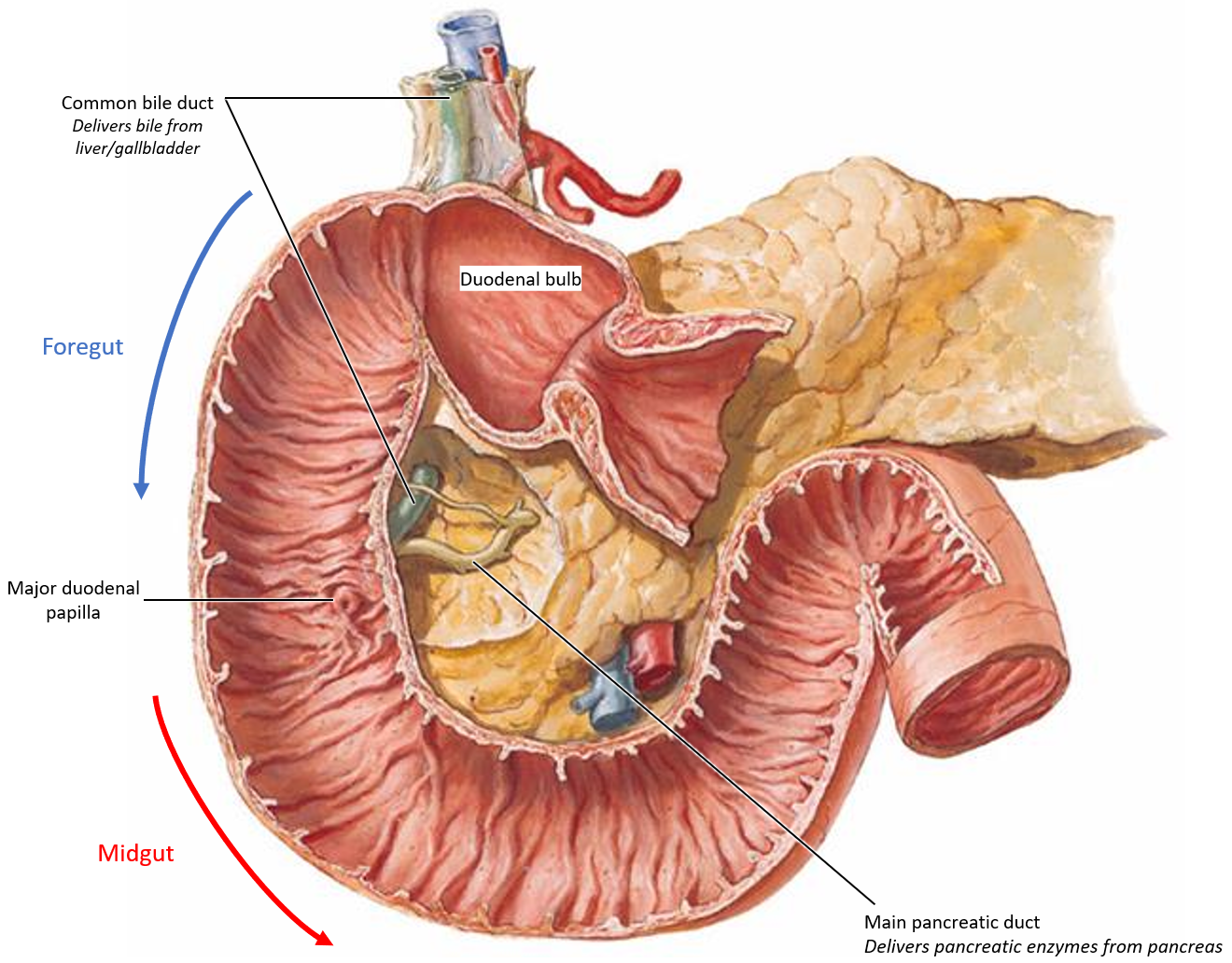
What controls the release of bile and pancreatic enzymes?
The sphincter of Oddi, a circular smooth muscle, regulates the flow of bile and pancreatic enzymes into the duodenum.
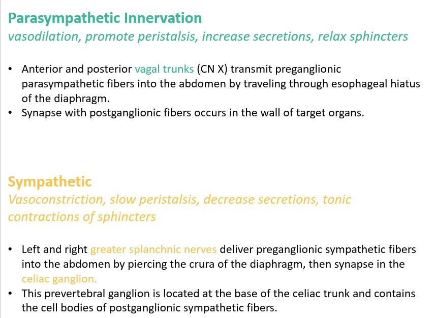
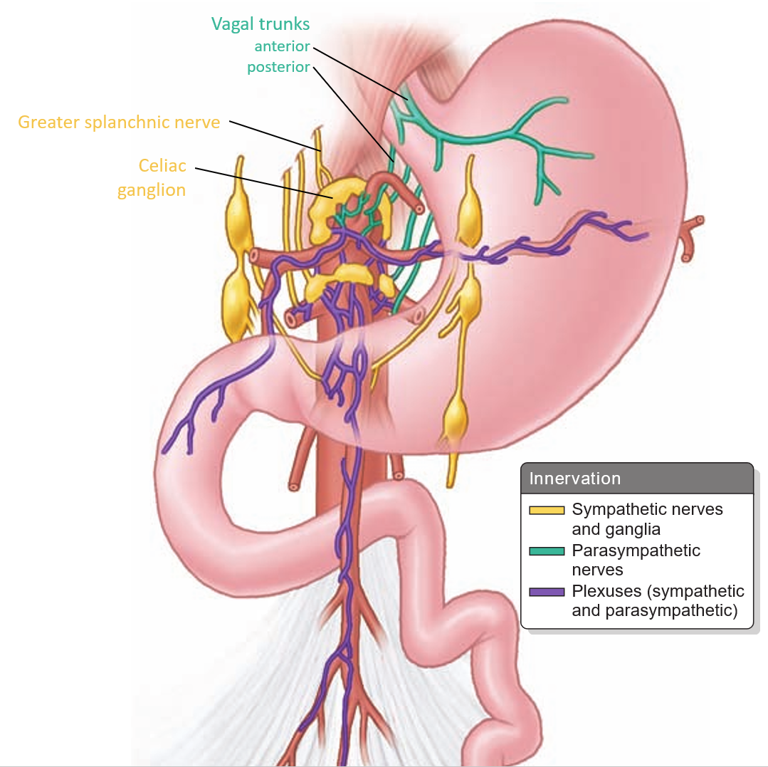
The foregut is innervated by which nerves? What are their functions?
Parasympathetic Innervation (via vagus nerve):
Promotes digestion by increasing peristalsis, secretions, and relaxing sphincters.
Sympathetic Innervation (via greater splanchnic nerves):
Slows digestion by causing vasoconstriction, reducing secretions, and contracting sphincters.
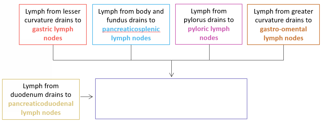
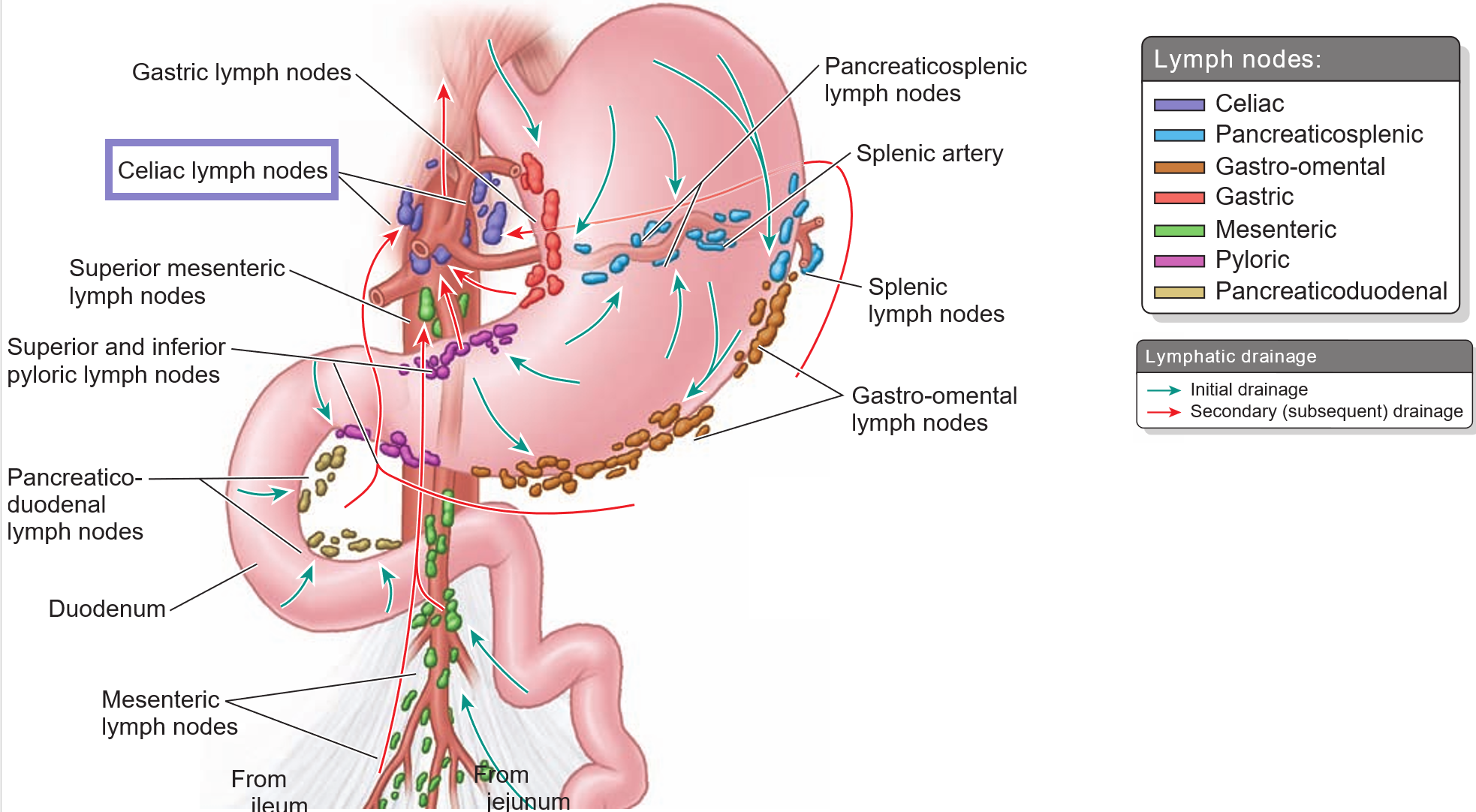
Lymph from different parts of the stomach and duodenum drains into specific lymph nodes (i.e. gastric, pyloric, pancreaticoduodenal LNs, etc.). Where do all these lymph nodes eventually drain into?
Celiac lymph nodes, which are located at the base of the celiac trunk and receive lymph from foregut structures.
What is the celiac trunk?
A short, unpaired artery that arises from the abdominal aorta at the T12 vertebral level and supplies blood to the foregut structures.
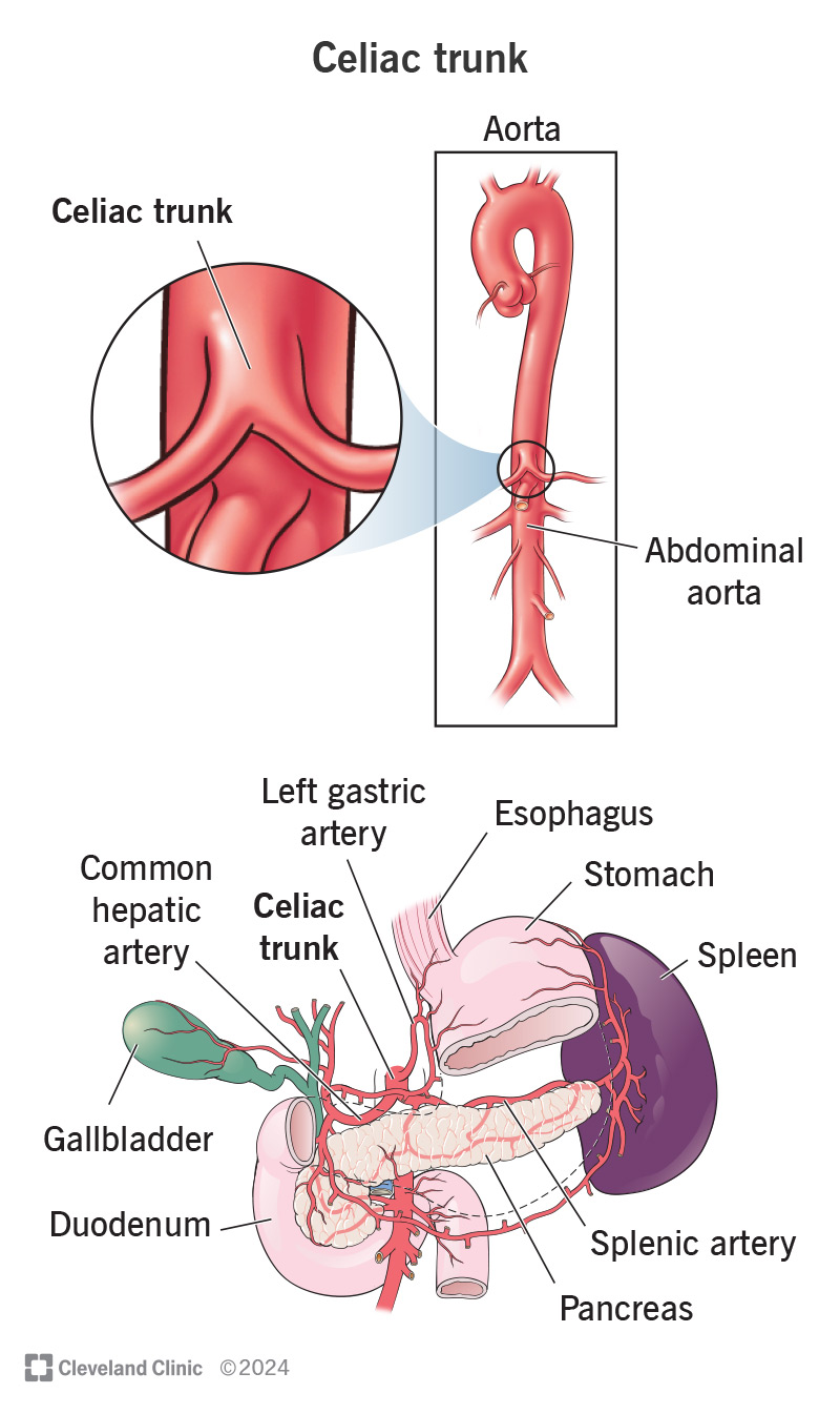
Which three main branches arise from the celiac trunk?
Left gastric artery (supplies the left part of the lesser curvature of the stomach).
Splenic artery (supplies the spleen, stomach, and pancreas).
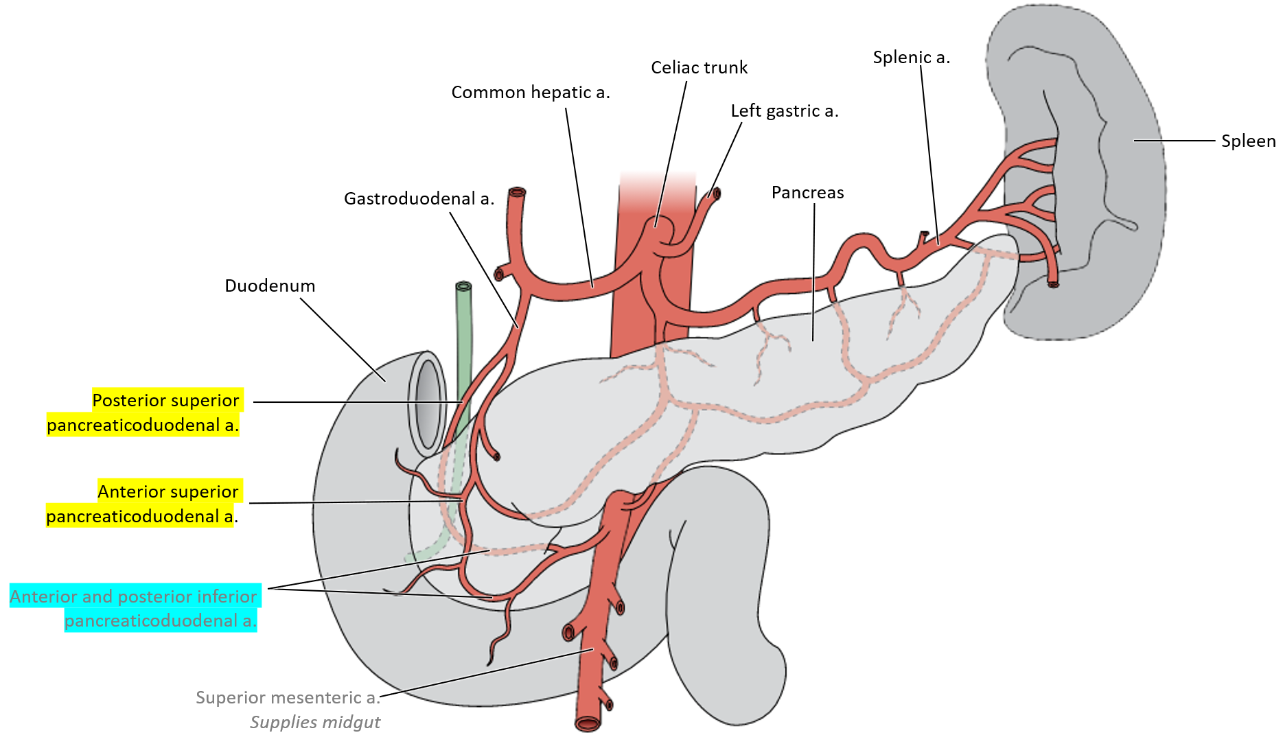
Common hepatic artery (divides into proper hepatic and gastroduodenal arteries).
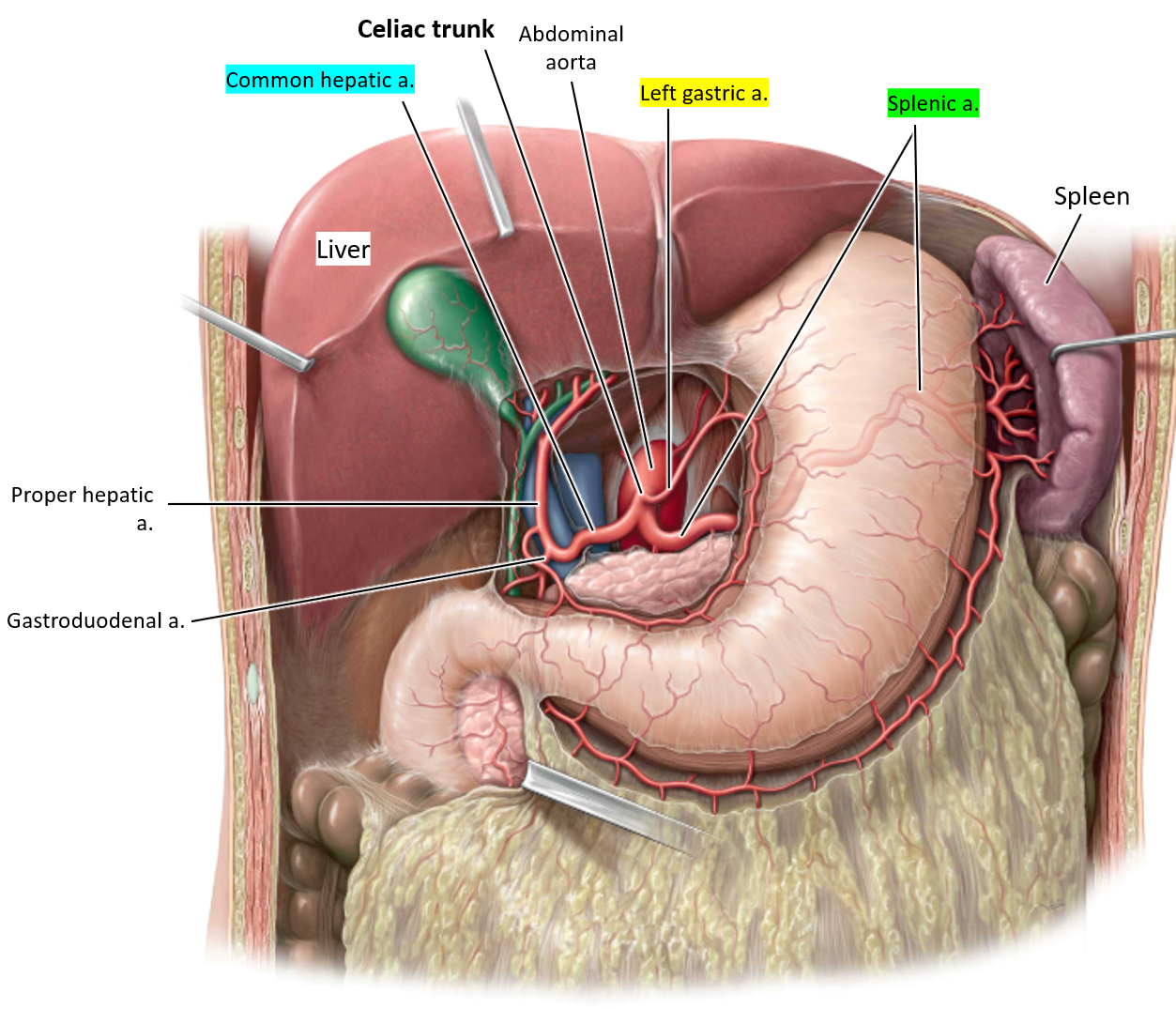
What does the common hepatic artery supply?
It travels toward the liver and divides into the proper hepatic artery (which supplies the liver) and the gastroduodenal artery (which supplies parts of the stomach and duodenum).


What supplies blood to the lesser curvature of the stomach?
The left and right gastric arteries
(right gastric a. will always go towards the right aspect of the curvature and vice-versa)
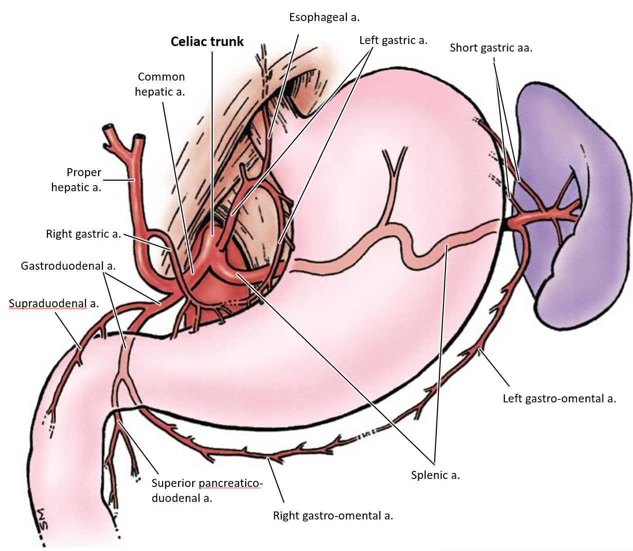
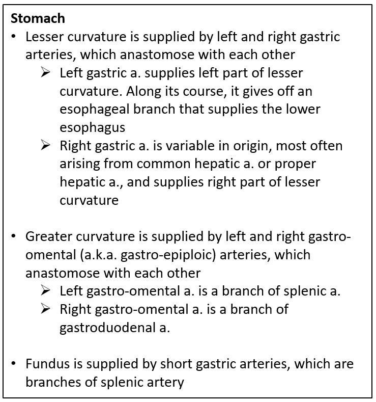
What supplies blood to the greater curvature of the stomach?
The left and right gastro-omental arteries

What supplies blood to the greater curvature of the stomach?
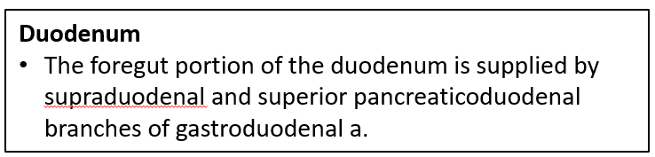
supraduodenal and superior pancreaticoduodenal branches of the gastroduodenal a.

What arteries supply blood to the head of the pancreas?
The superior pancreaticoduodenal arteries and the inferior pancreaticoduodenal arteries

Which major arteries form an anastomosis around the head of the pancreas?
The celiac trunk and the superior mesenteric artery
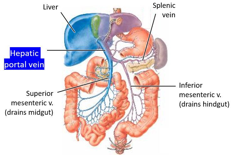
What vein collects blood from the foregut and transports it to the liver?
The hepatic portal vein collects blood from the foregut and delivers it to the liver for processing.

Where does venous blood go after being processed by the liver?
It enters the systemic circulation via the hepatic veins, which drain into the inferior vena cava.


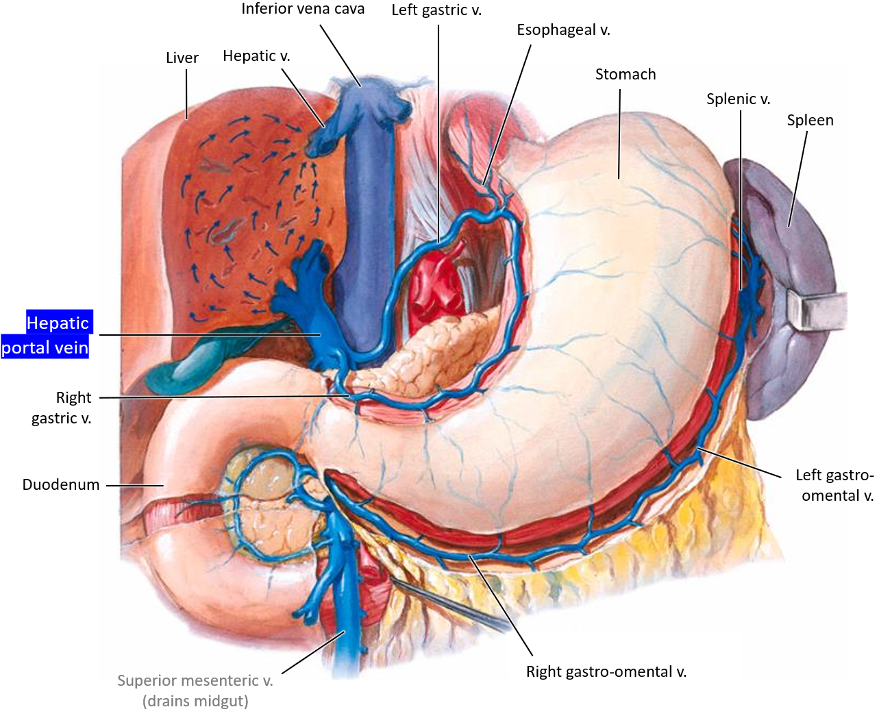
What happens when blood backs up (portal hypertension) in the hepatic portal vein and esophageal veins due to cirrhosis?
Esophageal varices


Why do esophageal veins become swollen in portal hypertension?
Blood is redirected through the left gastric vein into the esophageal veins, which cannot handle the extra volume, leading to swelling.
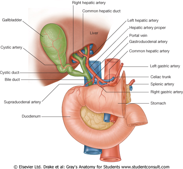
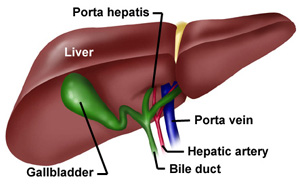
What is the portal triad?
A group of three key structures located in the hepatoduodenal ligament of the lesser omentum.

What structures does the portal triad consist of?
The proper hepatic artery (carries oxygen-rich blood to the liver)
The hepatic portal vein (brings nutrient-rich blood from the intestines)
The common bile duct (transports bile from the liver to the duodenum)
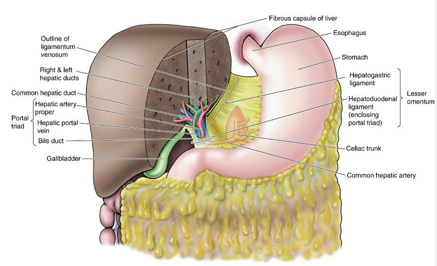
The portal triad enter and exit the liver through which opening?
The porta hepatis, which acts as the hilum of the liver.