lymphatic system anatomy
1/81
There's no tags or description
Looks like no tags are added yet.
Name | Mastery | Learn | Test | Matching | Spaced |
|---|
No study sessions yet.
82 Terms
Lymph
Interstitial fluid contained inside lymphatic cells, does not go out
Lymphatic capillaries
small closed-end vessels that collect and carry away excess fluid, in spaces between tissue cells, intertwined with blood capillaries
lymphatic vessels
similar to veins but with thinner walls, not influenced by pumping of the heart
lymphatic valves
prevent backflow of lymph, more quantity than veins to ensure undirectional flow
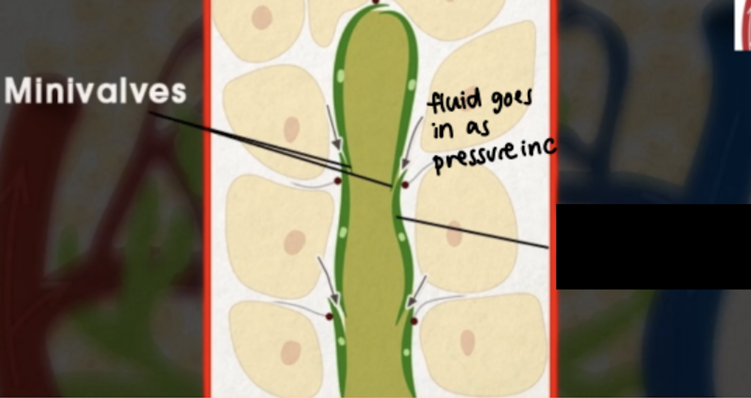
Minivalves
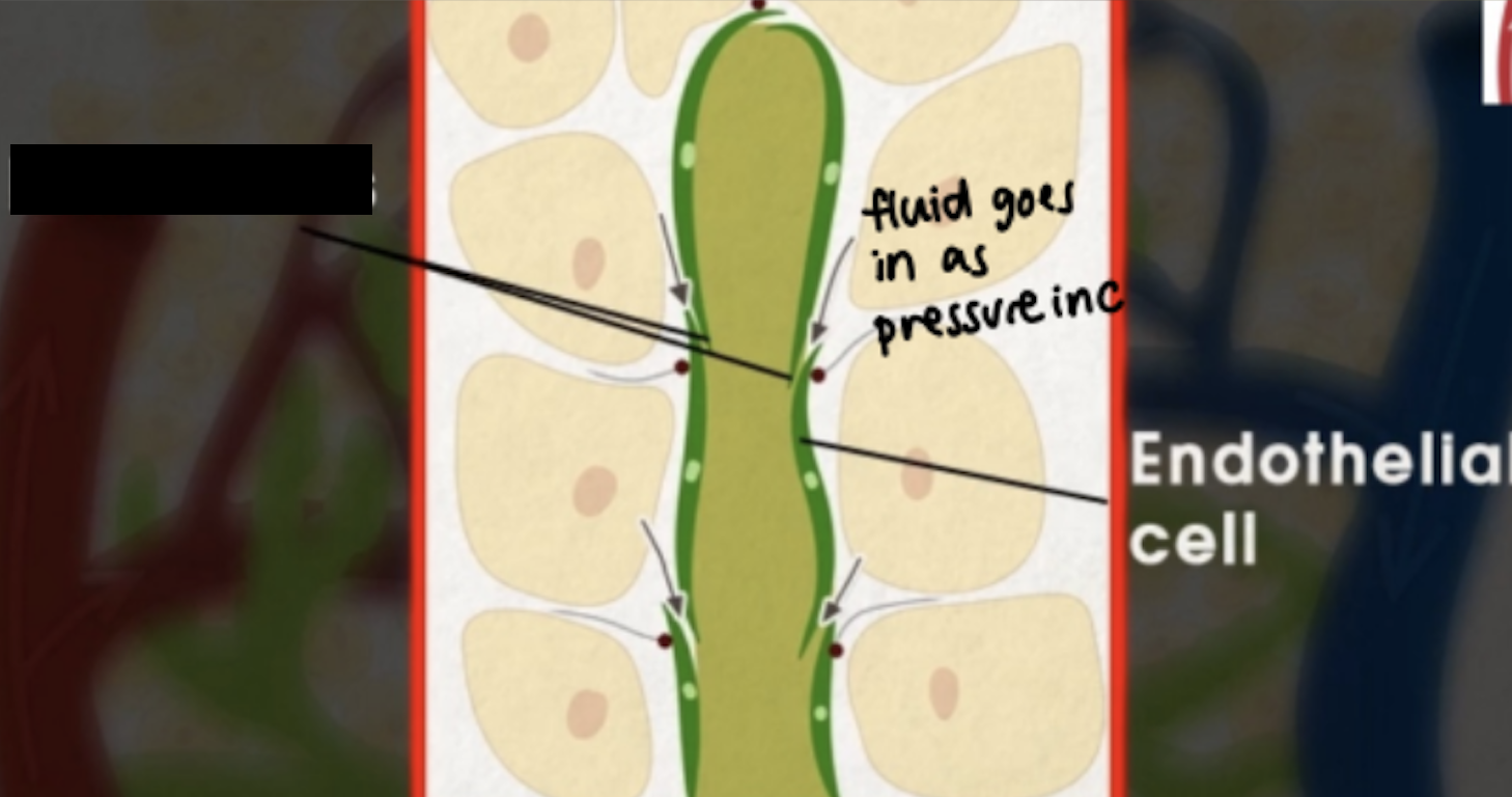
Endothelial cells
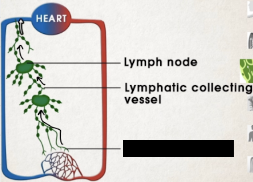
lymphatic collecting vessels
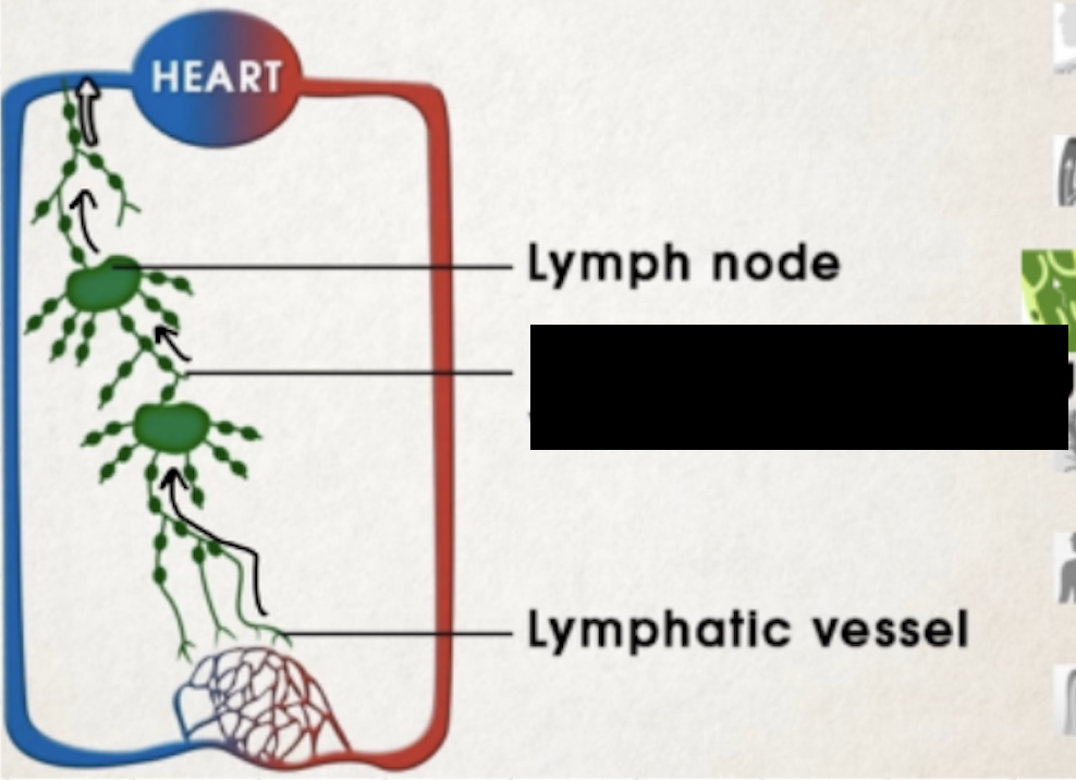
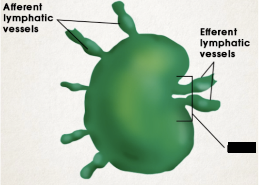
lymph node
Widely distributed in the body in clusters and groups
There are superficial and deep lymph nodes
Small and encapsulated bean-shaped organs about 1mm-2cm in length
Along lymphatic vessels, help filter out foreign substances and killing bacteria
Lymph enters via afferent lymphatic vessels and exit through 1-2 efferent lymphatic vessels at the hilum
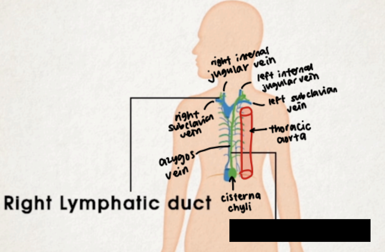
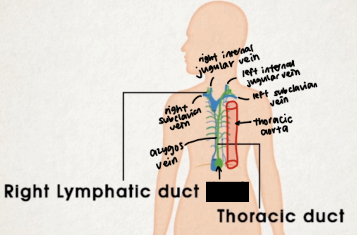
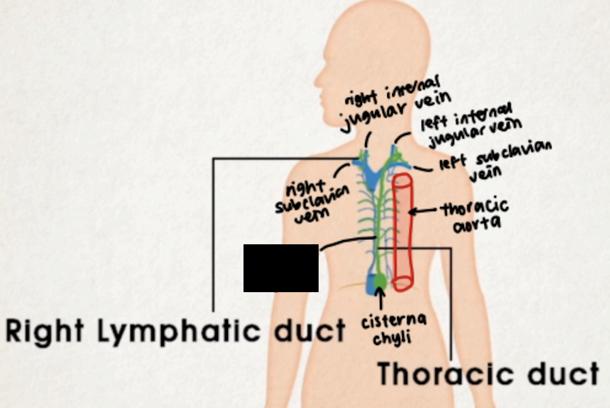
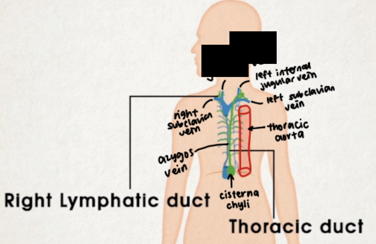
right lymphatic duct
short duct (1 cm)
drains from right upper part of body
accounts for ¼ of lymph drainage
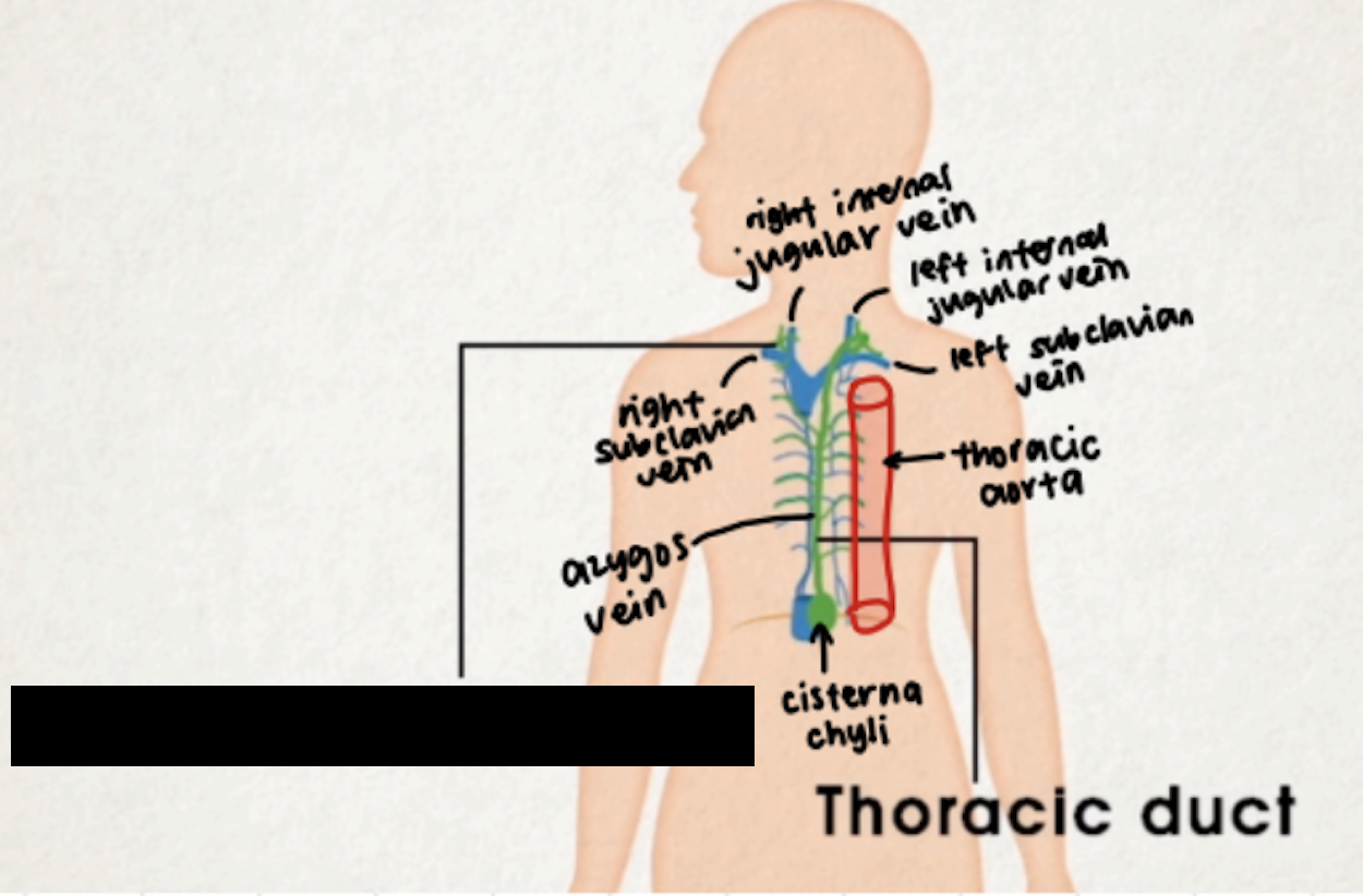
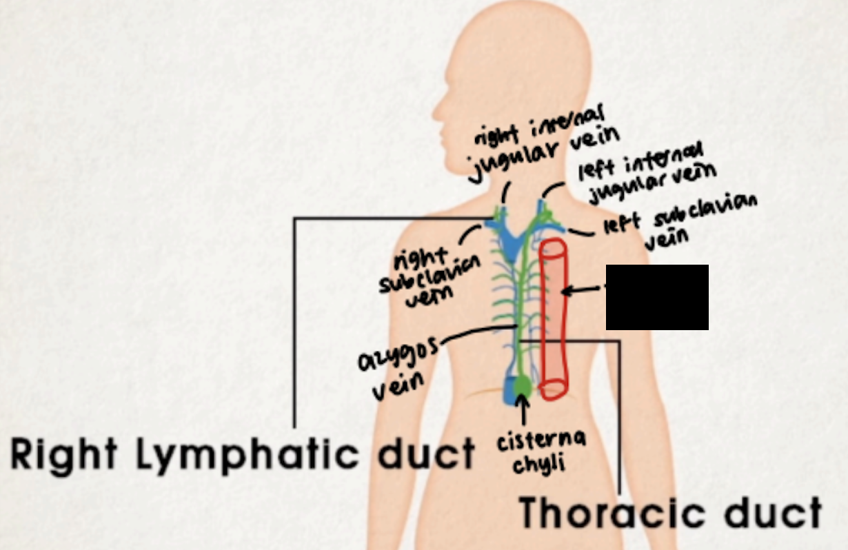
thoracic duct
long duct
between thoracic aorta and argygos vein
drains from right lower part and all of left part of body
cisterna chyli at 1st and 2nd lumbar level
cisterna chyli
azygos vein
thoracic aorta
right subclavian vein
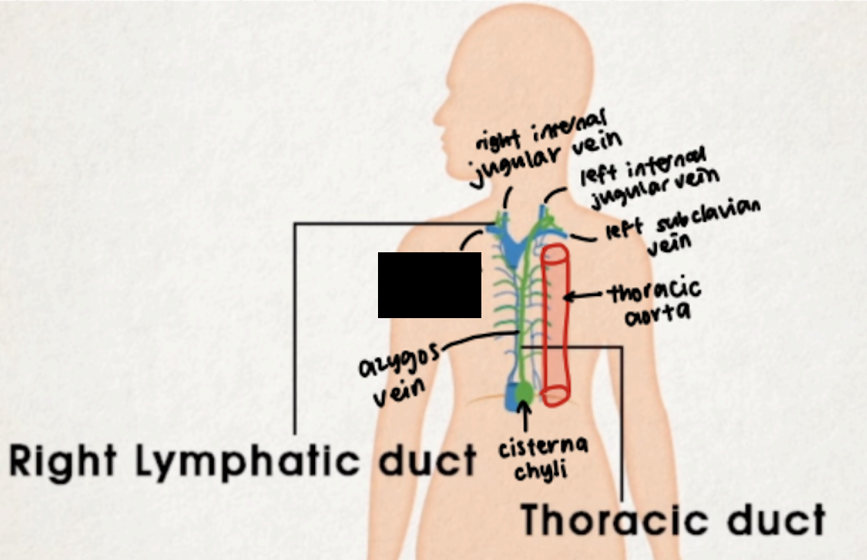
left subclavian vein
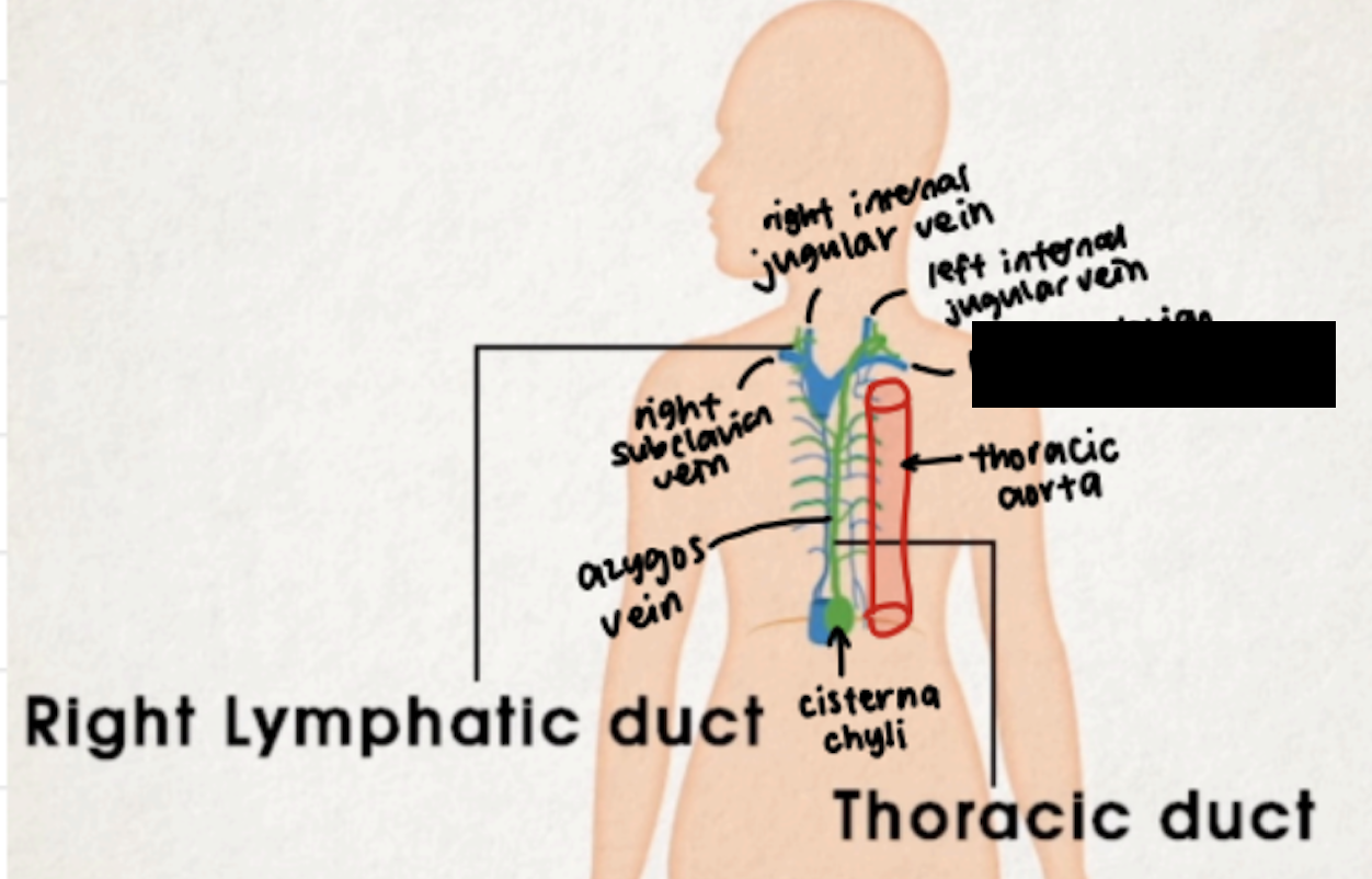
left internal jugular vein
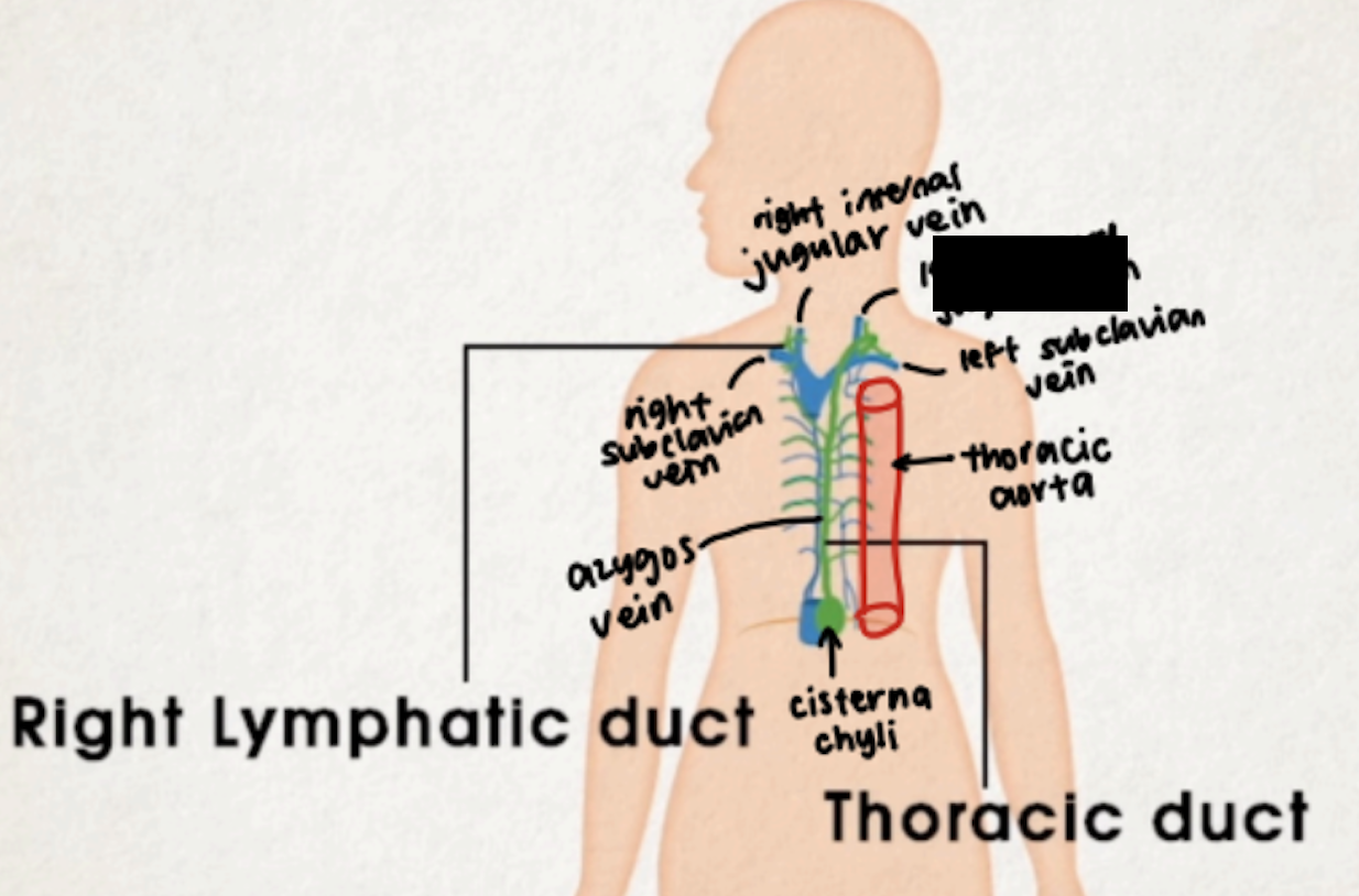
right internal jugular vein
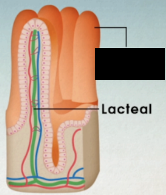
lacteals
specialized lymphatic vessels in small intestine
important for absorption/transport of lipids/chylomicrons
empties into nearby lymph nodes
if lymph in lacteals are fatty enough it turns white
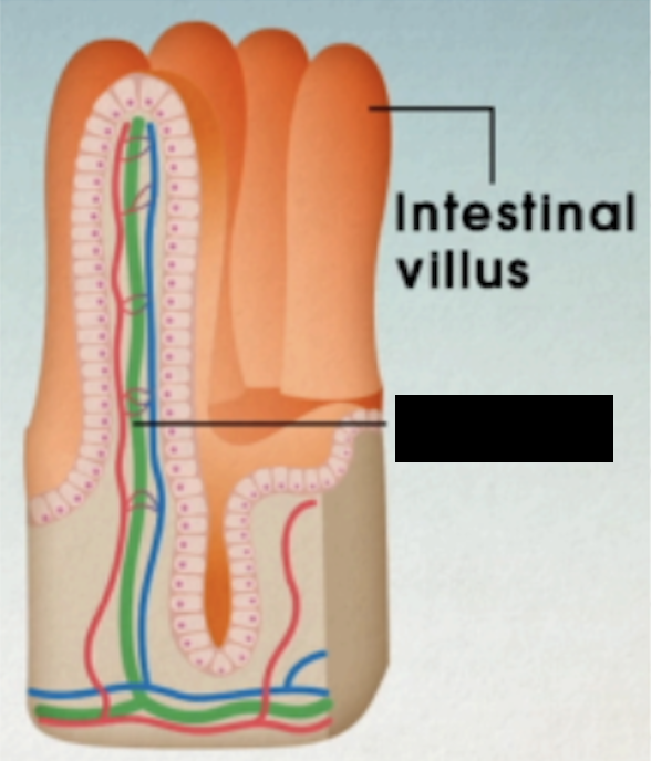
intestinal villus
primary section
red bone marrow and thymus
maturation of lymphocytes
B cells mature in bone marrow
T cells mature in thymus
secondary section
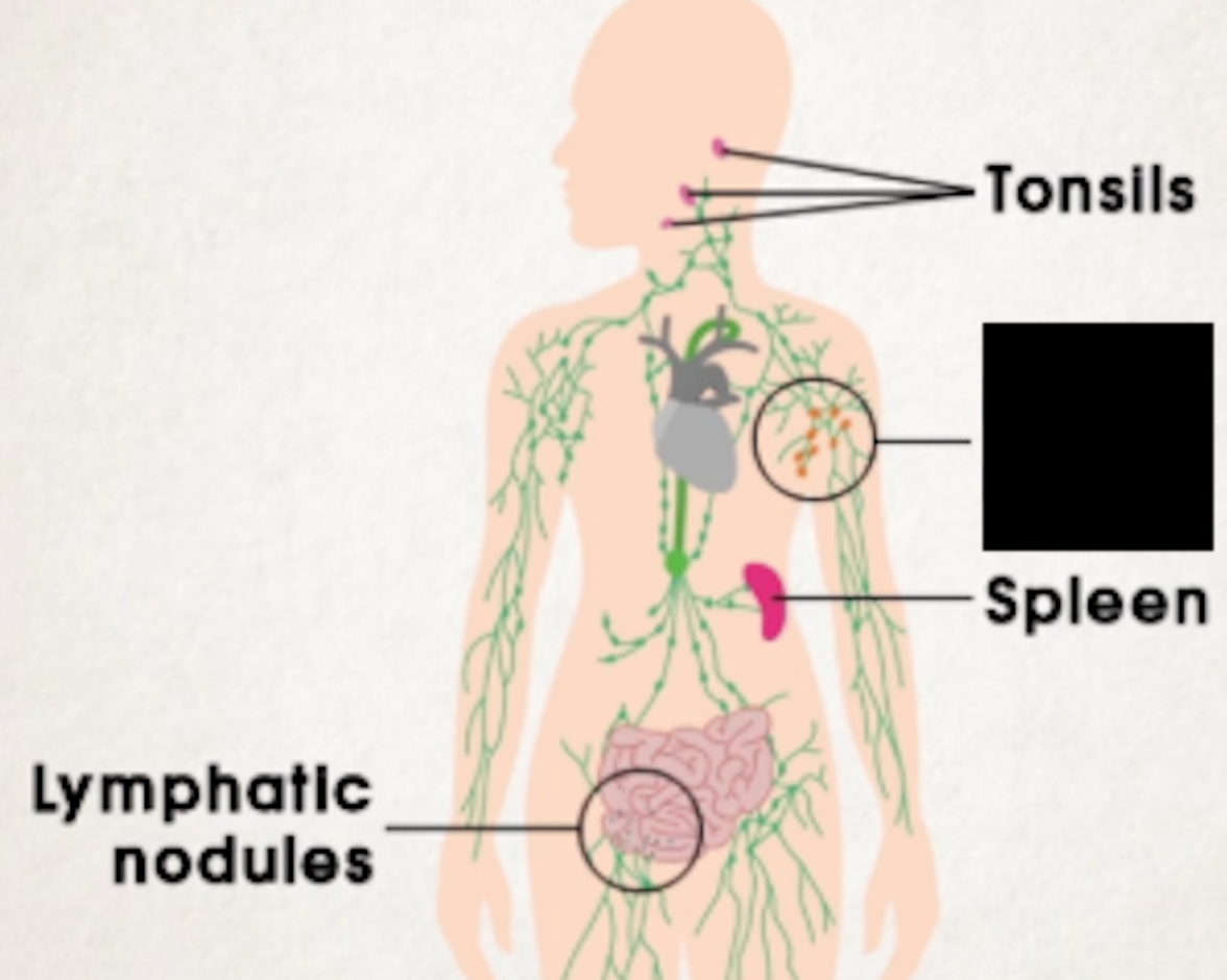
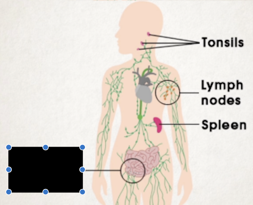
tonsils, lymph nodes, spleen, lymphatic nodules
capsules
provide support and form paths for blood vessels and nerves to course through
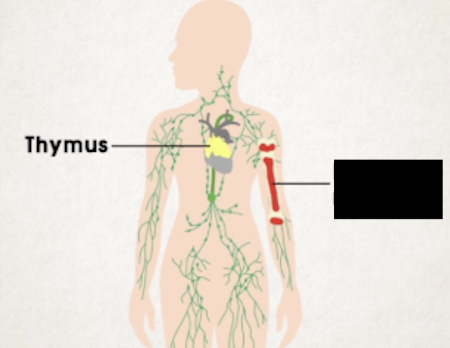
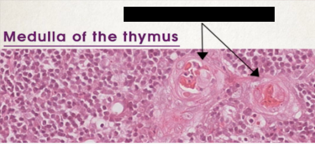
thymus
Bi-lobed organ with lumpy surface
Between sternum and upper part of heart
T cells that are produced in bone marrow are developed into mature T cells in thymus
Immature T cells populate outer cortex (densely packed) and move to inner medulla to mature
In medulla there are Hassall’s corpuscles, formed by flattened epithelial cells
Mature T cells continue to travel via blood to other lymphatic organs
Active in youth and degenerates slowly after puberty
Fat accumulates inside and turns yellow at old age
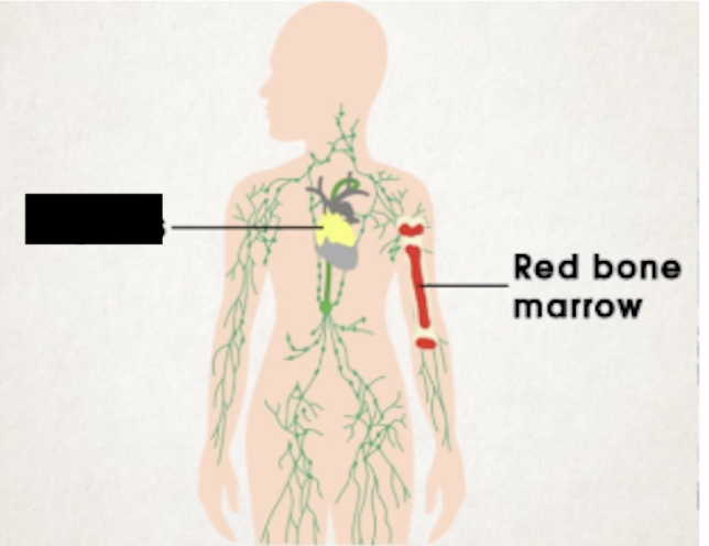
red bone marrow
tonsils
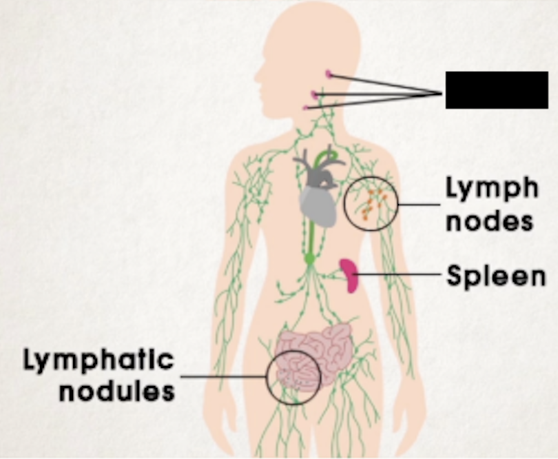
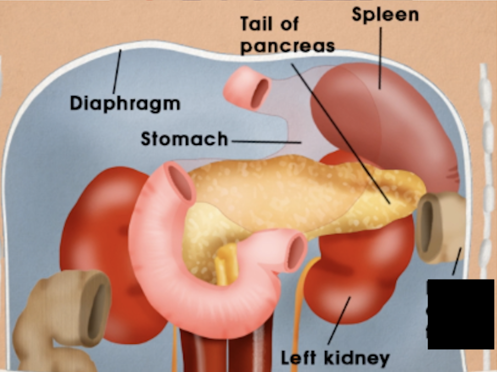
spleen
Largest lymphatic organ in the body
In upper left abdomen close to diaphragm
Smooth and convex, dark-purplish color
Holds a lot of blood that comes from celiac trunk of aorta to splenic artery to spleen
Where aged, damaged, and broken RBC are broken down and recycled
Blood reserve, can pump up to 100mL into circulation
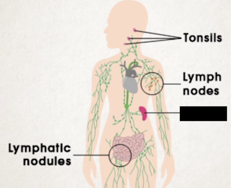
lymphatic nodules
medulla
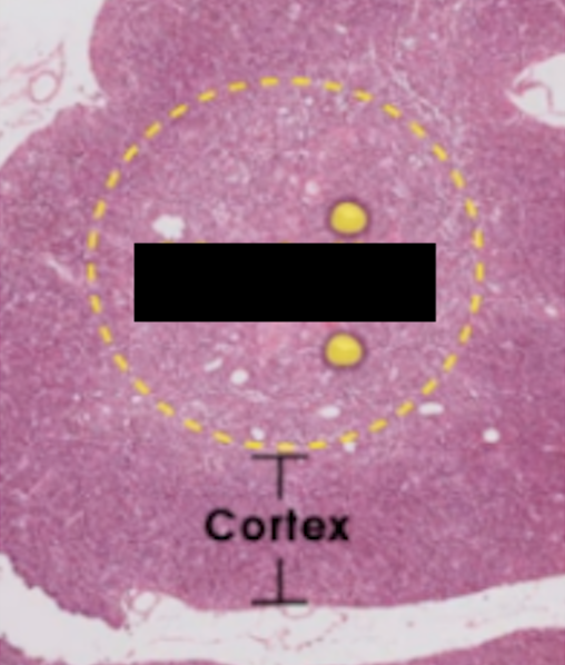
Thymus cortex
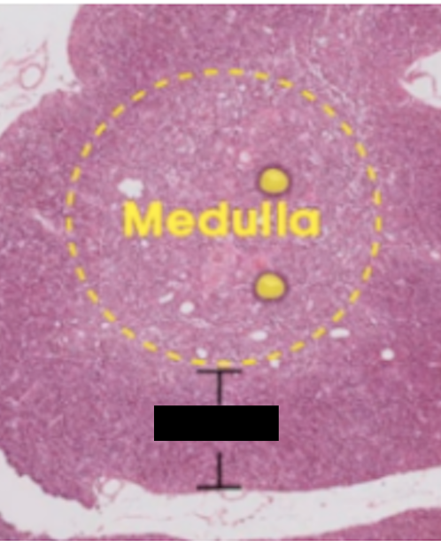
hassall’s corpuscles
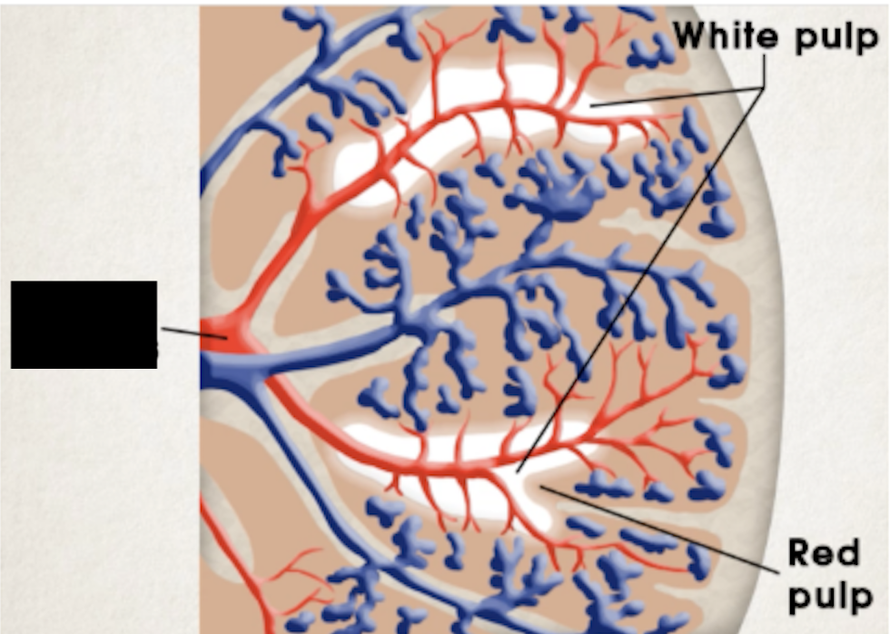
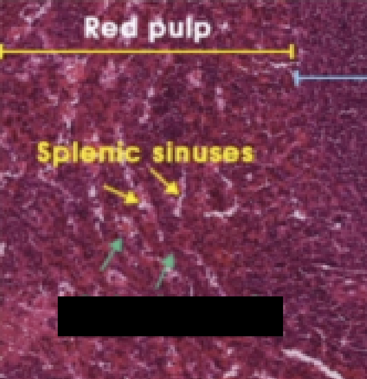
red pulp
Blood-filled splenic sinuses and cords
Houses macrophages, lymphocytes, plasma cells, and others
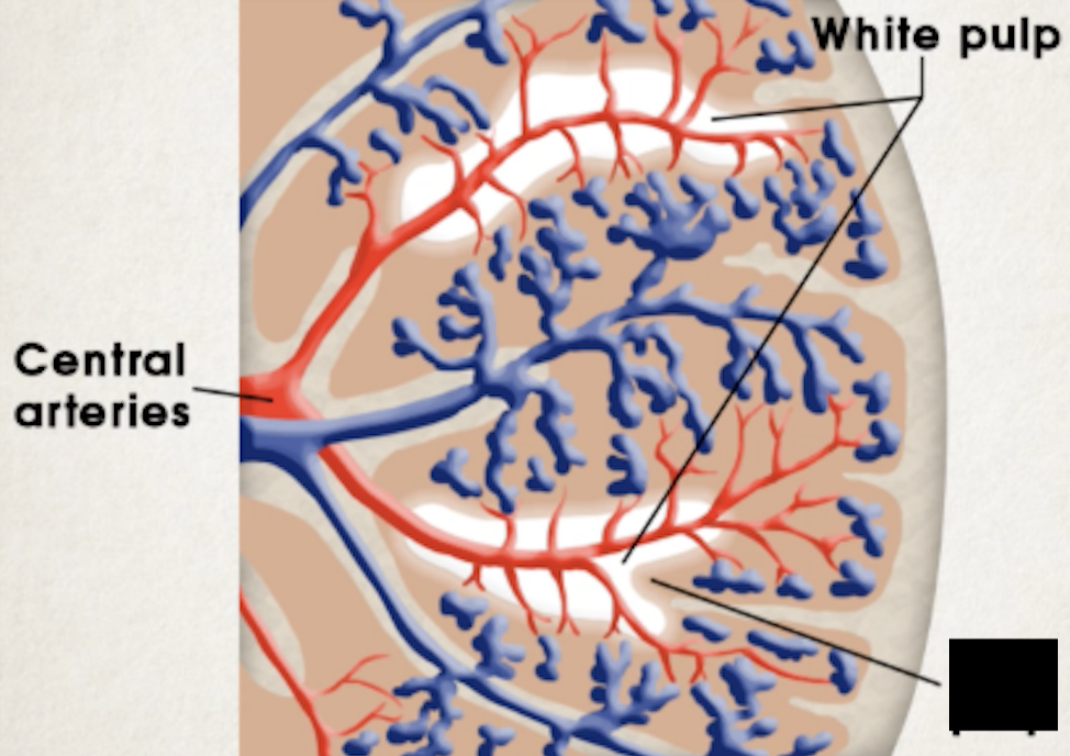
white pulp
Lymphoid tissue formed by clumps of lymphocytes around central arteries (branches of splenic artery)
Allow for rapid immune response against antigens in blood transported to spleen
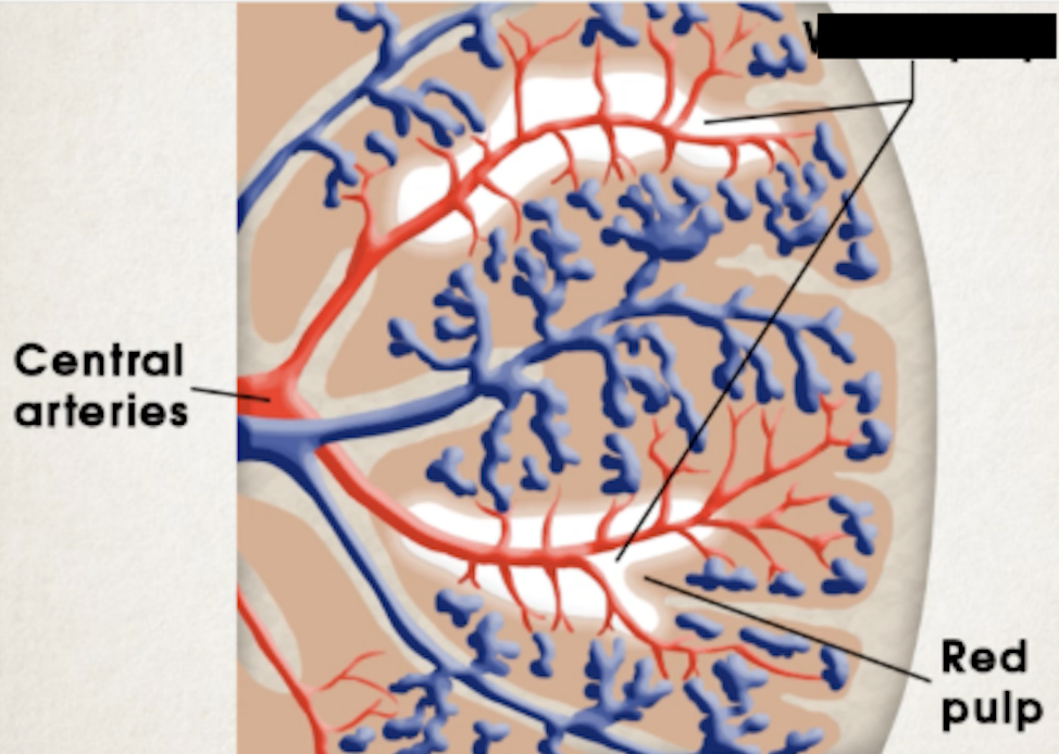
central ateries
tail of pancreas
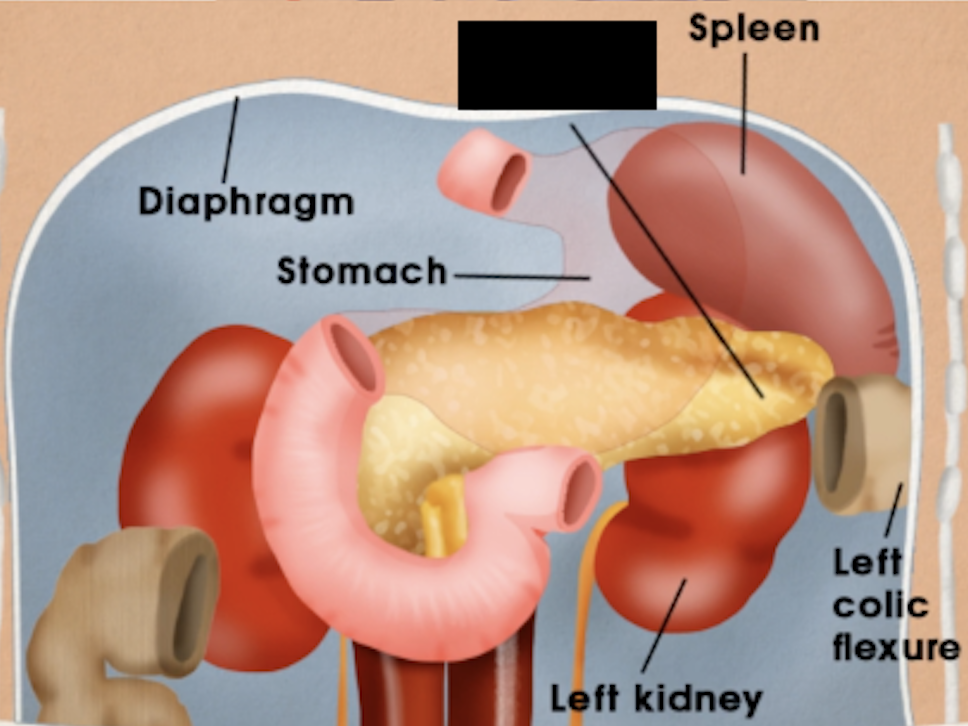
diaphragm
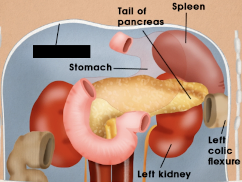
stomach
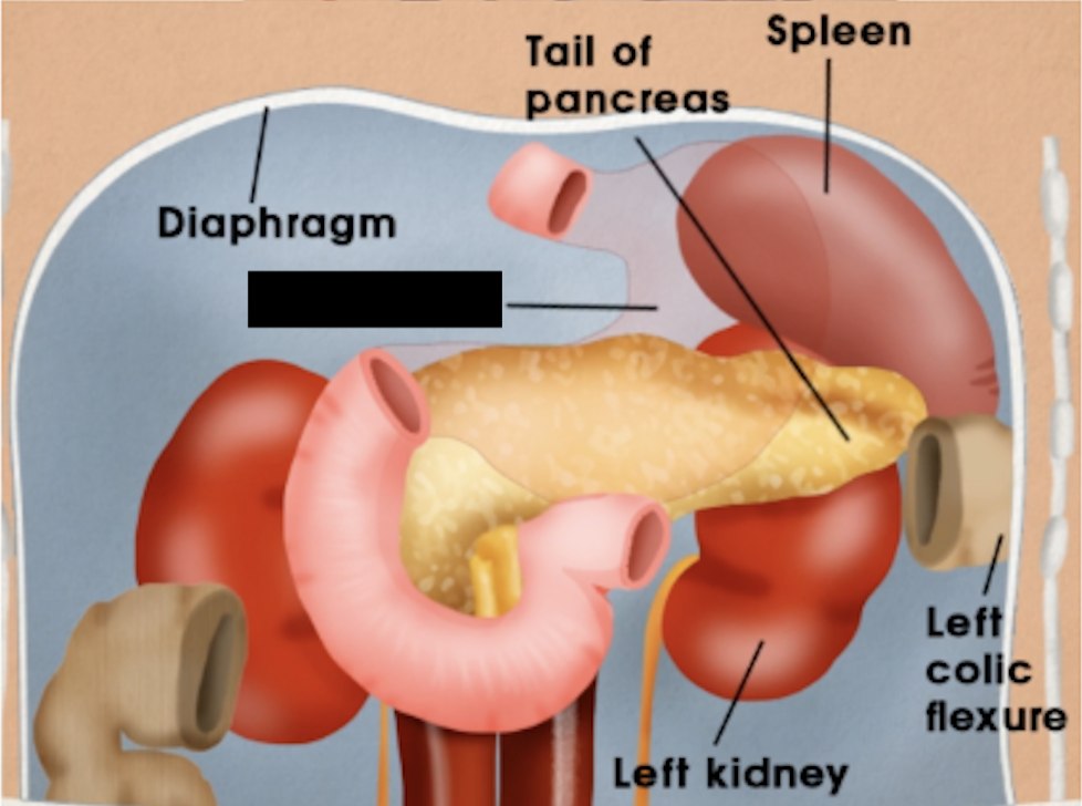
left kidney
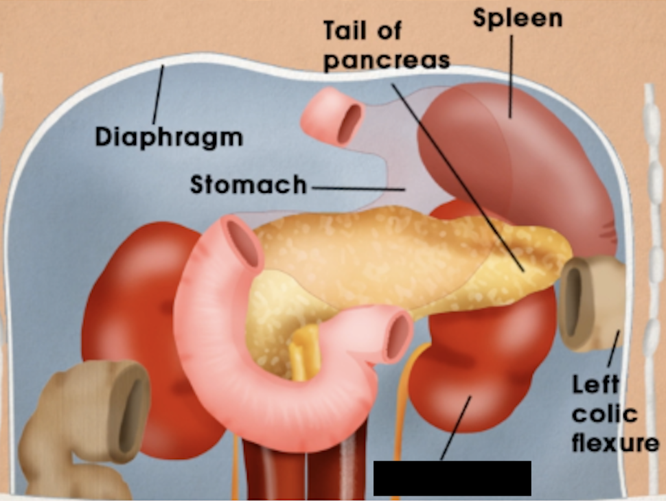
left colic fixture
splenic sinuses
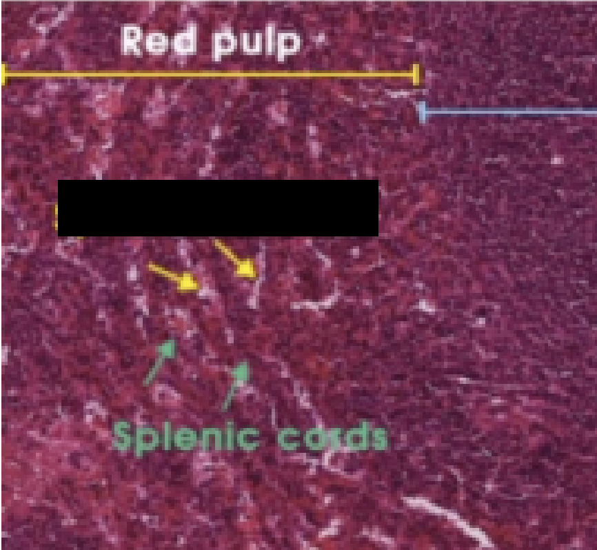
splenic cords
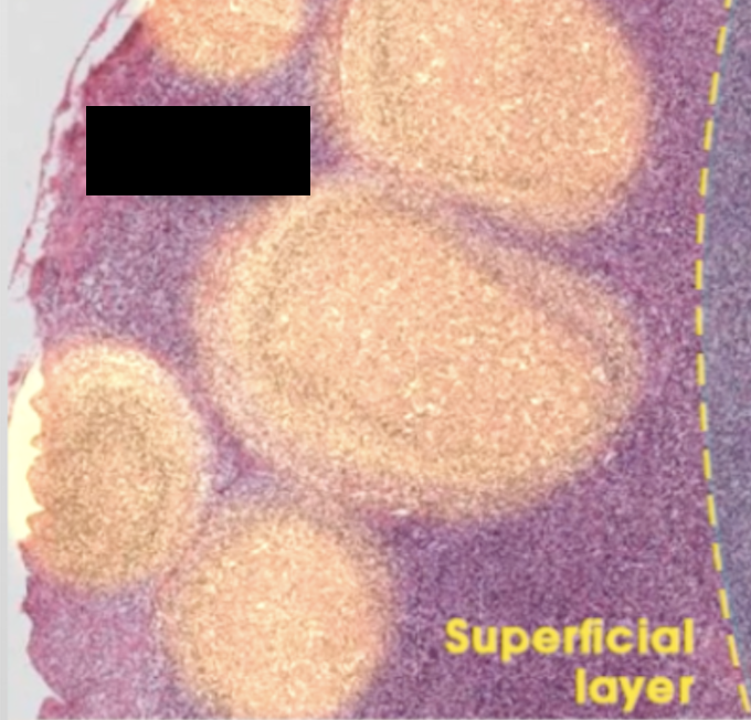
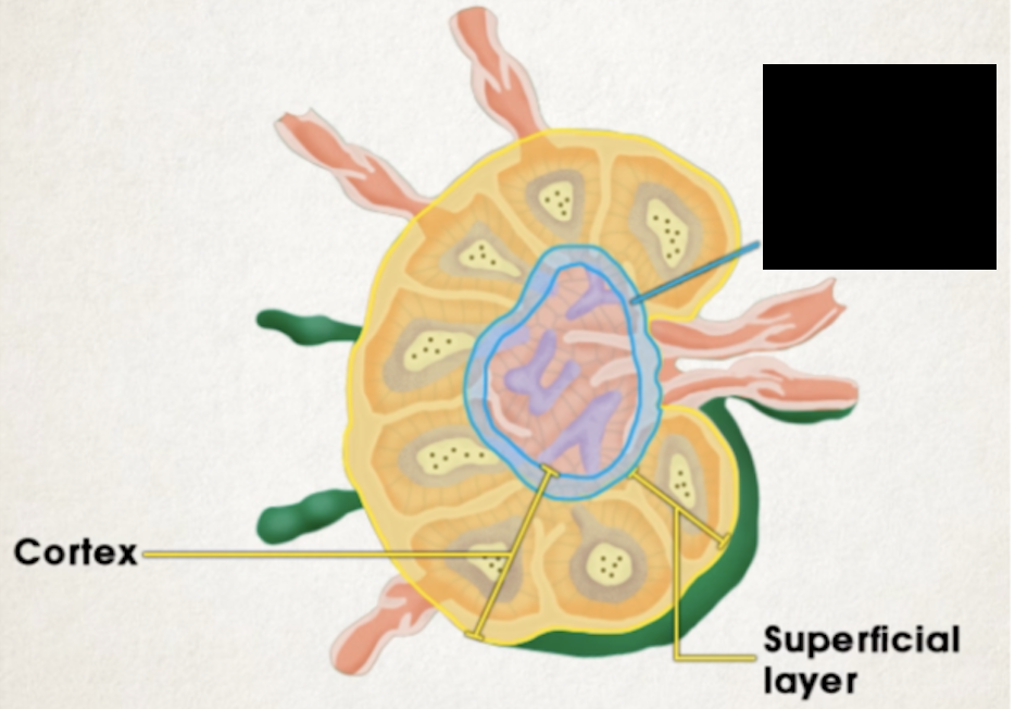
Lymph node cortex
has superficial and deep (paracortical) layers
Deep layer contains mostly T cells

Lymphatic nodule
contain B-cells and macrophages
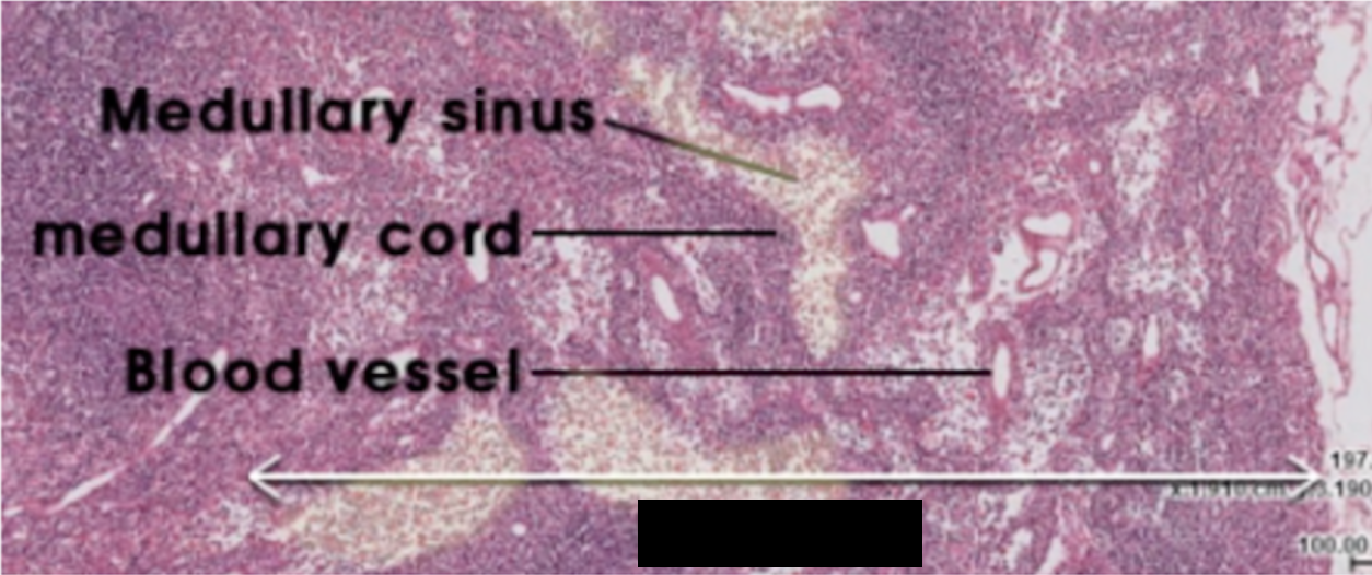
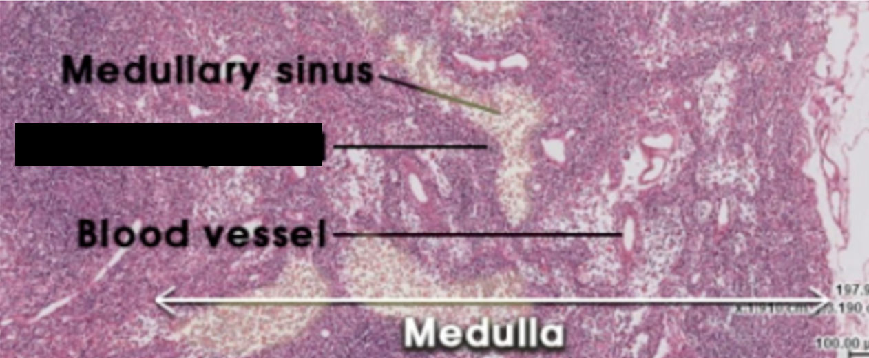
Lymph node medulla
has cords of lymphatic tissue containing B-cells, macrophages, antigen presenting cells, and plasma cells
Trabeculae:
supportive fibers between nodules
Reticular meshwork
composed of reticular cells and fibers
Lymph node medullary cord
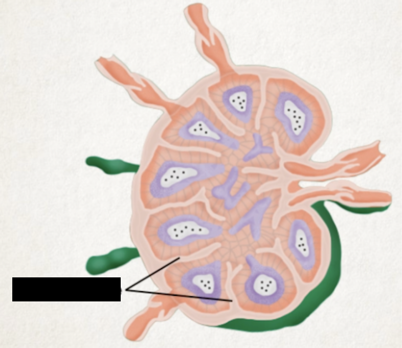
Subcapsular sinus

Trabecular sinus
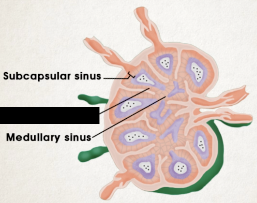
Lymph node medullary sinus

Superficial layer
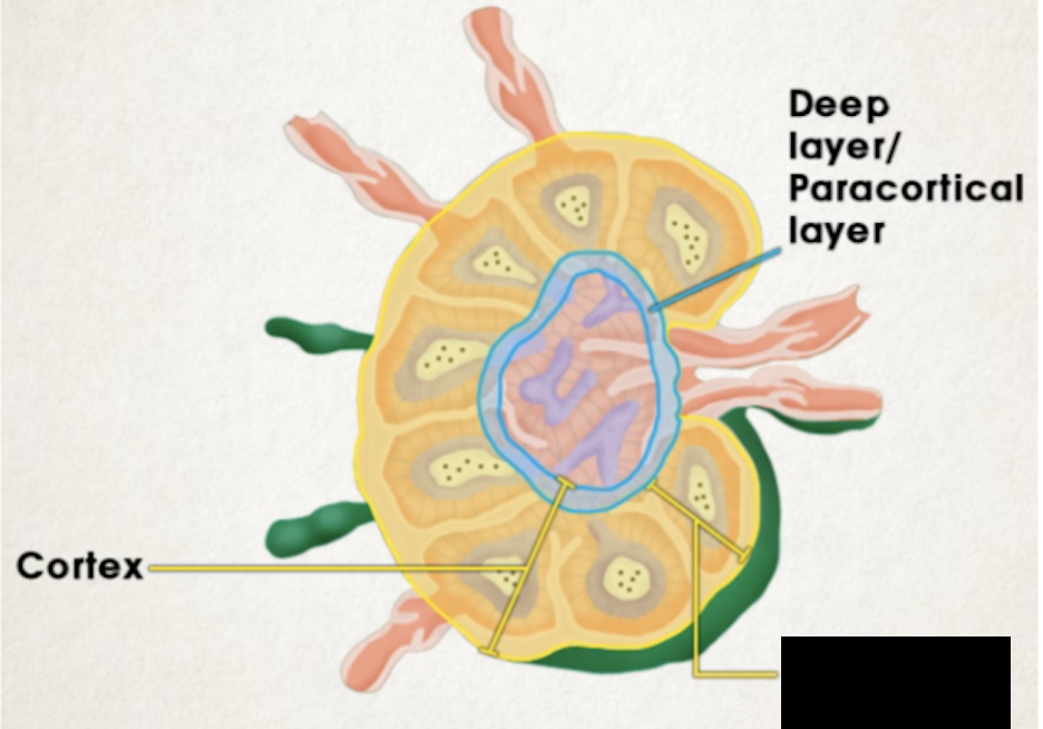
Deep layer
Afferent lymphatic vessels
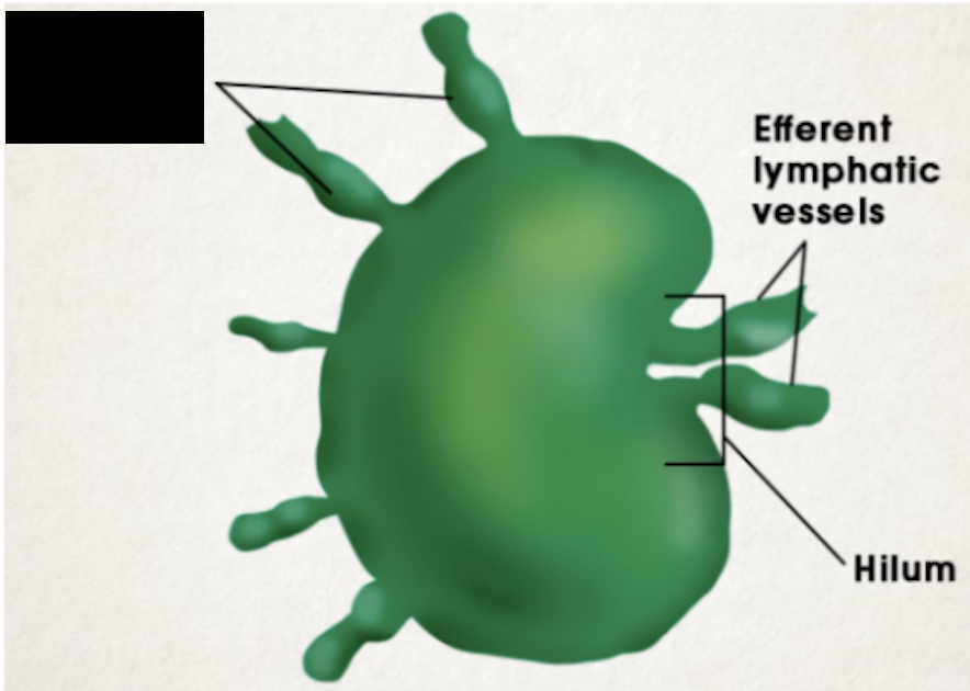
Efferent lymphatic vessels
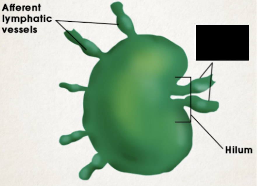
hilum
macrophages
can expose fragment of antigen on surface
release helper T-cells which will trigger B-cells (via interleukin-2) to divide into plasma and memory B-cells
plasma cells
produce antibodies that bind to antigens, attracting macrophages to engulf them
Helper T-cells
trigger cytotoxic T-cells to attack antigen cells directly by creating pores, causing apoptosis
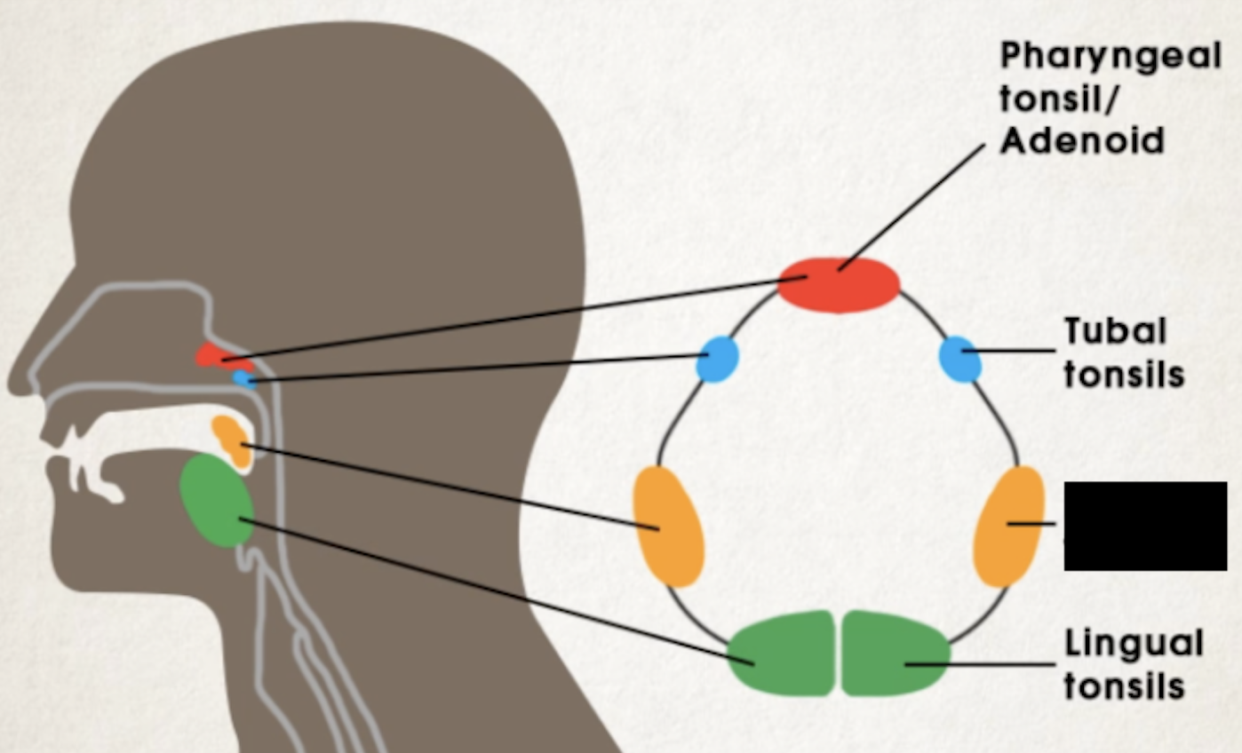
Pharyngeal tonsil/adenoid
roof of pharynx posterior to nasal cavity
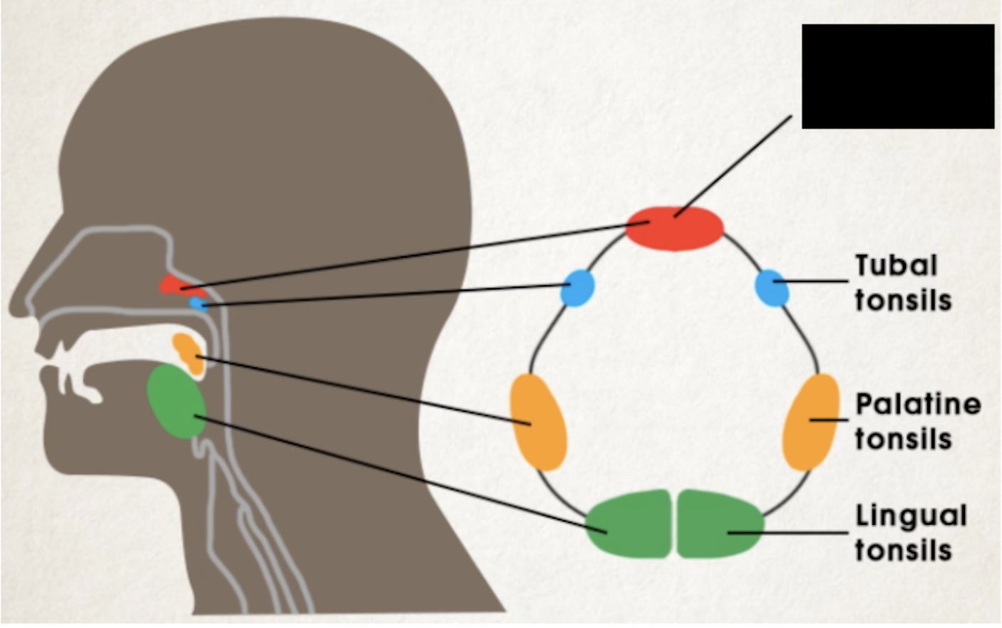
Tubal tonsils
slightly below adenoid, behind 2 eustachian tube openings
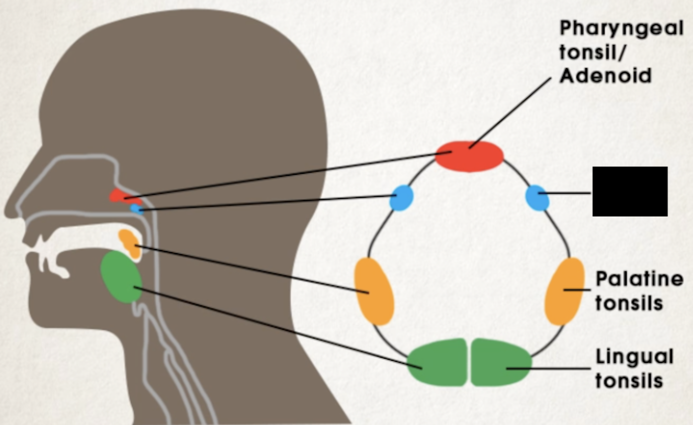
Palatine tonsils
lie on the lateral pharyngeal wall directly behind mouth and palate
Largest and most often infected in childhood
Tonsillectomy
surgical removal of tonsils
Lingual tonsils
posterior surface of the tongue

Tonsils
Group of lymphoid organs in pharyngeal region at top of the throat
mucosal swellings deep to the epithelium of pharyngeal wall
Inside swellings are spherical lymphoid nodules with lymphocytes
Tonsillar crypts
help trap bacteria and foreign matter, which activates the lymphocytes
lymphoid nodule
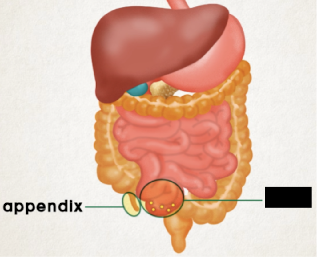
High concentration can be found in ileum of small intestine and appendix because small intestine always attacked by bacteria in swallowed food
generate a diverse range of memory lymphocytes from sampling different antigens, important in immune response

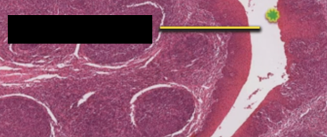
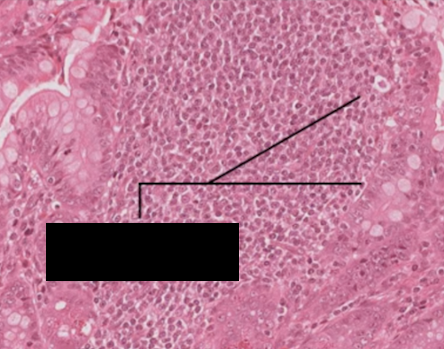
Peyer’s patches
lymphatic nodules aggregated in ileum, large densely stained spherical structures
lymphocyte nuclei
appendix
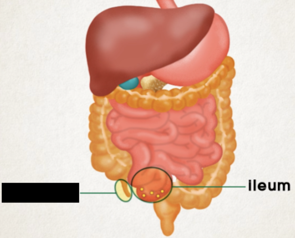
ileum