Histoslides (C2)
1/43
Earn XP
Description and Tags
credit 2 - taken from "Credit II short" 2019. Alt. Name, description, etiology, pathogenesis
Name | Mastery | Learn | Test | Matching | Spaced |
|---|
No study sessions yet.
44 Terms
This is?
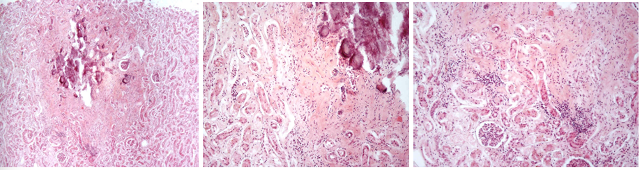
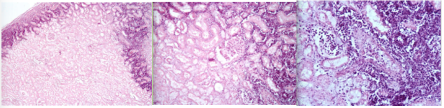
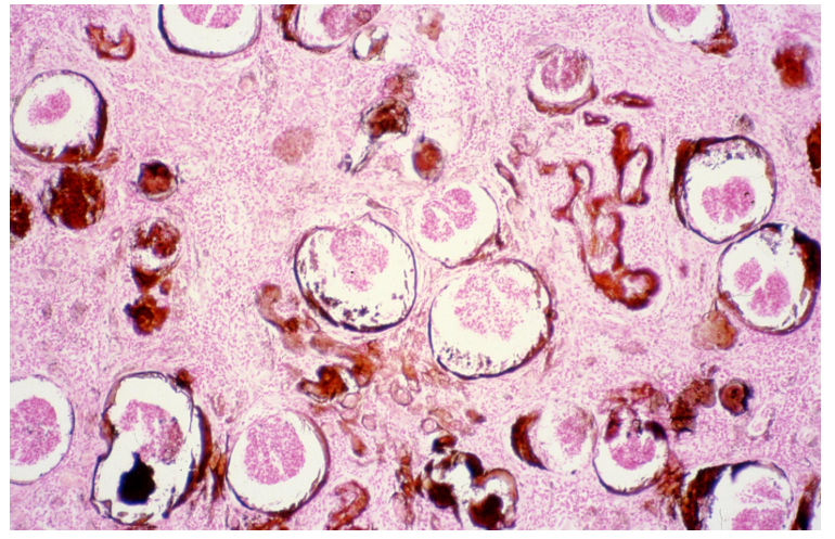
285 - Dystrophic calcification (infarctus renis anemicus)
Description: No regular oval focus in cortex of kidneys, red-violet center (calcification). Relatively mature granulation tissue of fibrocytes and collagenous fibers. At margin we can observe infiltration by lymphocytes and histiocytes.
Etiology: Caused by deposition of calcium into the tissue. Prominent in chronic tissue destructive lesions such as tuberculoid granulomas in cattle (Tuberculosis)
Pathogenesis: Dystrophic calcification implies tissue damage, degeneration, death of cells or denaturation of proteins in tissue, typically occurring in areas of necrosis.
This is?
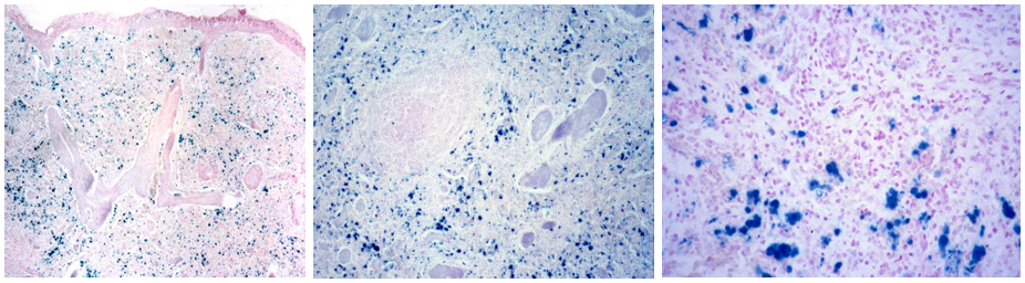
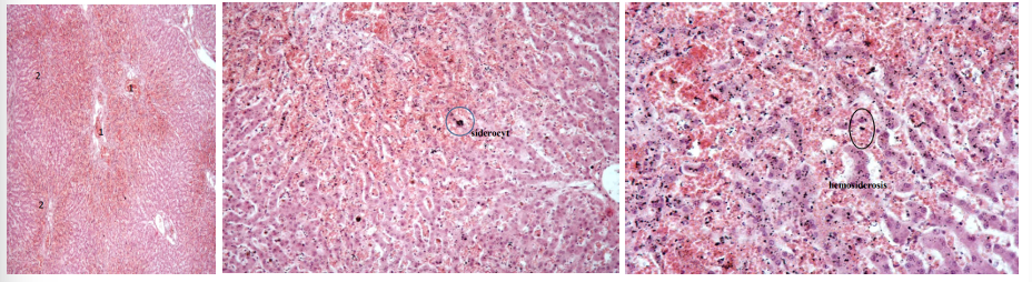
Hemosiderosis of spleen (Liesegang)
Description: Stained by Liesegang to prove hemosiderin. In the red pulp of the spleen, excess hemosiderin is stained blue. It is present as lumps in macrophages (siderophages, siderocytes) or encrusted in fibers of connective tissue.
Etiology: Occur during excess intravascular destruction of erythrocytes (infectious, parasitic diseases and blood poisoning). Increased concentration in one area is also an indication of previous hemorrhages.
Pathogenesis: Hemosiderin is a iron-containing pigment found in macrophages and is prominent as a lesion (hemosiderosis). If excess iron is absorbed, or a lot is released from hemolysis, the levels in macrophages will increase and accumulate. In older dogs, there are often brown, raised, granular lesions on the edge of the spleen. These contain iron, ceroid and hemosiderin, and probably represent previous hemorrhages.
This is?
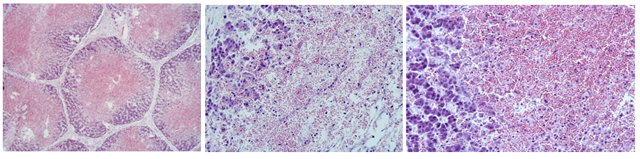
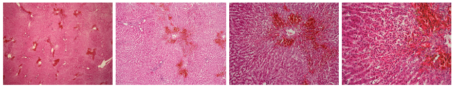
Necrosis hepatitis centrolobularis
Description: All liver lobules exhibit alterations. Branching is normal only at the periphery, and the center is filled with erythrocytes. Necrosis in a particular zone of tubules results in dilatation and congestion of sinusoid and affected zone appear red. Normal framework is lost.
Etiology: Hypoxia from viral infections (e.g. canine adenovirus) or ischemia, toxins
Pathogenesis: Zonal necrosis is divided into pariacinar (centrilobular), midzonal and periportal (centroacinar). Zone 1 (centrilobular) necrosis is the most common form as this area receives the least amount of oxygen and nutrients, making it susceptible to hypoxia.
this is?
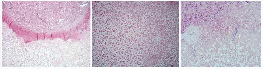
Necrosis hepatis focalis
Description: At one end there is a necrotic nodule stained eosinophilic. Complete karyolysis is observed, and inflammatory cells are on the edge of the nodule. These cells undergo necrobiosis. In the necrotic tissue there are some basophilic stained bacteria.
Etiology: Infectious agents (Toxoplasma), viruses and bacteria (Salmonella, listeria)
Pathogenesis: Focal hepatocellular necrosis presents as multifocal accumulations causing pale or dark red foci. “Sawdust liver” results from gut organism entering the blood at slaughter of cattle.
This is?
Infarctus renis anemicus
Description: Wedge shaped area with its base at capsular part and apex at the site of vascular obstruction. Necrotic tissue stains bright pink. In areas of coagulative necrosis, cellular outlines and tissue structure are present. Area is surrounded by a zone of neutrophilic granulocytes and red line (erythrocytes).
Etiology: Caused by the same events which cause ischemia, commonly secondary to thrombosis or thromboembolism.
Pathogenesis: Infarct means an obstruction of blood supply to tissue (complete ischemia), causing the tissue to undergo coagulative necrosis. Usually occur in areas where there is inadequate collateral circulation to maintain life in tissues after blockage of vessels. Therefore, well-supplied areas (skin, thyroid, lungs, liver) are rarely the site of infarcts, while it occurs most commonly in tissues with end-arteries.
this is?
Chronic passive hyperemia of liver
Description: Strong congestion and dilation of blood vessels (1). Sinusoids (2), mainly in the centrilobular area, are widened and filled with erythrocytes. Increased presence of hemosiderin in macrophages testifies about the chronic character of the process.
Etiology: Caused by obstruction in return of blood to the heart, leading to increased venous pressure. Due to heart failure or physical obstruction.
Pathogenesis: Obstruction in return of blood or in its passage through lungs to heart result in increase in venous pressure, slower blood flow and consequent increase of blood volume in many organs (liver, spleen, kidneys). Blood accumulates in dilated capillaries and venules, and tissue appear blue because of lack of oxygen (cyanotic).
this is?
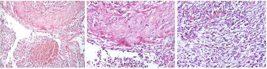
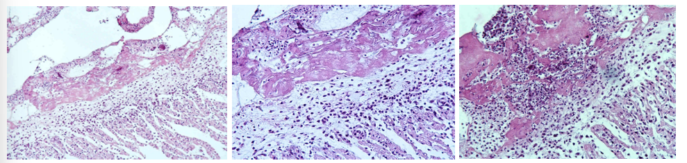
Pleuritis fibrosa/fibrinosa (repair by organization)
Description: On thoracic pleural surface. Pink-stained fibrin is infiltrated by macrophages (histiocytes), lymphocytes and neutrophils. Beneath there is young granulation tissue proliferating fibrin. Spaces between capillaries are filled with macrophages, lymphocytes, and elongated spindle-shaped fibroblasts.
Etiology: Pneumonia (lung parenchyma), thromboendocarditis (heart), fibrinous pleurisy (pleura) and other body tissues which have been sites of inflammation.
Pathogenesis: Repair by organization occurs when there’s an incomplete resolution of inflammation. Unabsorbed fibrin forms a scaffold for fibroblasts and capillary loops, so fibrosis develops in the area. Area doesn’t return to normal, but remains in a scarred state.
this is?
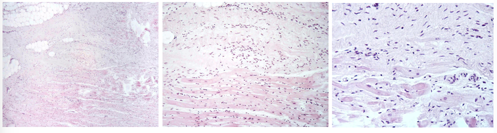
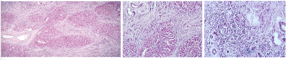
Fibrosis myocardii
Description: Myocardium of the heart is locally substituted by mature fibrous tissue with bright staining (some places fatty tissue is present). Substitution of damaged myocardium (infarct, necrosis, inflammation) by repair, when granulation tissue mature to highly-differentiated connective tissue (healing process).
Etiology: Damage from infarct, necrosis of different etiology and inflammation
Pathogenesis: Healing process begins with macrophages removing dead cells and debris, eliminating inflammatory exudate and dead tissue. The myocardial defect is filled with granulation tissue- fibroblasts, collagen-producing cells, and new blood vessels. This tissue matures into a fibrous scar, depositing collagen fibers for structural support. This restores structural integrity, but there may be a loss of function due to the replacement of specialized cells, determined by extent of loss.
this is?
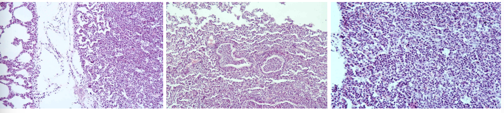
Bronchopneumonia purulenta
Description: Enlargement and engorgement of capillaries with blood and oozing of erythrocytes can be seen. Alveoli are filled with extensive infiltrate, containing different cells (leukocytes, lymphocytes, desquamated pneumocytes and macrophages, but predominantly neutrophils). Cell infiltrate lie in a blank amount of gray-pinkish homogeneous fluid exudate. Bronchial spaces are also filled with neutrophils.
Etiology: Purulent inflammation occurs when damage is more severe and large numbers of neutrophils are present. Can be caused by pyogenic bacteria such as staphylococcus, corynebacterium, streptococci (diphtheria in pigs)
Pathogenesis: Purulent inflammation is characterized by pus and large number of neutrophils. Neutrophils, pathogen or a combination of the two cause necrosis of surrounding tissue.
compensatory interstitial emphysema
red hepatization
overloading of air (alveolar emphysema) - compensatory
normal alveoli
this is?
Pericarditis fibrinosa
Description: A layer of thickening edematous subepicaridal tissue and remnant endothelium on the surface of myocardium, covered by fibrinous mass with admixture of lymphocytes and neutrophils. Process demonstrate acute stadium of serosal fibrinous inflammation.
Etiology: Microorganisms such as diphteria, salmonella, sphaerophorus necrophorus
Pathogenesis: Fibrinous inflammation is characterized by exudate containing large amounts of fibrinogen which clots so fibrin is the most conspicuous agent. Fibrin exudation indicates severe acute vascular injury. Occur mostly on mucous and serous membranes. Fibrin can sometimes be reddened by blood and since it is chemotactic, it attracts neutrophils. In cattle, high plasma fibrinogen levels make fibrous exudate more common.
this is?
Cirrhosis hepatis (Hepatitis interstitialis chronica)
Description: Proliferated fibrinous tissue found in the whole section. It is infiltrated by chronic inflammatory cells (lymphocytes, plasma cells, macrophages). Fibrinous tissue originates from interlobular spaces and spreads into the center of the lobules, causing press atrophy of liver parenchyma. Regeneration of bile ducts can be seen in new fibrinous tissue.
Etiology: End-result of necrosis or apoptosis and active inflammation with chronic fibrosis.
Pathogenesis: End-stage diffuse hepatic disease characterized by nodular regeneration with fibrovascular bridging scars containing shunts. Regeneration of tissue leads to formation of variable sized nodules between fibrous connective tissue, distorting the entire liver. In the end, the liver is unable to perform its normal function, and cirrhosis can lead to ascites and hypoproteinemia.
this is?
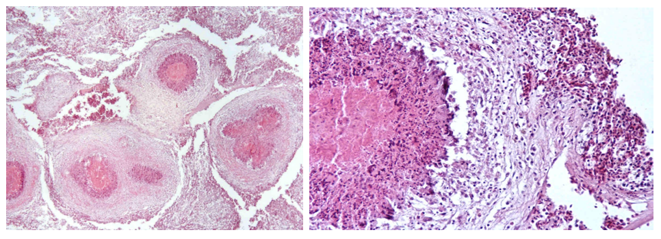
Actinomycosis
Description: We can see actinomycotic nodules. In the center there are bacterial colonies consisting of intertwined radiating filaments (rays), capped by eosinophilic hyaline material (clubs), creating a sunburst pattern- sulphur granules, surrounded by disintegrated neutrophils- pus. An outer area of large mononuclear cells with abundant, often foamy cytoplasm- histiocytes and plasma cells, surround neutrophils. Non-specific granulation tissue border nodules.
Etiology: Caused by Actinomyces bovis
Pathogenesis: Hard, irregular, enlargement result from infection of mandible or maxilla, leading to the disease of lumpy jaw.
this is?
Pneumonia TBC miliaris - poultry
Description: Pathological content fills lung fistulas. It is formed by round foci with central necrosis surrounded by specific granulation tissue. Close to necrotic parts of granuloma it is predominated by macrophages (epithelioid cells) and lymphocytes, which are numerous at the periphery. Start of giant cell formation can be observed in some granulomas.
Etiology: Caused by mycobacterium avium which is transmitted by ingestion or inhalation.
Pathogenesis: Basic form is the tubercle which appears as a pin-head sized focus on tissue. Large masses arise by coalescence of smaller tubercles. Bacteria can spread to lymph nodes and other organs by lymphatic system and bloodstream. Generalization may be sudden and massive if large numbers of bacilli enter the bloodstream.
this is?
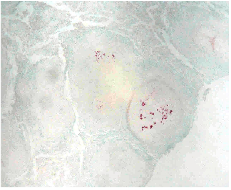
Pneumonia TBC miliaris- poultry (ZN) same as nr. 263
Ziehl-Nielsen staining is used for special proof of mycobacterium. Positive acid-fast rods bacilli and their clumps of red color are mainly found in the necrotic center. During the generalized TBC process, the lungs are the last organ with dissemination of tubercles.
this is?
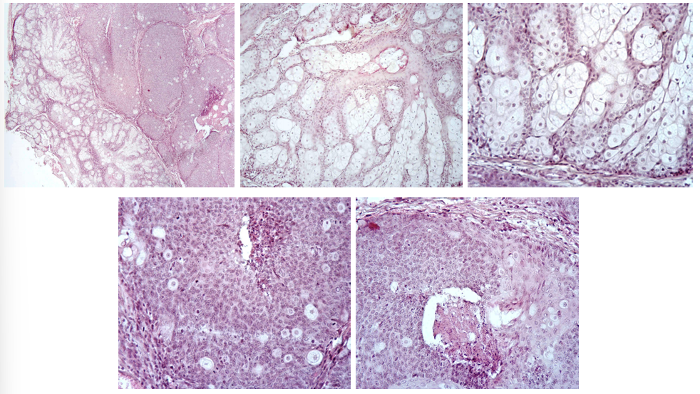
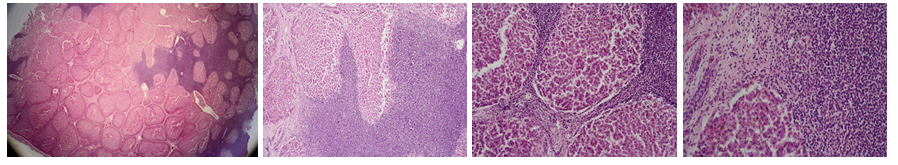
Adenoma et adenocarcinoma sebaceum
Description: 2 sections on slide. Adenoma is smaller, adenocarcinoma larger. Epidermis has relatively normal structure, in some places it intrudes into the tumor with acinar structure. It consists of many gland clusters lined by narrow interstitial tissue. Tumor is relatively large, light cells with vacuolated foamy cytoplasm and large, light nuclei. In some nuclei there are necrotic changes. In adenocarcinoma proliferated glandular cells are more basophilic and pathological mitosis can be observed in nuclei. Alveolar centers undergo regressive changes (dystrophy, necrosis)
Etiology: Exact causes are unknown, but can have a genetic and environmental component
Pathogenesis: Adenoma is a benign epithelial tumor occurring in various glands (circumanal, sebaceous, mammary) and also adrenal and thyroid glands. Lesions can be difficult to differentiate from nodular hyperplasia. A common group is the basal cell group, deriving from basal layer of squamous epithelium and can differentiate into tumors of any adnexa of skin. Adenocarcinoma is a malignant gland-forming neoplasm. They can grow in papillary form and in retention of secretion (cystic hyperplasia). Tumors can be well or poorly differentiated.
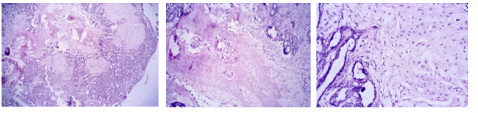
this is?
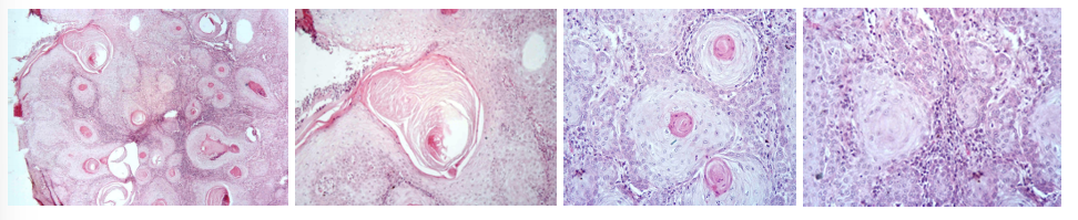
SCC (Canceroid- Squamous cell carcinoma)
Description: Cells resemble those of normal stratum spinosum, but have vesicular nuclei with one or more prominent nucleoli. We can observe eosinophilic stained formations growing into the dermis. Tumor cells spread throughout the dermis as slender anastomotic cords, with some cells falling off and remaining as isolated islands in the dermal stroma. Keratinization within islands results in laminated keratin pearls surrounded by tumor cells. At the periphery cells resemble basal cells following differentiated cells of stratum spinosum, and keratinization in the center.
Etiology: UV radiation (sunlight). Some breeds are more at risk than others.
Pathogenesis: Carcinomas are malignant epithelial neoplasms and SCC are tumors of squamous epithelium. They are common on eyelid and conjunctiva of cattle and horses, ear pinna of cats and sheep, vulva of cattle, goats and recently sheared sheep.
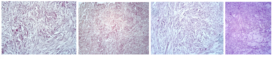
this is?
Fibroma molle
Description: Consists of fibrinous material, predominated by nuclei and plumb cell bodies. Cells with few fibrils run aimlessly in many directions. Mitosis is seldom. Tumor tissue is edematous permeated (no stained slots among individual cells) or infiltrated by heterophils, lymphocytes and macrophages (body reaction).
Etiology: Unknown
Pathogenesis: A benign mesenchymal tumor arising from fibrous connective tissue. Has 2 forms: Fibroma durum which is predominated by fibrous material, causing it to be hard. And fibroma molle, where cell bodies and nuclei predominate, which is softer due to fewer fibrils.
this is?
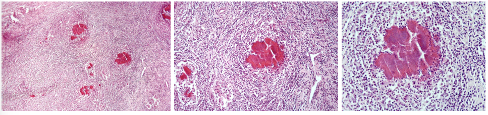
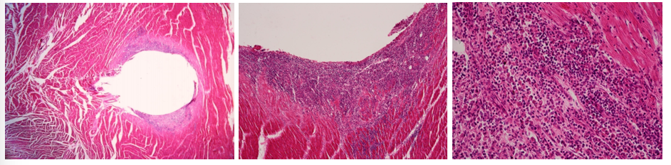
248. Post traumatic focal inflammatory reaction of myocardium
Description: Round hole surrounded by expressive reaction tissue consisting of neutrophils, macrophages and lymphocytes. Process intrude into deeper areas locally.
Etiology: Trauma, foreign object penetrating the tissue (e.g. bullet)
Pathogenesis: Localized immune response. Trauma causes loss of tissue, and the body responds by inflammatory reaction (neutrophils, macrophages, lymphocytes)
this is?
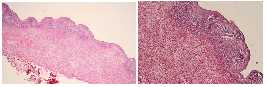
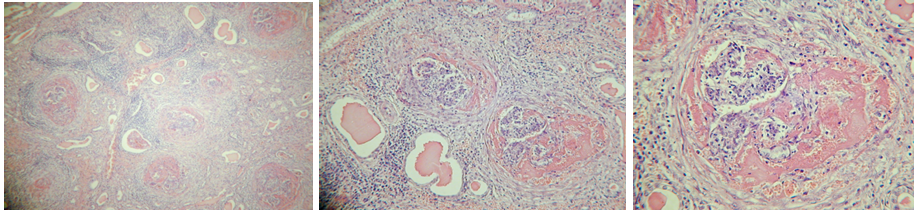
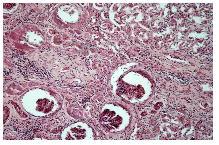
Vasculitis chronica (Mediocalcinosis- H&E staining)
Description: Thicker tunica media of aorta. Consists of native fibrous tissue which proliferates. Evidence is the increased number of fibroblasts with production of intercellular collagen fibers. Infiltration by lymphocytes and macrophages can also be observed, showing a chronic inflammatory process in the blood vessel wall.
Etiology: Characteristic of uremic syndrome in dogs. Can be caused by infectious agents, immune-mediated mechanisms or as a result of inflammatory processes in adjacent vessels.
Pathogenesis: An inflammation of blood vessels. Developed by complex mechanisms which depend on causative agents. The immune system mistakes components of vessel walls as foreign and attacks them with immune cells. Continuous inflammation leads to damages and thickening of the wall, narrowing the lumen and sometimes blockage which compromise the blood flow.
this is?
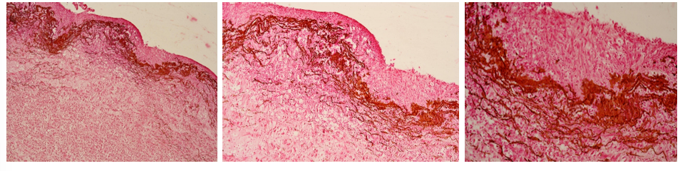
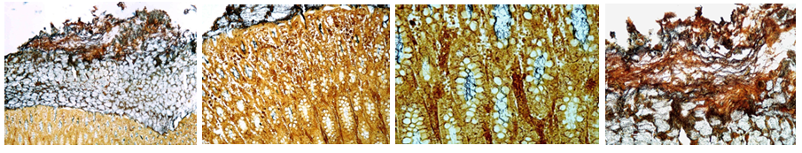
Mediocalcinosis (van Koss)
Description: Same as 261, stained with Van Koss to prove deposition of calcium (dark brown staining). Between t.intima and t.media among the cells, there is also observed brown diffuse staining in intercellular mass.
This is?
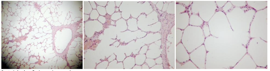
6. Emphysema alveolare chronicum (we see that septa is broken and air area increased in picture)
Description: Overinflation with air. Alveoli are too large and have wide openings to each other or into a common space due to rupture of alveolar walls. Blunt ends of broken alveolar walls often persist and become thickened and hyperplastic rounded knobs. Some walls are slightly thickened and inelastic, but others are stretched and very thin. Blood-filled capillaries are scarce.
Etiology: Component of disease conditions, related to obstruction of outflow of air, or in agony such as slaughter.
Pathogenesis: Emphysema is an abnormal, permanent, enlargement of airspaces, accompanied by destruction of alveolar walls. All species can be affected by alveolar emphysema which appears as small air bubbles. Other types is interstitial which is found mainly in cattle and consist of migration of air from alveoli to interstitium, and bullous which is a large accumulation of air. All 3 can co-exist.
this is?
200. Endobronchiolotis obliterans et peribronchitis nodusa
Description: In atelectatic lung tissue we observe areas of round or irregular shape. There are bronchioles with obstructed lamina, filled with fibroblastic granulation tissue. In some parts, granulation tissue can be seen in the periphery of bronchioles. On one part of the lung, granulation tissue proliferates into the alveoli (calcification).
Etiology: Caused by pulmonary injury by viral infection or pulmonary toxicity. Common in cattle with atypical interstitial pneumonia, but also in other species under various conditions.
Pathogenesis: Inflammation in bronchioles results in thickening of the walls which can lead to an increase in respiratory resistance. Physical obstruction occurs more readily in bronchioles than in bronchi as the walls collapse more easily and small size permit occlusion of exudate. The result is accumulation of exudate and delayed repair. Around, peribronchial fibrosis and neutrophils or eosinophils are present.
this is?
228. Crupous (fibrinous, lobular) pneumonia and chronic interstitial pneumonia
Description: Trapezium with basophilic margins can be observed. Margin is created by polymorphus leukocytes inside of lung alveoli. The first stage of gray hepatization can be seen. Alveoli are filled with pinkish mass containing fibrinous fibers. Dilated lymph vessels and two foci of fungal hyphae in these areas can be seen and mycotic infection can be considered as the cause. At one margin, there is a cord created by maturated fibrous tissue. Alveoli wall next to it and bronchioles are thickened by fibrous hyperplasia- chronic perilobular, intralobular and peribronchial interstitial pneumonia.
Etiology: Lobar penumonia is caused by viral or bacterial infections. Interstitial pneumonia can be caused by various agents such as bovine respiratory syncytial virus (cattle), maedi (sheep), influenza (horses), distemper (dogs) and toxins from bloodstream.
Pathogenesis: Lobar pneumonia involves one or more, or all lobes. Distribution implies acute or severe pneumonia in ruminants and pigs. Can be hemorrhagic, fibrinous, fibrinopurulent, necrotizing or gangrenous. Most of them are fibrinous and it can be characterized by fibrinous exudation. Development stages are congestion, red and gray hepatization, and resolution. Interstitial pneumonia are inflammatory conditions where there’s predominantly exudative and proliferative responses involving the alveolar wall.
this is?
270. Epulis fibromatosa
Description: We can observe islands of proliferated gingival squamous epithelium separated by marked matured dense fibrous stroma with blood vessels.
Etiology: Occur in dogs 7 years of age or older, increased frequency in Boxer breed. Probably caused by irritation or trauma.
Pathogenesis: Tumor-like masses on the gingiva and supporting tissue in mouth, usually around the molars and premolars, and adjacent to carnassial teeth. They are abnormal tissue growth due to inflammatory response. They are usually solitary, pedunculated and rounded or cauliflower surfaced which can be pink or ulcerated. If it has a pronounced squamous epithelial component it is called a acanthomatous epulis, other types are foreign body, fibromatous or ossifying epulis.
177. Colitis fibrinosa (Levaditi- Treponema dysentery)
Description: Masses of fibrin on the yellow floor base of the large intestine. Blackish round curve, often distinctly undulating sticks (over 10 um) can be observed in exudate. Sticks are agents of disease (Serpulina hyodysenteriae). Agents are present also deeper in the mucous membrane, sometimes at the base of crypts.
Etiology: Swine dysentery is a contagious disease of young pigs caused by Serpulina (Treponema) hyodysenteriae
Pathogenesis: The disease is characterized by diarrhea and heavy morbidity and mortality rates. Organisms colonize the mucosal surface and lumen of colonic glands. Liberation of toxins may result in necrosis.
this is?
9. Lymphadentis acuta simplex
Description: Conspicuous follicular structure of lymph node is obscured as a result of moderate follicular hyperplasia and edema. Blood vessels are dilated and filled with blood. Subcapsular sinuses are also dilated and filled with leukocytes (mainly neutrophils).
Etiology: The most common form of lymph node inflammation. Caused by localization of irritant or drainage of inflammatory products into the node.
Pathogenesis: An infection in the vicinity causes an inflammatory response, mobilizing immune cells to the infected site. In cases of pyogenic organisms abscessation, while in cases of infections (Salmonella, toxoplasma) necrosis is an attribute
this is?
238. Infectious bursal disease
Description: Changes in bursa of fabricius during bursal disease
Extensive hyperemy of folds and edema of interstitial tissue
Nearly complete lymphocytes lack in lymph follicles and proliferation of reticuloendothelial stroma. In some follicles only individual lymphocytes occur and undergo apoptosis.
Some follicles include minute eosinophilic stained necrosis.
Desquamation of epithelial cells of folds.
Fibrinous exudate leakage from bursa (Cross section)
Etiology: Also called gumboro disease. Caused by birna virus.
Pathogenesis: Disease targets the bursa of fabricius in birds, an organ responsible for the development of B-lymphocytes. This can lead to decreased immune system response, cell death and immunosuppression
this is?
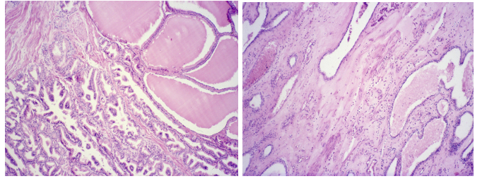
27. Cholangitis et pericholangitis chronica hyperplastica (Coccidiosis hepatis cuniculorum)
Description: In normal liver parenchyma, irregularly shaped areas bordered by fibrinous tissue are seen. Areas indicate hyperplasia of bile duct wall. Proliferation of epithelium, which is forced into papillary folds, simulates adenomatous hyperplasia.
Etiology: Coccidiosis is an infection by protozoan parasite eimeria or isospora.
Pathogenesis: Inflammation of the biliary tract. Coccidia are obligatory intracellular parasites which develop within the cytoplasm of epithelial cells, which result in death of affected cells. Loss of epithelial cells result in villus atrophy in the intestine. Chronic infection results in hyperplastic epithelium.
this is?
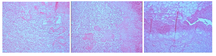
268. Hepatitis purulenta acuta
Description: In some lobules between branches of hepatocytes there is conspicuous extravasation and increased infiltration of parenchyma by neutrophils. This is evidence of an early stage of purulent inflammation arising in liver parenchyma. Later stages are characterized by formation of neutrophilic granulocyte accumulations (abscess formation).
Etiology: Traumatic reticuloperitonitis in cattle. Probably originates by transfer of purulent bacteria from the reticulum.
Pathogenesis: Purulent inflammation is characterized by pus and a large number of neutrophils in the tissue. It can occur on mucosal, serosal or other types of surfaces, or in solid organs. Within a solid tissue, purulent exudate can be confined into an abscess. Neutrophils, pathogen or a combination can cause necrosis of surrounding tissue. Acute hepatitis is associated with hepatocellular necrosis, and neutrophils, lymphocytes and macrophages infiltrate the necrotic area.

this is?
196. Leucosis lymphadenoidea hepatitis (Lymphosarcoma)
Description: At one end there is basophilic stained tumor tissue formed by slightly differentiated lymphocytes. Tumor tissue arises from portal and bile tracts (sign of liver lymphoid leukemia) gradually invades the center of the lobules, and may substitute them. In some parts of normal tissue, hemorrhagic tracts arise after migration of parasitic larvae.
Etiology: Viral infection, such as Marek’s disease in young birds.
Pathogenesis: Leukosis is a tumor of lymphocytes, but when the tumor is malignant the proper term is lymphosarcoma. Leucosis originates either in lymph nodes or in bone marrow, and metastasis is a characteristic feature. Tumor gradually replaces normal liver tissue which can lead to the death of the animal.
This is?
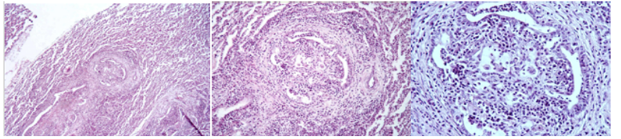
164. Glomerulonephritis chronica
Description: Most glomeruli are morphologically changed. They show thickened Bowman’s capsule which press on glomeruli, causing atrophy (periglomerulitis chronica). Many vessels of glomeruli are hyalinized. Some glomeruli are fibrotic (sclerosis). A large number of tubules have atrophy and others are dilated and become filled with eosinophilic casts. Interstitium is dilated and infiltrated by chronic inflammatory cells (lymphocytes, plasma cells, macrophages, rarely neutrophils).
Etiology: Chronic glomerulonephritis is caused by a long-term inflammation of glomeruli. The initial cause can be viral, bacterial, parasites, neoplasms, autoimmune disease or genetic predisposition.
Pathogenesis: The formation of immune complexes stimulate production of proteins which are chemotactic to neutrophils. These neutrophils infiltrate and damage the basement membrane and afterwards, monocytes infiltrate the glomeruli and continue damaging the glomeruli. When the lesion is severe and prolonged, the glomeruli turns hypocellular and non-functional (glomerulonecrosis)
this is?
259. Nephrocirrhosis (Nephritis interstitialis chronica) Renal fibrosis/scarring
Description: Differentiated granulation tissue causing atrophy of parenchyma. Tubular epithelium and glomeruli undergo atrophy and necrosis, interstitium is infiltrated by lymphocytes. Lumen of some tubules is filled with hyaline. Similarly, walls of vessels show marked fibrotic thickening. Thickened wall of Bowman capsule is observed as finely basophilic stained amorphous mass.
Etiology: Viral, bacterial or parasitic infections, toxins or nephrotoxic substances, immune-mediated disease, chronic urinary tract obstruction, certain medications, underlying conditions affecting the kidney. Most common in aging cats and dogs.
Pathogenesis: Occur following primary inflammation, degeneration or necrosis of glomeruli, tubules or interstitial tissue. It leads to scarring (fibrosis) and structural changes. Severe fibrosis and loss of nephrons can manifest as renal failure and uremia
this is?
260. Nephrocirrhosis (Van Koss) Same as nr. 259
Description: Sample from dog suffering from uremic syndrome, prepared for proof of calcium salts (Van Koss). Basophilic amorphous mass of Bowman’s capsule thickened walls is stained dark brown (positive calcia reaction)
this is?
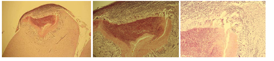
206. Meningitis spinalis purulenta
Description: Meninges (dura mater, arachnoidea, pia mater) are dilated and infiltrated by cells. Cell infiltrate consist of neutrophils. Tissue of the spinal cord is not seriously damaged. Dilated lymph and blood vessels, and purulent infiltrate. White areas represent some form of edematous reaction.
Etiology: Meningitis is caused by viral (non-purulent) and bacterial (purulent) infections. Purulent inflammation is most common in domestic animals, especially neonates.
Pathogenesis: Purulent meningitis is of hematogenous origin. The bacteria spread by blood and upon reaching the meninges, provoke an inflammatory response. Immune system reaction lead to accumulation of pus and inflammation of the spinal meninges, resulting in increased pressure of spinal cord and surrounding tissue.
this is?
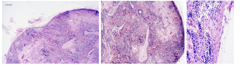
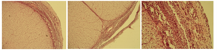
159. Cysticercus ovis (brain)
Description: At the periphery, under the meninges, there is a cyst intruding into the brain. In the center there is necrosis, mostly infiltrated by eosinophils. Surrounding the necrosis is granulation tissue (with giant cells), which form a capsule of the host organism. Death of parasites probably results from the development of an immune reaction.
Etiology: Intermediate stage of Taenia ovis, a tapeworm of dogs. Found in sheep after ingestion of eggs from contaminated food or water.
Pathogenesis: Eggs hatch in the intestine and migrate through the bloodstream to various tissues in the body, including the brain. There, the larvae form a cyst (cysticercus) and form a condition known as cerebral cysticercosis
this is?
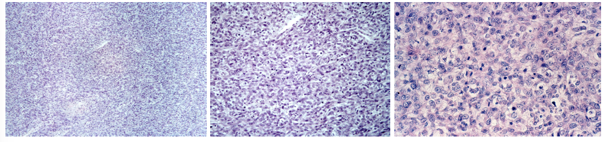
207. Fibrosarcomna uteri
Description: Tumor tissue composed of bundles of fibrous cells whirled in different orientations and directions. There is an abundant capillary framework in the tissue. Cells are slender, prolonged, and typically spindles with oval, bright nuclei. Ratio of mitotic figures in nuclei is relatively high. Tissue is poor from collagen fibers. At the margin there is extensive necrosis bordered with neutrophils with breaking nuclei.
Etiology: Malignant tumor arising from fibrous tissue. Cause is not fully known, but genetic factors, environmental causes and chemical exposure can contribute to the development.
Pathogenesis: Uncontrolled growth of cells within the uterine connective tissue leads to the formation of the tumor.
this is?
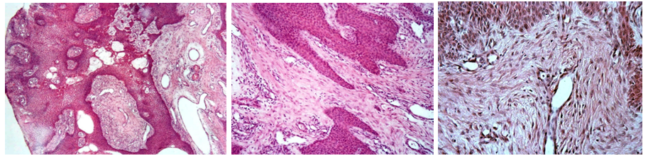
242. Hypertrophy of prostate
Description: We can see fibroelastic capsule with smooth muscle cell bundles. Fibrotic septae divide the parenchyma into lobules. Glandular epithelium is papillomatous proliferated. Under the capsule there are extensive cysts including fluid that arise by enlargement of lumens of prostatic glands. On the other side there is expressive proliferation of fibromuscular stroma, as well as different large cysts with gentle atrophic epithelium.
Etiology: Enlargement is common in older dogs and is hormone related, but the exact cause is unknown. Does not occur in castrated dogs. Hormone imbalance is divided into 2:
Acinar with formation of cysts - Overproduction of androgens
Fibromuscular - Overproduction of estrogens
Pathogenesis: Increased volume of the prostate serves as a protective mechanism, which can be due to compensatory or adaptive hypertrophy or hormonal influences. As animals age, hormonal fluctuations can lead to enlargement of the prostate gland. Consequences: Infection and obstruction of urinary bladder, hydronephrosis, constipation.
this is?

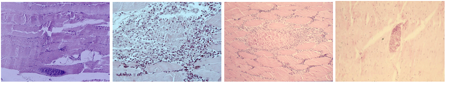
163. Dystrophia musculorum /146. Hyaline dystrophy
Description: Diffusely, you can see normal and degenerated myofibers. The sarcoplasm of degenerated fibers is homogenized (hyaline dystrophy) and damaged by large shreds or tiny granules, in others fibers also sarcolemma is ruptured (Zenker’s necrosis). The surroundings are infiltrated by leukocytes. Lymphatic capillaries are dilated
Etiology:
Nutritional myopathy of calves, lambs, piglets and foals: Deficiency of vit. E and selenium
Other types of myopathy: Monday morning disease
Gene defect of ryanodine receptor: Predisposition of bigger breeds
Pathogenesis: Muscle dystrophy means loss of muscle mass. It is characterized by changes in the morphology of cells caused by metabolites or other substances in a damaged or overloaded cell. It manifests as progressive muscle weakness and degeneration
this is?
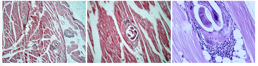
151. Trichinellosis musculorum
Description: Larvae of Trichinella spiralis are located in the tissue sections. Some are capsulated and others are without capsules. Surroundings show focal myositis characterized by the presence of neutrophils, lymphocytes and eosinophils.
Etiology: Affects humans, dogs, cats and pigs. T. spiralis is most common. Animal is infected when they eat meat containing cysts.
Pathogenesis: Larvae is released from cysts by digestive juices after they are ingested and turn into adults in the small intestine. These then produce larvae which migrate to the myofibers of the muscles, primarily masseter, tongue, eye and respiratory muscles (e.g. diaphragm). This results in focal myositis. The larva then forms a cyst and is ready to infect new hosts
this is?
109. Myositis sarcosporidica (Sarcocystosis of the muscles)
Description: Sarcocysts inside skeletal muscle with sarcosporidia. Some are stained by eosinophilic color, meaning they contain dead parasites. Other sarcocysts can be seen as calcified (dark red violet color). Cysts are bordered by inflammatory cells- Macrophages, lymphocytes, eosinophils, and rarely by fibroblasts (formation of capsule)
Etiology: 2-host protozoan parasite using carnivores as final host, and birds, reptiles, small rodents, herbivores and swine as intermediate host. Affect muscles of tongue, esophagus, diaphragm, masseter, etc.
Pathogenesis: In IH they penetrate endothelial cells and later leukocytes. They then migrate to muscles where they form cysts. FH then eats infected meat, and the parasite becomes an adult in the small intestine and reproduces. Eggs are released into the environment where they are ingested by IH.
this is?
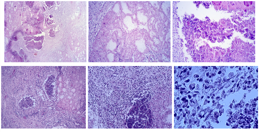
273. Mastitis apostematosa (Infectious sheep mastitis)
Description: Necrotic remnants of native glandular structure of mammary gland (simple necrosis). This necrotic area are bordered by remnants of purulent exudate (necrotized neutrophils), followed by formation of non-specific granulation tissue (characteristic of chronic process). Consists of macrophages, lymphocytes and fibroblasts. Violet stained homogeneous round mass are corpora amylacea.
Etiology: Caused by an infection of staphylococcus aureus and pasteruella haemolytica, often due to poor hygiene.
Pathogenesis: Virulence of bacteria is associated with the ability to adhere to mammary epithelium. It initially affects the tissue lining the milk collecting ducts and cisterns before entering small ducts and alveolar areas where they produce toxins and irritants which causes swelling and death of alveoli. In case of fast elimination, things go back to normal in a few days, but if the infection persists, the destruction of alveoli can be more severe due to accumulation of milk.
this is?
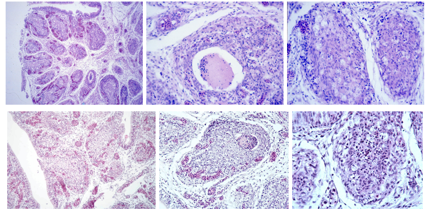
240. Tumor mixtus mammae
Description: Tumor characterized by proliferation of epithelial and fibrous tissue. Epithelial tissue preserved glandular structure with different size and formation of lumina. Connective tissue is mottled and includes fibroid, myxoid and chondroid structure with precipitation of minerals. Tumors grow slowly and may become malignant.
Etiology: Most common mammary growth in dogs. More than half are benign, while ⅓are malignant. Usually occurs in older female dogs. Causes are poorly understood.
Pathogenesis: Benign tumors have a slow and gradual growth, and recurrences or metastases can take a long time to become apparent. Sometimes, a carcinoma can arise from a benign mixed tumor. Malignant tumors grow usually more rapidly and the epithelial and myoepithelial cells preponderate with a considerable variation in cell size and shape.
this is?
258. Dermatitis eosinophilica (Scabies of rabbits)
Description: Dermis and excessive hyperemic subcutis on the inner side of the ear are abundantly infiltrated by eosinophils. A lot of erythrocytes can be observed free in tissue (extravasation in subcutis and skin surface).
Etiology: Ectoparasite Psoroptes cuniculi, which causes scabies in rabbits.
Pathogenesis: Non-burrowing mite living superficially in the rabbit’s ear. They feed on tissue and create an inflammatory reaction. They are permanent parasites but can be treated by ivermectin.
this is?
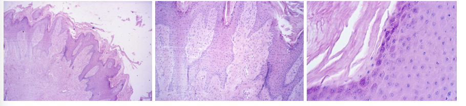
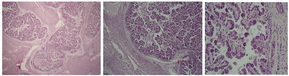
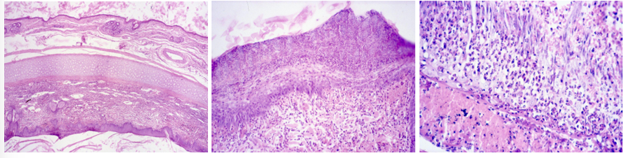
237. Fibropapilloma (Papillomatosis in cattle)
Description: Proliferation of fibrous tissue is as great or greater than that of epithelial tissue. Fibrous tissue forms whorls of fibers and has plumb, stippled nuclei with few mitotic figures. Epithelial rete pegs penetrate deep into fibrous moiety. On the surface of epidermis there is excessive hyperkeratosis and down also parakeratosis. Epidermal cells under hyperkeratosis or parakeratosis undergo dystrophy, and cytoplasm contain smaller and larger eosinophilic clumps.
Etiology: Virus-induced tumor commonly found on vulva and glans penis of younger animals. It is usually caused as a reaction to bovine verrucae virus. They can also be found in the periocular region and on the ventral part of the abdomen.
Pathogenesis: Virus infecting the keratinized bovine skin provokes the formation of the papilloma of relatively scant connective tissue core and abundant epidermal overgrowth. The result is one or multiple wart-like growths.