67. Benign and malignant bone tumours
1/49
There's no tags or description
Looks like no tags are added yet.
Name | Mastery | Learn | Test | Matching | Spaced |
|---|
No study sessions yet.
50 Terms
Primary bone tumor vs bone metastasis?
Primary bone tumors are considerably less common than bone metastases
4 subgroups of primary bone tumors?
Bone-forming tumors
- Osteoma
- Osteoid osteoma
- Osteoblastoma
- Osteosarcoma
Cartilage-forming tumors
- Osteochrondroma
- Chondroma
- Chondrosarcoma
Fibrous and fibroosseous tumors
- Fibrous cortical defect/nonossifying fibroma
- Fibrous dysplasia
Tumor-like lesions
- Aneurysmal bone cyst
Others
- Giant cell tumor
- Ewing sarcoma
Characteristic for bone-forming tumors?
They form osteoid, the unmineralized portion of bone
Osteoma?
- Benign tumor
- Exophytic masses of bone
- Commonly occurs on the surface of facial bones, paranasal sinuses or the skull
- Associated with Gardner syndrome
Gardner syndrome?
FAP + osseous and soft tissue tumors
Osteoid osteoma?
Benign tumor of osteoblasts which affects young adults (10-30)
- Characteristically forms a nidus of osteoid which is surrounded by dense bone
- Osteoid itself can be calcified to some extent
Where does osteoid osteoma occur?
In the metaphysis of long bones
- Especially the femur
Characteristic symptom of osteoid osteoma?
Precise pain to the affected site of the tumor
- Pain typically becomes stronger during the night
- Is effectively treated with NSAIDs, as the pain comes from prostaglandins produced by the tumor
Osteoid osteoma om X-ray?
There is a radiolucent central nidus which is rarely larger than 1,5cm
- May be surrounded by a radiopaque ring
Osteoblastoma?
Benign tumor of osteoblasts
Very similar to osteoid osteoma in many ways:
- Both affect young adults
- Produce a radiolucent nidus
Difference between osteoblastoma and osteoid osteoma?
- Osteoblastomas are larger (>2cm)
- Often occur in parts of the vertebrae
- Pain does not respond to NSAIDs, and is not worse in the night
- On X-ray; the radiolucent nidus is not surrounded by radiopaque bone
Osteosarcoma?
Highly malignant tumor of the osteoblasts
- Most frequent in teenagers and elderly
Risk factors osteosarcoma?
- Paget disease of bone
- Hereditary retinoblastoma
- Irradiation
- Benign bone processes
• Osteochondromatosis
• Chondromatosis
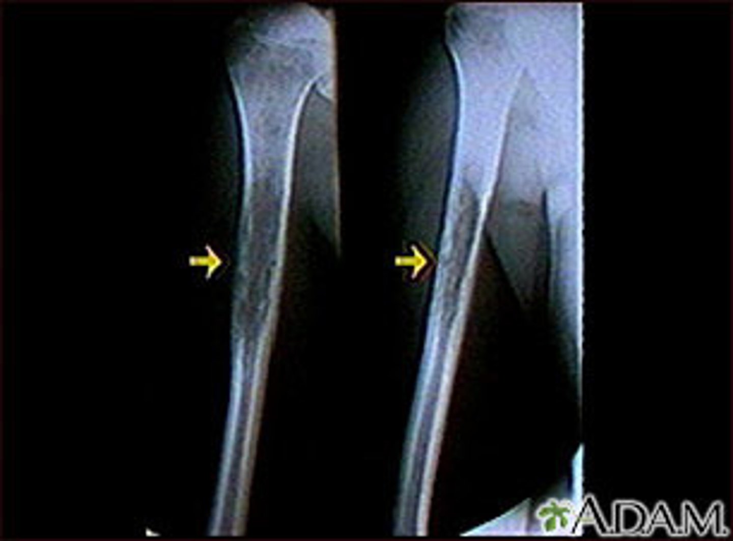
How does osteosarcoma often present?
Arises in the metaphysis of long bones
- Distal femur
- Proximal tibia
= Meaning that it often occurs around the knee joint
Manifest as a pathological fracture
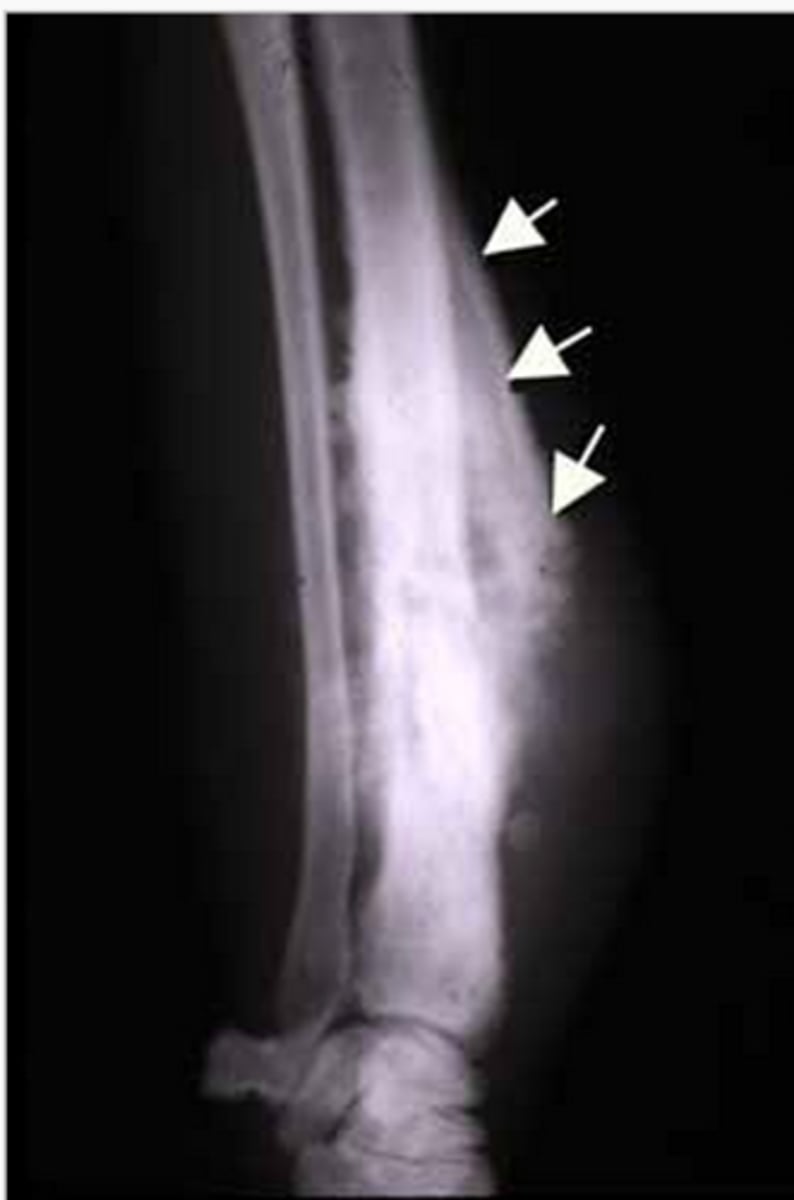
Where does osteosarcoma originate from?
Originates from the medulla of the bone
- From which they frequently break through the cortex & "lift" up the periosteum
= This forms a characteristic X-ray picture called Codman triangle
Codman triangle?
the triangular area of new subperiosteal bone that is created when a lesion, often a tumour, raises the periosteum away from the bone.
Infiltration and metastasis of osteosarcoma?
- Frequently invade surrounding soft tissue
- Metastasize to the lung in almost 100% of cases
Treatment osteosarcoma?
Surgical removal and aggressive chemotherapy
Cartilage-forming tumor?
These tumors form cartilage instead of bone, and are often visible as radiolucent masses inside bones on the X-ray
Osteochondroma?
Most common benign tumor of bone
- It produces a growth that grows laterally out of the epiphyseal growth plate
- The growth has a cartilage cap on it

Chondroma?
Benign tumor of hyalin cartilage
- Occur in bones of endochondral origin
- Usually arises in the medulla of small bones of hand and feet
= Especially proximal phalanges
Chondroblastoma?
Very rare benign tumor of cartilage
Chondrosarcoma?
Malignant tumor of cartilage
- Affects adults in 30-60y/o
- Is much more rare than osteosarcoma
Where does chondrosarcoma usually arise?
- Medulla of pelvic bones
- Ribs
What can be seen on X-ray - chondrosarcoma?
Osteolytic lesions with calcifications
(spots of bone damage that result from cancerous plasma cells building up in your bone marrow.)
Morphology of chondrosarcoma?
Ranges from well-differentiated to poorly differentiated
- The well-differentiated can be hard to distinguish from chondroma
Treatment chondrosarcoma?
Surgical removal
- Can be difficult due to bones are commonly involved
Fibrous and Fibroosseous Tumors?
- Fibrous cortical defects
- Fibrous dysplasia
Fibrous cortical defects?
Not true neoplasms
- Rather developmental malformations of the bone
- Form demarcated radiolucencies in bones - often in epiphysis
- Small lesions (0,5cm)
Nonossifying fibromas?
The name of larger lesions of "Fibrous cortical defects"
- Up to 5-6 cm
- Often incidental findings & asymptomatic
- Rarely requires treatment
Fibrous dysplasia?
Benign tumor comprised of immature bone structures
How can fibrous dysplasia manifest?
Three different ways:
1. Monostotic -affecting only a single bone - 70% of cases
2. Polyostotic - affecting multiple bones - 27% of cases
3. As part of McCune-Albright syndrome - 3% of cases
McCune-Albright syndrome?
Condition characterized by:
- Café au lait skin pigmentations
- Endocrine abnormalities
- Multiple fibrous dysplasia
Other bone tumors?
- Giant cell tumor
- Ewing sarcoma
Giant cell tumor (osteoclastoma)?
Arises in the epiphysis of long bones
- Distal femur
- Proximal tibia
- Is locally infiltrative
- Almost NEVER metastasize
Cell types in giant cell tumor?
Comprised of two cell types:
- Neoplastic "stromal cells"
- Reactive osteoclast-type giant cells
How does giant cell tumor look in X-ray?
Soap bubbles
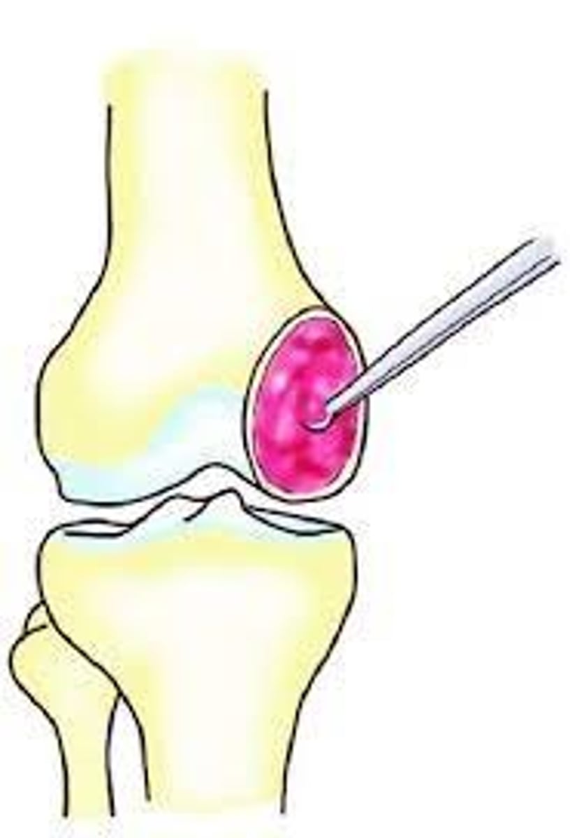
How are Giant cell tumor treated?
By curettage
- However, around 40-60% recur locally
Ewing Sarcoma?
Aggressive malignant proliferation of poorly differentiated cells from neuroectoderm
- Type of "primitive neuroectodermal tumor" PNET
- Arises from the diaphysis of long bones, usually in male children
What mutation is common in Ewing sarcoma?
t(11;22) translocation
- Can be important during diagnosis
Ewing sarcoma - histology?
Tumor cells are PAS positive, as they contain glycogen
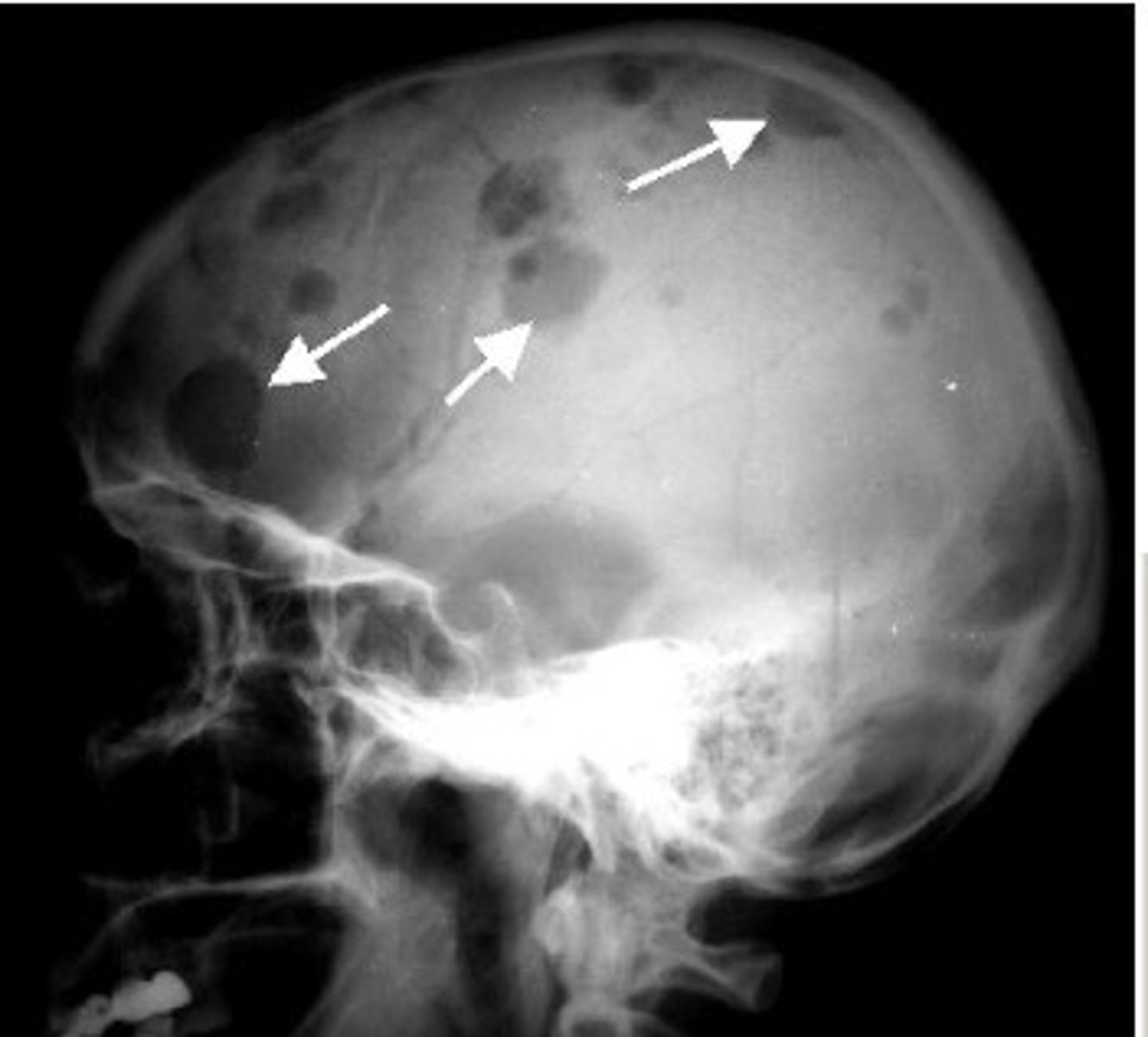
How is Ewing sarcoma on x-ray?
Characteristic onion-skin pattern
Metastasis of ewing sarcoma?
Frequently metastasize into lung or other bones
Treatment ewig sarcoma?
- High-dose irradiation
- Chemotherapy
- Surgical removal
Tumor-like lesions - bone?
Aneurysmal bone cysts
- Blood-filled cysts with septa that form osteolytic lesions
- Often occur in the spine & flat bones
- Cysts are lined by fibroblasts and myofibroblasts
- Septa is comprised of osteoid and bone
Metastatic tumors of the bone?
Metastatic tumors in bone are much more common than primary bone tumors
- Bones: 3rd most common site of metastasis, after lung and liver
- They are often multiple rather than solitary
Where does bone metastases often originating from?
- Breast
- Lung
- Prostate
- Thyroid
- Kidney
(Breast, lung & prostate account for 80% of cases)
What does metastases to bone often form?
Osteolytic lesions (decreased bone density) in the bone
What does bone metastases from prostate cancer form?
They form osteoblastic lesions (increased bone density)
Frequent symptom of bone metastasis?
- Strong bone pain
- Pathological fractures