MOD 6 - Thoracic Cage Fractures: Video Notes
1/20
Earn XP
Description and Tags
Flashcards covering thoracic cage functions, rib and sternum fractures, imaging views, potential complications, and technologist considerations.
Name | Mastery | Learn | Test | Matching | Spaced | Call with Kai |
|---|
No analytics yet
Send a link to your students to track their progress
21 Terms
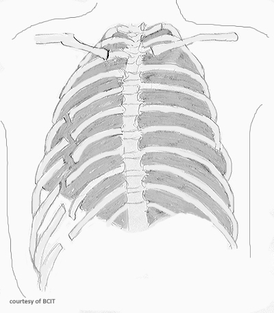
What are the two main functions of the thoracic cage?
To protect vital organs and to serve as an attachment site for muscles of the neck, back, upper abdomen, and shoulder girdle.
What holds the ribs together and maintains stability even if a rib is fractured?
Intercostal muscles.
What is a common symptom of thoracic cage fractures, especially when lying down?
Painful and laboured breathing.
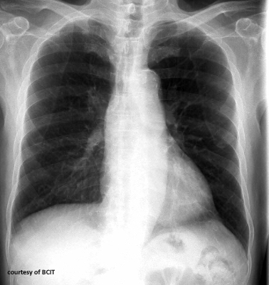
Which ribs are most commonly fractured?
Ribs 3–10.
Where is the weakest point of the rib?
Just anterior to the rib angle, along the axillary border.
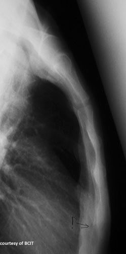
Which radiographic view best visualizes rib fractures?
Oblique rib views.
What are the two main causes (etiologies) of rib fractures?
External force (e.g., falls, car accidents) and internal force (e.g., excessive coughing in elderly or osteoporotic patients).
How are non-displaced rib fractures usually treated?
They typically heal without intervention.
Name three possible complications of rib fractures.
Pneumothorax, cardiac perforation, flail chest.
What is flail chest?
A condition where three or more ribs are fractured in multiple places, causing unstable thoracic pressure and independent rib movement during breathing.
What are the three parts of the sternum?
Manubrium, body, and xiphoid process.
Where do most sternal fractures occur?
In the mid-body of the sternum.
What is the most common cause of sternal fractures?
Direct trauma from impacts or crushing injuries, often in motor vehicle collisions.
Give one example of a sternal stress fracture cause.
Overuse in athletes (e.g., wrestlers).
How are non-displaced sternal fractures usually treated?
Conservatively, without surgery.
Name three possible complications of sternal fractures.
Thoracic organ injury; Cardiac injury; Pneumothorax or punctured mediastinum.
What factors influence patient positioning for imaging a thoracic cage fracture?
Pain level, breathing difficulty, fracture location, suspected injury mechanism, and clinical signs.
Why are oblique rib views often used?
They better visualize rib fractures compared to standard views.
What should a technologist monitor for in thoracic cage fracture patients?
Respiratory distress and complications.
When should urgent intervention be sought?
If there are signs of pneumothorax or cardiac involvement.
What is the technologist’s priority when positioning a patient with suspected thoracic cage fractures?
Minimize discomfort while maximizing image quality.