Exam #4
1/21
There's no tags or description
Looks like no tags are added yet.
Name | Mastery | Learn | Test | Matching | Spaced | Call with Kai |
|---|
No analytics yet
Send a link to your students to track their progress
22 Terms
Function of the NS
Sensory receptors monitor changes inside and outside the body
Processes and interprets sensory input
Socrates a response by activating effector organs
Divisions of the NS
CNS = brain and spinal cord
PNS = cranial nerve, spinal nerves, ganglia
special somatic senses
hearing -soundwaves
balance - position
vision - light
Proprioception
series of senses that monitor the degree of stretch in muscles, tendons, and joint capsules
-senses the positions and movements of our body parts
-body sense - position and movement of body in space (time and space)
Nissl Bodies
Clusters of rough E R and free ribosomes; also called chromatophilic bodies
–Function to renew membranes of the neuron
Vesicles and Neurotransmitters
▪Neurotransmitters are released from vesicles at the terminal boutons
Neurotransmitters
Chemical released by neurons that may, upon binding to receptors on neurons or effector cells, stimulate or inhibit them
Application Q about Glial cells and general cell biology and replication and tumors
In clinical and research applications, the intersection of glial cells, cell biology, and replication is where we find the origin of most primary brain tumors. Because mature neurons generally do not replicate (they are post-mitotic), the vast majority of "brain" tumors actually arise from glial cells.
Diseases and Conditions in the NS
-MS = autoimmune disease in which myelin in the CNS is destroyed, leading to neuronal dysfunction.
-Characterized by periods of relapses and remissions. Common symptoms include visual disturbances, muscle weakness, fatigue, and depression
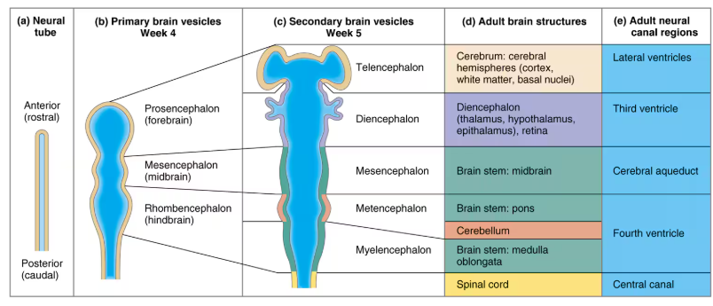
Hindbrain development
primary brain vesicles (wk 4) → Hind brain = rhombencephalon
secondary brain vesicles (wk 5)→ The early hindbrain has become metencephalon and myelencephalon
Metencephalon = brain stem: pons & cerebellum
Myelencephalon =brain stem: medulla oblongata
all is in fourth ventricle

Diencephalon development
primary brain vesicles (wk 4) → prosencephalon (forebrain)
secondary brain vesicles (wk 5) → diecephalon
adult brain strutures = diencephalon = thalamus, hypothalamus, epithalamus, retina
all in 3rd ventricle
gray matter in the CNS
In the spinal cord, it forms a butterfly-shaped region with interneurons in the dorsal half and motor neurons in the ventral half.
In the brain, gray matter is found both internally around ventricles and externally as cortex layers on structures like the cerebrum and cerebellum.
Thalamus
Structure: The thalamus is an egg-shaped, paired structure that constitutes about 80% of the diencephalon. It contains approximately a dozen major nuclei divided into three groups by the internal medullary lamina: anterior nuclei, medial group (including the medial dorsal nucleus), and large lateral nuclear group.
Location: Situated in the superolateral walls of the third ventricle, the thalamus is deeply embedded within the brain. The right and left parts are typically connected by a small midline structure known as the interthalamic adhesion.
Function: Acting as a "gateway" to the cerebral cortex, the thalamus relays sensory impulses from all conscious senses except olfaction. It processes these signals, organizing them and either amplifying or toning them down before they reach the cerebral cortex. This function allows for focused attention amidst distractions.
Homunculus
The term "homunculus" refers to a visual representation of the human body mapped onto the brain's sensory and motor cortices. There are two types: the sensory homunculus and the motor homunculus.
Sensory Homunculus: This map is located on the postcentral gyrus of the somatosensory cortex. It represents how different parts of the body send sensory information to specific regions of the brain. The size of each body part in this map corresponds to its sensitivity, not its physical size. For example, lips and fingertips appear larger because they have more sensory receptors, making them highly sensitive [1].
Motor Homunculus: Found on the precentral gyrus of the primary motor cortex, this map illustrates how different areas control voluntary movements. Like the sensory version, it shows an exaggerated representation for areas requiring precise control, such as hands and facial muscles [2].
Both maps exhibit contralateral projection, meaning each hemisphere controls or receives input from the opposite side of the body.
Visual Association Areas
he visual association areas are part of the cerebral cortex involved in processing visual information. They extend from the occipital lobe into the posterior association area, which includes parts of the temporal and parietal lobes. These areas process complex visual stimuli through two main pathways:
Dorsal Stream ("Where" Pathway): This pathway extends through the posterior parietal cortex to perceive spatial relationships among objects, helping identify their location.
Ventral Stream ("What" Pathway): Extending through the inferior temporal lobe, this pathway is responsible for recognizing objects, words during reading, and faces.
These pathways work together to integrate visual information with other sensory inputs, contributing to a unified perception of our environment and aiding in tasks like language comprehension and spatial awareness.
Blood-Brain Barrier
The blood-brain barrier (BBB) is a critical structure that protects the brain from harmful substances in the bloodstream while allowing essential nutrients to pass through. It consists of tightly joined epithelial cells in the brain's capillaries, making them the least permeable capillaries in the body. This tight junction prevents toxins like urea and bacterial toxins from entering brain tissue.
However, the BBB is not an absolute barrier. Nutrients such as glucose and oxygen are transported through specialized mechanisms, while fat-soluble molecules like alcohol and anesthetics can diffuse freely across it. During prolonged stress, these tight junctions may open, potentially allowing harmful substances to enter, which has been linked to conditions like Gulf War syndrome.
Understanding how to manipulate this barrier could aid in delivering drugs directly to the brain for treating diseases or tumors.
RAS
Reticular Activating System (RAS)
The Reticular Activating System (RAS) is a network of neurons located in the brainstem that plays a crucial role in regulating wakefulness and sleep-wake transitions. Axons from major sensory tracts synapse on RAS neurons, which helps keep these neurons active and enhances their arousing effect on the cerebrum. This explains why stimuli such as visual, auditory, and touch can help maintain alertness, making environments like crowded rooms conducive for studying due to increased stimulation.
The RAS also influences sleep and arousal from sleep. Substances like general anesthesia, alcohol, tranquilizers, and sleep-inducing drugs can depress the RAS, leading to decreased alertness or consciousness. Severe injury to the RAS may result in coma. Additionally, descending pathways from the reticular formation influence somatic motor neurons affecting posture and muscle tone during sleep.
medulla
Medulla Oblongata
The medulla oblongata, often simply called the medulla, is the most caudal part of the brain stem. It connects directly to the spinal cord at the foramen magnum of the skull. This region is crucial as it contains major fiber tracts and brain nuclei that facilitate communication between the brain and spinal cord.
Structure: The medulla features two longitudinal ridges known as pyramids, formed by large fiber tracts called pyramidal tracts. These tracts carry voluntary motor signals from the cerebrum to the spinal cord.
Function: A significant function of these pyramidal fibers is their crossover in the caudal medulla, known as decussation. This crossover ensures that each cerebral hemisphere controls movements on the opposite side of the body.
Relay Stations: The medulla also houses important relay nuclei like nucleus gracilis and nucleus cuneatus, which process sensory information before sending it to higher brain regions.
Additionally, four pairs of cranial nerves attach to this area, playing roles in both sensory input and motor output.
Diseases and Condition of the CNS
The central nervous system (CNS) can be affected by various disorders, including injuries and diseases. Here's a brief overview:
Spinal Cord Injuries: Severe injuries to the spinal cord can lead to paralysis or loss of sensation below the injury site. The extent of these effects depends on the location and severity of the injury.
Concussions: These are traumatic brain injuries that may cause temporary confusion, headaches, or memory issues. Symptoms vary based on the severity of the concussion.
Strokes: Occur when blood flow to a part of the brain is interrupted, leading to potential brain damage and symptoms like weakness, speech difficulties, or vision problems.
Alzheimer’s Disease: A progressive disorder causing memory loss and cognitive decline due to changes in brain structure.
Congenital Disorders: Conditions such as anencephaly, cerebral palsy, and spina bifida affect CNS development from birth.
Understanding these conditions helps in recognizing symptoms early for better management and treatment outcomes.
Effectors of the ANS
Effectors of the ANS
The Autonomic Nervous System (ANS) is a crucial part of the peripheral nervous system that controls involuntary bodily functions by innervating smooth muscle, cardiac muscle, and glands. These effectors are responsible for regulating vital functions such as heart rate, blood pressure, digestion, and urination. The ANS operates largely below the level of consciousness to maintain homeostasis.
The ANS is divided into two main divisions: the sympathetic and parasympathetic systems. Both divisions often target the same organs but have opposite effects:
Sympathetic Division: Prepares the body for "fight or flight" responses by increasing heart rate and energy availability.
Parasympathetic Division: Promotes "rest and digest" activities by conserving energy and facilitating digestion.
These divisions differ in their neurotransmitter release at effector organs: acetylcholine for parasympathetic fibers and norepinephrine for sympathetic fibers. This dual control allows precise regulation of organ function to meet varying physiological demands.
Enteric Nervous System
The enteric nervous system (ENS) is often referred to as the "brain in the gut" due to its extensive network of over 100 million neurons, comparable to the spinal cord. It operates independently within the walls of the alimentary canal, forming reflex arcs with sensory neurons, interneurons, and motor neurons. These arcs control muscular and secretory activities in response to stimuli from the digestive tract's contents.
The ENS consists of two main nerve plexuses:
Myenteric Nerve Plexus: Located between circular and longitudinal muscle layers, it controls peristalsis and segmentation.
Submucosal Nerve Plexus: Found within the submucosa, it signals glands for secretion and muscularis mucosae for contraction.
While capable of independent function, the ENS is influenced by the central nervous system through visceral sensory fibers and autonomic nervous system inputs. Parasympathetic stimulation enhances digestive functions, whereas sympathetic input inhibits them
Hypothalamus
The Hypothalamus
The hypothalamus is a crucial part of the brain located below the thalamus, forming the inferolateral walls of the third ventricle. It plays a significant role as the main visceral control center of the body, regulating various activities of visceral organs.
Autonomic Nervous System Control: The hypothalamus directs autonomic neurons that regulate heart rate, blood pressure, digestive tract activity, and glandular secretions.
Temperature Regulation: It acts as the body's thermostat by monitoring blood temperature and initiating cooling or heating mechanisms like sweating or shivering.
Hunger and Thirst Regulation: By sensing nutrient and salt concentrations in the blood, it mediates hunger and thirst sensations to maintain balance.
Sleep-Wake Cycles: The suprachiasmatic nucleus within the hypothalamus regulates circadian rhythms based on light-dark information from the optic nerve.
Endocrine System Control: It influences hormone secretion by controlling the pituitary gland.
Emotional Responses and Motivational Behavior: As part of the limbic system, it affects emotions like pleasure, rage, fear, and motivational behaviors such as feeding and sexual behavior.
Injuries to this area can lead to disorders affecting weight, sleep, hydration, and emotions.