systems unit review
1/129
There's no tags or description
Looks like no tags are added yet.
Name | Mastery | Learn | Test | Matching | Spaced | Call with Kai |
|---|
No analytics yet
Send a link to your students to track their progress
130 Terms
Types of circulation
Pulmonary circulation – pathway of blood from heart to the lungs and back
Cardiac circulation – route of blood WITHIN the heart
Systemic Circulation – route of blood from the heart to the body. It includes all the blood vessels other than those associated with the lungs
The circulatory (or cardiovascular) system has several functions:
Transportation of O2, CO2, wastes, nutrients, and hormones
Maintain body temperature
Maintain body fluid levels
Parts of the Mammalian Circulatory System
The Heart: a muscular organ that continuously pumps blood through the body, generating blood flow.
The Blood Vessels: a system of hollow tubes through which the blood moves.
The Blood: The fluid that transports nutrients, O2, CO2 and many other materials throughout the body.
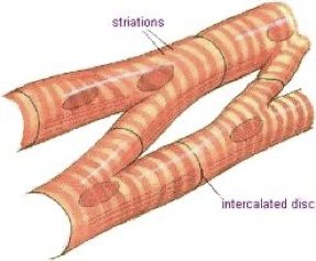
Human Heart Anatomy
Cardiac muscle cells are arranged in a network that allows the heart to contract and relax rhythmically and involuntarily without becoming fatigued.
Location of the Heart
The heart is located in the chest cavity
Slightly behind and to left of sternum
Rib cage protects it
Human Heart Anatomy
Has four chambers
Atria: the two top chambers that fill with blood returning from the body or the lungs (singular atrium).
Ventricles: two bottom chambers that receive blood from the atria and pump it out to the body or the lungs.
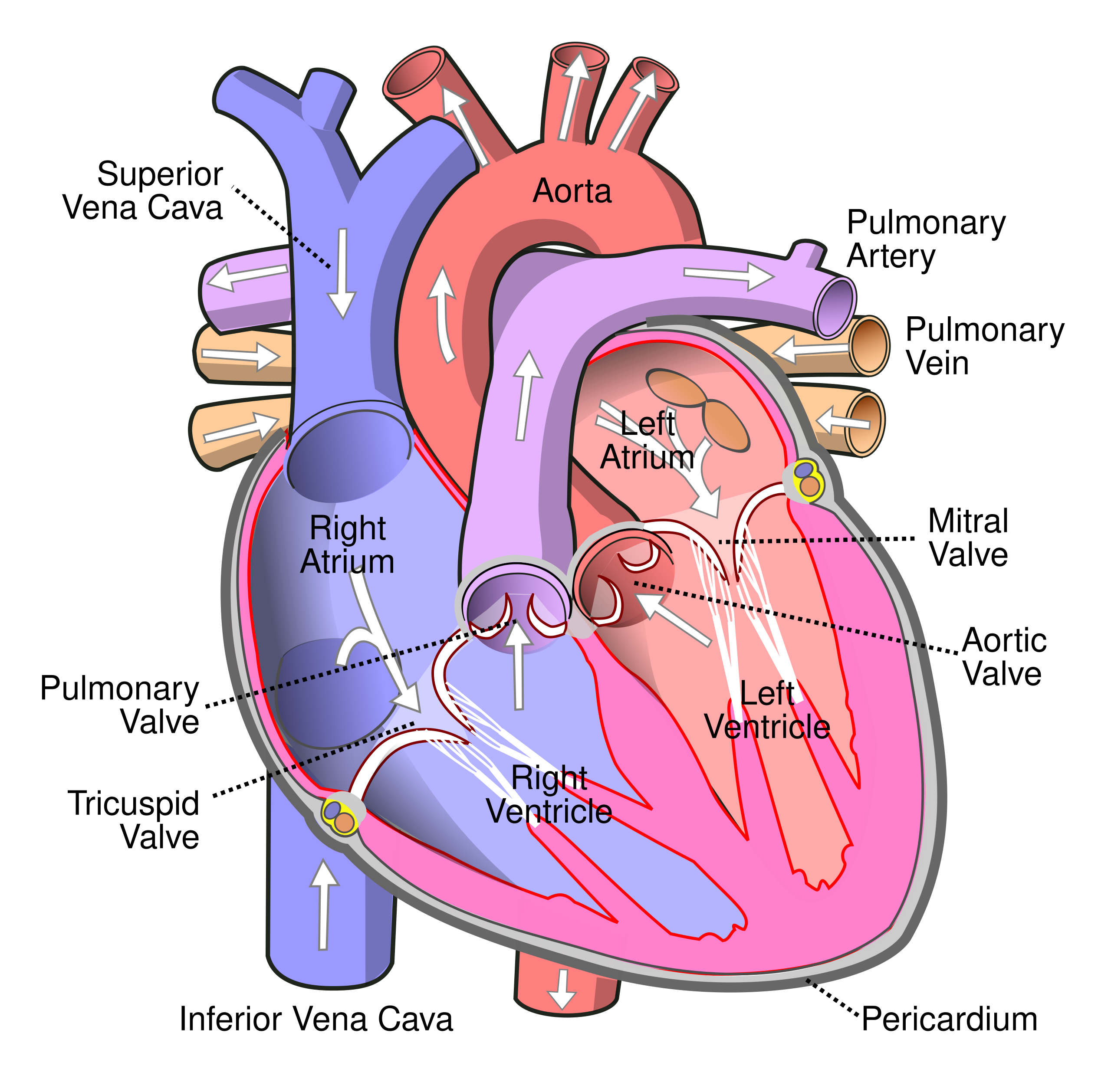
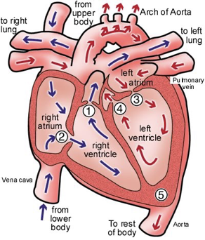
Blood Flow in the Heart
The vena cavae bring oxygen-poor blood from the body to the right atrium.
The oxygen-poor blood flows from the right atrium into the right ventricle.
The right ventricle pumps the oxygen-poor blood to the lungs through the pulmonary arteries.
The pulmonary veins bring oxygen-rich blood from the lungs back to the heart through the left atrium.
Oxygen-rich blood flows from the left atrium to the left ventricle.
The left ventricle pumps the oxygen-rich blood to the body through the aorta.
Heartbeat “lub-DUB”
Valves prevent the blood from flowing backwards.
The “lub” sound is caused by the closing of the atrioventricular (AV) valves as blood is pumped from the atria to the ventricles.
The “DUB” sound is caused by semilunar valves, as blood is pumped from the ventricles into the arteries
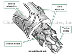
Blood Vessel Layers
Vasa vasorum
“vessels of vessels”
Small blood vessels that supply oxygen to the layers of large blood vessels (i.e. aorta, vena cava)
Nervi vasorum
Nerve supply to large blood vessels
Allows regulation by the sympathetic nervous system
Major Veins & Arteries
Internal jugular vein
Subclavian vein and artery
Cephalic vein
Basilic vein
Inferior vena cava
Common iliac artery and vein
Great saphenous vein
Femoral vein
Renal vein
Renal artery
Common carotid artery
Brachial artery
Radial artery
Ulnar artery
Femoral artery
Aorta
Heart
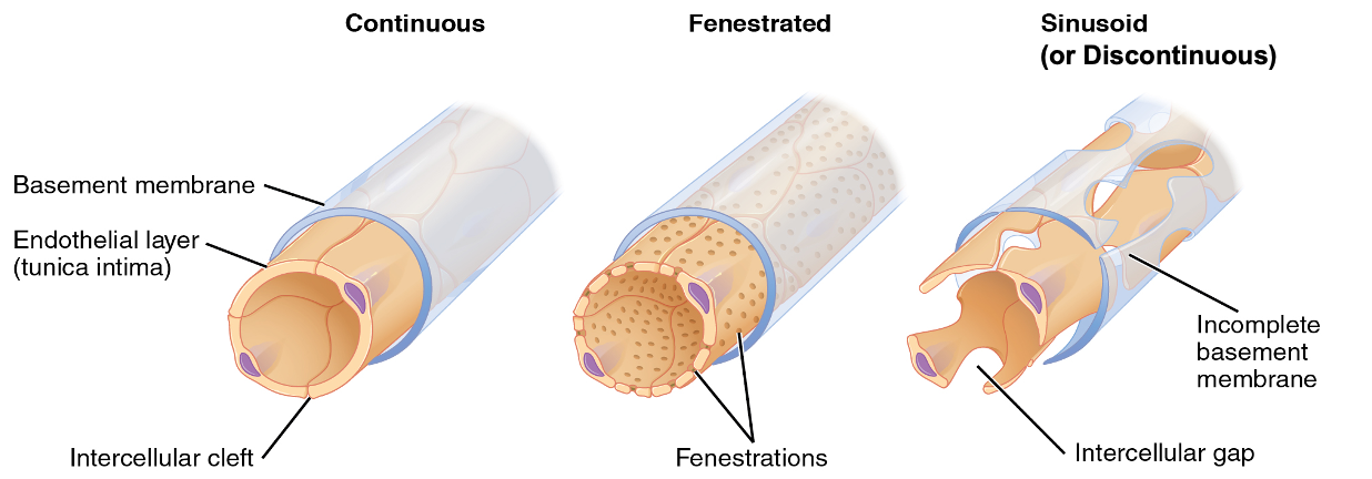
3 Types of Capillaries
Least permeable - continous
Found in: fat, muscle, nervous tissue (most common)
More permeable - fenestrated
Found in: intestinal villi, endocrine glands, kidney glomeruli (tissues that specialize in fluid/molecule exchange)
Highly permeable - sinusoid
Found in: liver, bone marrow, spleen (where there is exchange of large proteins between blood and tissue)
Blood Introduction
Blood is a collection of cells that have been specialized to perform a set of tasks within an organism.
For this reason, doctors and scientists consider blood a tissue and not a fluid.
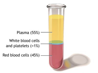
Blood consists of two distinct elements:
1.Plasma: the fluid portion of the blood (55% of blood)
2.Cells: the solid portion of blood (45% of blood)
Plasma
Fluid portion of the blood that carries blood cells.
Made up of 90% water, the other 10% made up of blood proteins, glucose, vitamins, minerals, dissolved gases, waste products of cell metabolism.
Also transports CO2.
Red Blood Cells
Erythrocytes
Make up about 44% of blood.
Specialized to transport oxygen; without RBCs, plasma could only carry about 2% of the normal oxygen load.
Have a biconcave disc shape, which increases surface area for oxygen exchange.
They contain no nucleus, live for about 120 days, and are constantly reproduced.
Average count: approximately 5.5 billion RBCs per mL of blood in males and 4.5 billion per mL in females.
Each RBC contains roughly 280 million hemoglobin molecules.
Hemoglobin is an iron-containing protein with four globin chains and one iron molecule, giving it a high affinity for oxygen.
Hemoglobin plus oxygen forms oxyhemoglobin.
RBCs lose their nucleus to maximize space for hemoglobin.
White Blood Cells
Make up about 1% of blood's volume.
Produced in bone marrow.
White blood cells contain nuclei and appear colourless.
They play many roles in fighting off infection and protecting the body from pathogens.
The number of WBC may increase by double when you are fighting off an infection.
Pus: fragments of remaining protein of the WBC and the invader.
Leukocytes and Lymphocytes
White Blood Cells:
Two major disease-fighting types are leukocytes and lymphocytes.
Leukocytes (Macrophages):
Engulf and digest pathogens.
Part of the innate immune response, which provides a general defense against infection.
Can pass through capillary walls to reach infected tissues.
Lymphocytes:
Part of the acquired (specific) immune response.
Recognize and remember particular pathogens.
Provide targeted defense and respond more effectively if the same pathogen invades again.
Platelets
Are not cells.
Fragments of larger cells that broke apart in the bone marrow.
They contain no nucleus and break down relatively quickly.
They help the blood to clot and protect the body from excessive blood loss after an injury.
Blood Clotting
What stimulates a blood clot?
Injury to the vessel lining and contact of the blood with tissues outside the vessel
1. Blood will NOT clot until a blood vessel is broken. This is the trigger!
2. Substances released by broken blood vessels attract platelets to the site
3. As platelets collect, they rupture and release special chemicals that combine with other clotting agents to produce thromboplastin
4. As long as calcium ions are present in the blood, thromboplastin reacts with prothrombin to produce thrombin
5. Thrombin reacts with fibrinogen to produce fibrin
6. Fibrin forms mesh strands around the area of injury. This traps the escaping material and forms a clot.
What are white blood cells?
White blood cells, a.k.a. leukocytes, are responsible for protecting your body from infection via pathogens
WBC circulate in your blood and respond to injury or illness
What do white blood cells do?
Locate the site of infection and locate other white blood cells to come and help defend your body
Many white blood cells carry out phagocytosis – cell eating (engulf invader to protect you)
Once this WBC army arrives and fights, they produce antibodies for protection.
LOCATION of WBC
WBC are found all around in your blood and are able to travel through blood vessel walls
Appearance and size of wbc
-white blood cells are colorless --can appear as a very light purple to pink color when examined under a microscope and colored with dye -round shape with a distinct nucleus
Microscopic
14-16 µm in diameter
How are white blood cells formed?
White blood cell formation occurs in the soft tissue inside of your bones (bone marrow).
Types of WBC/Leukocytes- The macrophage
Innate immune response
Not specific
Respond FAST
Initiate other responses
Types of WBC/leukocytes
Neutrophils
•Help protect your body from infections by killing bacteria, fungi and foreign debris.
•Lymphocyte
Consist of T cells, natural killer cells and B cells to protect against infections and produce immune response proteins to help you fight infection (antibodies).
Eosinophil
Identify and destroy parasites, cancer cells and assists basophils with your allergic response.
Basophil
•Produces an allergic response like coughing, sneezing or a runny nose. Main soldier in allergic response
Monocytes
Defend body by cleaning up damaged cells.
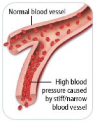
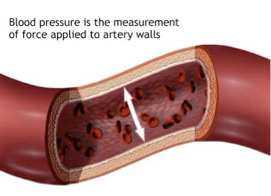
Blood Pressure
Force of the blood on the walls of the arteries.
Normal BP 120/80 mm Hg; decreases as you move away from the heart.
Stroke Volume: volume of blood leaving heart (L)
Heart Rate: number of beats (contractions) per minute (bpm)
Normal BP 120 mmHg/80 mmHg
The top number is your systolic blood pressure. (The highest pressure when your heart beats and pushes the blood around your body.)
The bottom one is your diastolic blood pressure. (The lowest pressure when your heart relaxes between beats.)
Two factors determine BP:
1.Cardiac Output (CO): amount of blood pumped from the heart each minute = Heart Rate (HR) x Stroke Volume (SV)
–⇡ CO = ⇡ BP
–increase CO by ⇡HR or ⇡ Stroke Volume (stronger heart)
2.Arteriolar resistance: diameter of the arteriole determines the amount of blood flow
–⇡ diameter = ⇣ BP
Blood Pressure Regulation
•Diameter of blood vessels regulated by the medulla oblongata.
•Vasoconstriction: nerve impulses cause muscle to contract, reducing diameter of vessel, reduces flow to tissue, increases pressure
•Vasodilation: nerve impulses cause muscles to relax, increasing diameter of vessel, increases flow to tissue, decreases pressure
The Cardiac Cycle
•A bundle of specialized muscle tissue, called the sinoatrial (SA) node, stimulates the muscle cells to contract and relax rhythmically.
•Also referred to as the pacemaker, because it sets the pace for cardiac activity
•Located in the wall of the right atrium.
•The SA node generates an electrical signal that spreads over the two atria and makes them contract simultaneously.
•As the atria contract, the signal reaches another node, called the atrioventricular (AV) node.
´1. Draw the correct passage of blood as it returns from the left leg through its journey all the way back
´2. What is the wall of muscle that separates the left and right side of the heart called
´3. What is the fluid filled membrane that surrounds the heart and protects from friction called?
´4. What side of the heart receives deoxygenated blood?
´5. What is the circuit called the carries blood to and from lungs and heart?
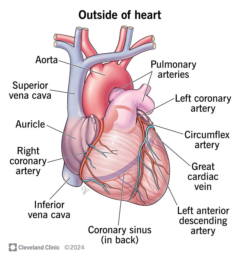
´6. These are able to supply heart muscle cells with oxygen and nutrients. If plaque builds up inside these, it causes chest pain/ angina which is treated with drugs or coronary bypass surgery. What structures are we discussing?
Left leg → inferior vena cava → right atrium → right ventricle → pulmonary arteries → lungs → pulmonary veins → left atrium → left ventricle → aorta → body
Septum
Pericardium
Right side
Pulmonary circuit
Coronary arteries
Pulmonary Circuit – Oxygenation Exception
Normally:
Arteries = oxygen-rich
Veins = oxygen-poor
Pulmonary circuit is the exception:
Pulmonary artery = oxygen-poor blood → lungs
Pulmonary veins = oxygen-rich blood → left atrium
Reason: lungs are where gas exchange occurs, so blood only becomes oxygenated after it reaches them.
Tunica Layers of Blood Vessels
1. Tunica Intima (or Tunica Interna)
Innermost layer
Smooth endothelium
Function: reduces friction for blood flow
2. Tunica Media
Middle layer
Made of smooth muscle and elastic fibers
Function: vasoconstriction and vasodilation (controls blood pressure and flow)
3. Tunica Externa (or Tunica Adventitia)
Outermost layer
Connective tissue
Function: protects vessel and anchors it to surrounding tissue
Organic Compounds
Compounds that contain CARBON are called organic.
Macromolecules are large organic molecules.
Also called POLYMERS.
Made up of smaller “building blocks” called MONOMERS.
Examples:
1. Carbohydrates
2. Lipids
3. Proteins
4. Nucleic acids (DNA and RNA)
Carbohydrates
Small sugar molecules to large sugar molecules.
Examples:
A. monosaccharide
B. disaccharide
C. polysaccharide
Monosaccharide
Monosaccharide: one sugar unit
Examples: glucose (C6H12O6)
deoxyribose
ribose
Fructose
Galactose
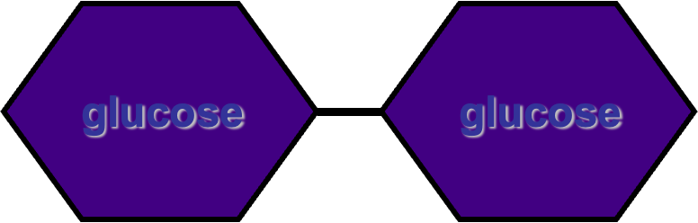
Disaccharide
Disaccharide: two sugar unit
Examples:
Sucrose (glucose+fructose)
Lactose (glucose+galactose)
Maltose (glucose+glucose)
Polysaccharide
Polysaccharide: many sugar units
Examples: starch (bread, potatoes)
glycogen (beef muscle)
cellulose (lettuce, corn)
Lipids
General term for compounds which are not soluble in water.
Lipids are soluble in hydrophobic (water hating) solvents.
Remember: “stores the most energy”
Examples: 1. Fats (Fatty acids)
2. Phospholipids
3. Oils
4. Waxes
5. Steroid hormones
6. Triglycerides
Six functions of lipids:
1. Long term energy storage
2. Protection against heat loss (insulation)
3. Protection against physical shock
4. Protection against water loss
5. Chemical messengers (hormones)
6. Major component of membranes (phospholipids)
Fatty Acids
There are two kinds of fatty acids you may see these on food labels:
1. Saturated fatty acids: no double bonds (bad)
2. Unsaturated fatty acids: double bonds (good)
Healthier fatty acids: unsaturated
State at room temperature: liquid (mostly)
Bonding: At least one double bond or more
Examples of food that contain FA’s:
Avocado, olive oil, natural PB
Proteins (Polypeptides)
Made of Amino acids (20 different kinds of aa) bonded together by peptide bonds (polypeptides).
Six functions of proteins:
1. Storage: albumin (egg white)
2. Transport: hemoglobin
3. Regulatory: hormones
4. Movement: muscles
5. Structural: membranes, hair, nails
6. Enzymes: cellular reactions
Four levels of protein structure:
A. Primary Structure
B. Secondary Structure
C. Tertiary Structure
D. Quaternary Structure
* Only in their tertiary and quaternary forms are they functional.
Nucleic acids
Two types:
a. Deoxyribonucleic acid (DNA double helix)
b. Ribonucleic acid (RNA-single strand)
Nucleic acids are composed of long chains of nucleotides linked
Nucleotides include:
phosphate group
pentose sugar (5-carbon)
nitrogenous bases:
adenine (A)
thymine (T) DNA only
uracil (U) RNA only
cytosine (C)
guanine (G)
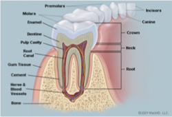
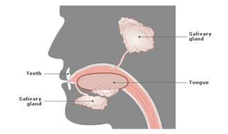
TEETH
Begin the process mechanical digestion (breaking the food down into smaller more manageable pieces) to assist in swallowing;
chopping, tearing and grinding
Each tooth is designed to complete a specific task ex. Canine as pointed and sharp to help tear, molars are flat to grind.
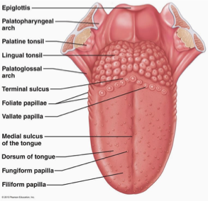
TONGUE
plays the role of moving
the food around the mouth
Taste - papillae (upper surface of the tongue, house the taste buds that allow us to taste food)
5 taste bud categories – salty, sweet, sour, bitter, umami
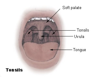
UVULA
hanging from the middle of the back edge of the soft palate
prevents food from entering the nasopharynx (or nose) during swallowing
SALIVA
clear liquid secreted into the mouth by the salivary glands and mucous glands of the mouth
moistens the mouth and lubricates food
assist in the chemical process of starch digestion by the enzyme amylase
BOLUS
round mass of food that has been chewed to the point of swallowing
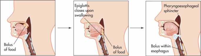
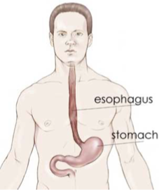
ESOPHAGUS
Tube connecting the pharynx to the stomach
approximately 24 cm long; lined with circular and longitudinal muscles which work to move food in ONE direction (down)
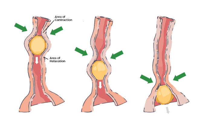
PERISTALSIS
symmetrical contraction of muscles which moves in a wave down the esophagus to help propel food through the digestive tract – uni-direction (one direction)
STOMACH
Muscular J shaped organ where food is temporarily stored while further chemical and mechanical digestion happens
walls are folded to allow distension
lined by gastric glands which secrete gastric juices that aid in chemical digestion (HCl, salts, enzymes, water and mucous) stimulated by the presence of food
mucus lines and protects the surface of the stomach from the acidic gastric juice
3 layers of muscle that relax and contract to churn stomach contents
CHYME
thick liquid of partially digested food mixed with gastric juices
CARDIAC SPHINCTER
muscular valve at the junction of the esophagus and the stomach; (top of the stomach)
controls the backflow of stomach contents back into the esophagus
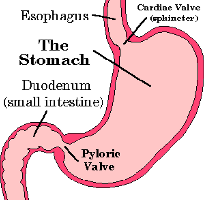
PYLORIC SPHINCTER
muscular valve at the lower end of stomach at the entrance of the small intestine.
when closed helps keep the food in the stomach

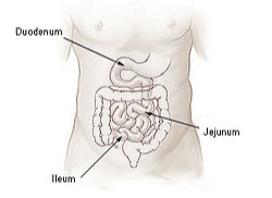
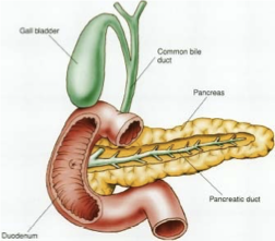
DUODENUM - SMALL INTESTINES
(the first part of the small intestine; “C” shaped)
Chemical digestion of chyme begins here
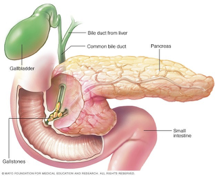
Bile from the gallbladder and digestive juices from the pancreas mix in here.
JEJUNUM- SMALL INTESTINES
Follows the duodenum, approximately 2.5m long, contains many more folds than the duodenum
The lining of the jejunum is specialized for absorption
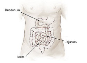
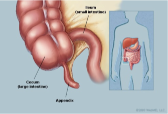
ILEUM SMALL INTESTINES
3m long, functions to absorb nutrients and to push undigested food into the large intestine
Mainly absorbs B12 and bile salts and anything left over that has not been absorbed
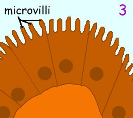
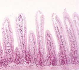
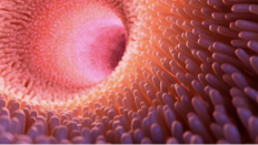
VILLI SMALL INTESTINES + MICROVILLI
tiny finger like projections that increase the surface area of the intestines.
MICROVILLI - each villi is in turn covered with many fine brush like microvilli that further increase the surface area
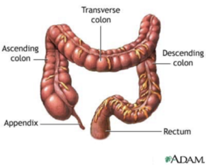
LARGE INTESTINE
absorb nearly 90% of water from the alimentary canal;
contains anaerobic bacteria to help digest undigested material;
leftover material is referred to as feces which is pushed by muscular contractions into the rectum for disposal
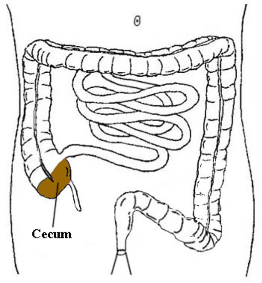
CECUM
the cavity in which the large intestine begins and into which the ileum opens; the appendix is an offshoot of the cecum
APPENDIX
finger like projection at the end of the cecum; no known function any longer
-vestigial feature
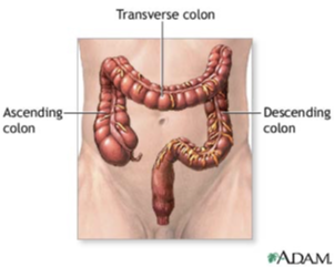
ACENDING, TRANSVERSE AND DESCENDING COLON
ASCENDING COLON - part of the large intestine that ascends from the cecum to the transverse colon
TRANSVERSE COLON - part of the large intestine that extends across the abdominal cavity and joins the ascending to the descending colon
DESCENDING COLON - part of the large intestine that descends from the transverse colon
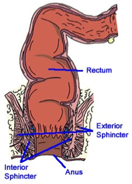
RECTUM, ANAL CANAL AND ANUS
RECTUM - the final part of the alimentary canal where waste is stored before being eliminated
ANAL CANAL - the terminal part of the large intestine
ANUS - the excretory opening at the end of the alimentary canal
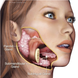
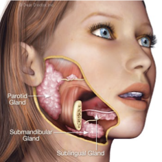
SALIVARY GLANDS` - Accessory Organs
PAROTID GLAND - the largest of the salivary glands located slightly below and in front of the 2 ears; a duct connects the gland to the oral cavity; produces the majority of saliva
SUBLINGUAL GLAND - small salivary glands located under the tongue that secrete saliva directly into the mouth through a series of pores
SUBMANDIBULAR GLAND -pair of glands located beneath the jaw which connect by a duct to the oral cavity;
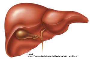
LIVER- Accessory Organs
Located in the right upper quadrant just below the diaphragm
Produces bile, an alkaline substance which aids in digestion of fats acting as an emulsifying agent (breaks fat down into smaller fat droplets that are more readily absorbed)
Some of the bile drains directly into the duodenum
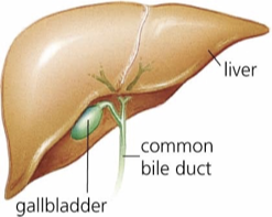
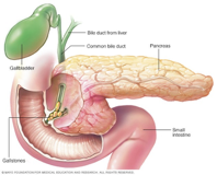
GALLBLADDER - Accessory Organs
Stores and concentrates bile produced by the liver and releases it through the common bile duct to the duodenum.
Attached on underside of liver
Humans can live without a gallbladder
PANCREAS - Accessory Organs
Glandular organ producing several important hormones, including insulin (used to move glucose from the blood into tissues) and glucagon (used to mobilize glucose from the tissues to the blood)
Secretes pancreatic juice containing digestive enzymes that pass to the small intestine and break down many macromolecules.
Enzymes
* Digestive enzymes help the body break down food.
* Different enzymes, each with specific functions, are produced in various parts of the digestive tract.
* Incomplete digestion can contribute to ailments such as:
* Flatulence, bloating, belching
* Food allergies, nausea, bad breath
* Bowel problems, stomach disorders
* Digestive enzymes mainly handle the chemical breakdown of food and make up a large part of digestive secretions.
* The human body produces around 22 different enzymes involved in digestion.
Mouth
Saliva contains the enzyme salivary amylase. This enzymes breaks starch into smaller sugars and is stimulated by chewing. It is important to chew food thoroughly as this is the first stage of the digestive process.
Stomach
The stomach is responsible for the digestion of protein and ionization of minerals. The parietal cells of the stomach secrete hydrochloric acid (gastric acid).
Pepsin is secreted by the stomach and breaks up proteins

Small Intestine
The small intestine has three segments and secretes various digestive substances.
It also receives enzymes and secretions from the pancreas, liver, and gallbladder.
Duodenum – primarily absorbs minerals.
Jejunum – absorbs water-soluble vitamins, proteins, and carbohydrates.
Ileum – absorbs fat-soluble vitamins, fats, cholesterol, and bile salts.
Pancreas
The pancreas produces digestive enzymes that act in the small intestine and play a major role in digestion.
It secretes about 1.5 litres of pancreatic juice per day.
Pancreatic enzymes include:
Lipases – digest fats, oils, and fat-soluble vitamins.
Amylases – break down starches and carbohydrates into smaller sugars like maltose.
Proteases – break down proteins into smaller amino acids.

Liver and Gall Bladder
The liver produces bile that is either stored by the gallbladder or secreted into the small intestine.
Bile emulsifies fats and fat-soluble vitamins.
It also helps keep the small intestine free from parasites.
The liver metabolises proteins, carbohydrates and cholesterol and is responsible for the detoxification of toxins, drugs and hormones.
Peptic Ulcers
Ulcers occur when stomach or duodenum tissues become inflamed due to a weakened mucous lining.
Most ulcers are caused by Helicobacter pylori, an acid-resistant bacterium that prevents mucus production.
Symptoms: abdominal pain, bloating, nausea, loss of appetite.
Treatment: antibiotics to eliminate bacteria and medications to reduce acid production.
Inflammatory Bowel Disease
Inflammatory bowel disease (IBD): general term for diseases causing intestinal inflammation.
Chronic conditions that cannot be cured but can be treated.
Crohn’s disease – can affect any part of the digestive tract
Symptoms: diarrhea, abdominal cramping, fatigue, blood in stool, reduced appetite
Treatment: medications to reduce signs and symptoms
Ulcerative colitis – affects the colon
Symptoms: loose/bloody stool, cramps, abdominal pain
Treatment: surgery to remove affected colon section, medications to reduce symptoms
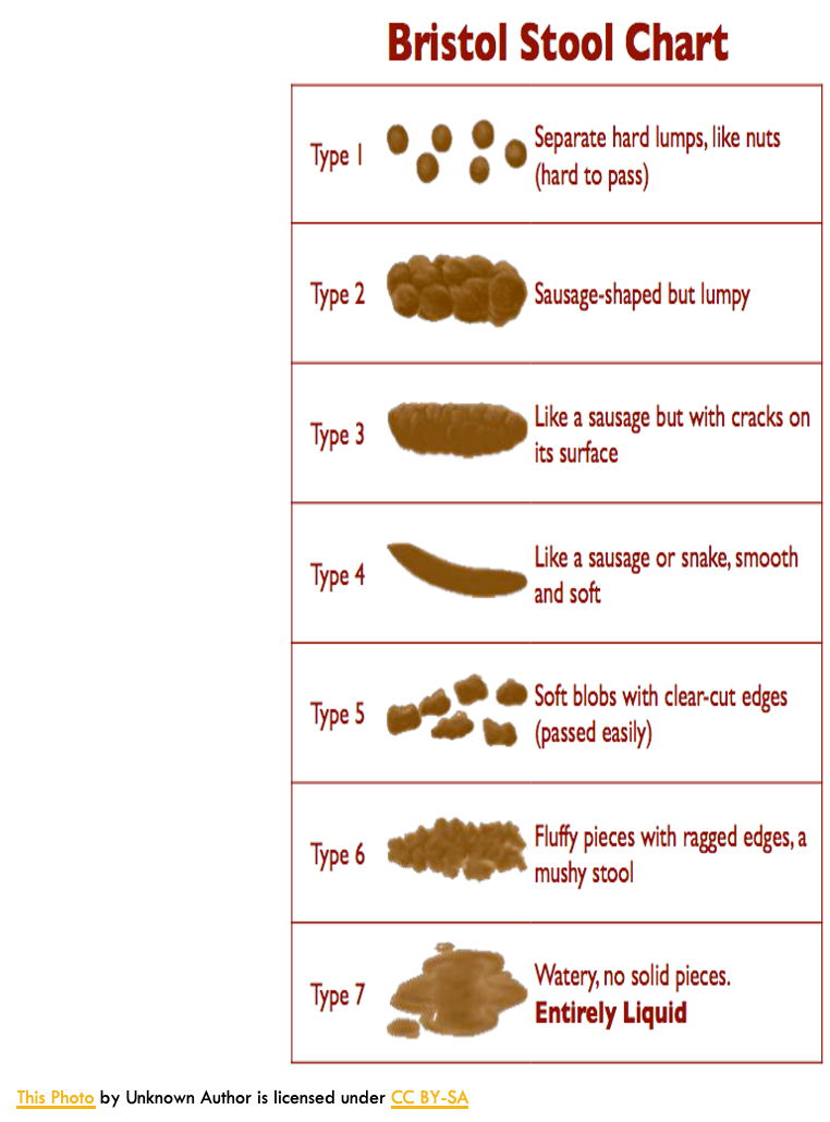
Constipation
Bowel movements are reduced to 3 or less per week. Stools are dry, small and difficult to eliminate.
Can be caused by inadequate water intake, lack of physical activity or healthy diet, and lack of good nerve and muscle function in the bowel
Treatment: Increase hydration and fiber
Hepatitis
Hepatitis: inflammation of the liver; three types – A, B, and C.
Symptoms: fever, malaise, loss of appetite, diarrhea, nausea, abdominal discomfort, dark urine, jaundice (yellowing of skin/eyes).fever, mal
Treatment: no specific cure; manage symptoms.
Hepatitis C:
Caused by the Hepatitis C virus, a bloodborne virus.
Transmission:
Sharing injection equipment (drug use)
Reuse/inadequate sterilization of medical equipment (syringes/needles)
Transfusion of unscreened blood products
Vaccine: none available
Hepatitis A:
Contracted from contaminated food or water.
Vaccine: available
Hepatitis B:
Potentially life-threatening liver infection.
Transmission: infected bodily fluids
Vaccine: available
Cirrhosis
Chronic disease of the liver that occurs when scar tissue replaces healthy liver tissue and prevents the liver from functioning properly.
Chronic alcoholism and hepatitis C are most common causes
Treatment: liver transplant
Gall bladder Stones
Cholesterol in the bile can precipitate out of the bile and form crystals. These crystals grow and become gall stones
Factors that can cause stones are obesity, heredity and alcohol intake
Can be treated with medications or ultrasound shock waves
If the gallstone problem is serious, the entire gall bladder may need to be surgically removed.
Common Procedures
Barium swallow:
X-ray using liquid barium to visualize the esophagus and upper digestive tract (throat, esophagus, stomach)
Helps detect swallowing issues, blockages, ulcers, or tumors
Endoscopy:
Medical procedure using an endoscope (thin, flexible tube with light and camera)
Can visualize:
Enzyme activity
Structure of digestive tract
Digestion in action
Can also remove polyps or take samples for biopsy
Maximizing the Efficiency of Respiration
All organisms need oxygen and must get rid of carbon dioxide.
Gas exchange requires special organs that connect the inside of the body to the outside and provide enough surface area for oxygen in and carbon dioxide out.
Respiration:
General meaning: taking in oxygen, releasing carbon dioxide.
Cellular respiration: oxygen is used by cells as the final electron acceptor to make ATP, producing carbon dioxide as a waste product.
Gas Exchange in Animals
Respiration & Gas Diffusion:
Gases diffuse across plasma membranes.
Oxygen must dissolve in water to cross membranes.
Plasma membranes are surrounded by water, so oxygen from air dissolves in this layer.
Primitive invertebrates:
No special respiratory organs.
Keep respiratory surfaces moist to allow gas exchange.
Example: Earthworm
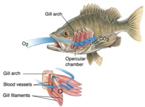
Gills as Respiratory Structures
Aquatic respiratory organs increase the diffusion surface area by extensions of tissue called gills
External gills increase surface area, but the organism must always be kept moving because oxygen in stagnant water can be quickly depleted.
The external gills are easily damaged
Gills
Evaginated structures (body outgrowths) with large surface area for gas exchange.
Circulatory system inside gills carries oxygen away and removes CO₂.
External gills: unprotected (e.g., Polychaete worms).
Internal gills: protected (e.g., fish).
Respiration in Amphibians
Gas exchange in unique organisms:
Two ways: lungs and skin.
Amphibians:
Lungs: force air in by creating higher pressure outside lungs.
Air fills buccal cavity → nose & mouth closed → floor of mouth raised → air pushed into lungs.
Skin: must stay moist for diffusion.
Important during hibernation, e.g., buried under mud in ponds.
Respiration in Mammals
Mammals have higher metabolic rates to produce and sustain their body temp and thus require a more efficient respiratory system
Lungs of mammals are packed with million of tiny air sacs called alveoli providing an enormous surface area for gas exchange
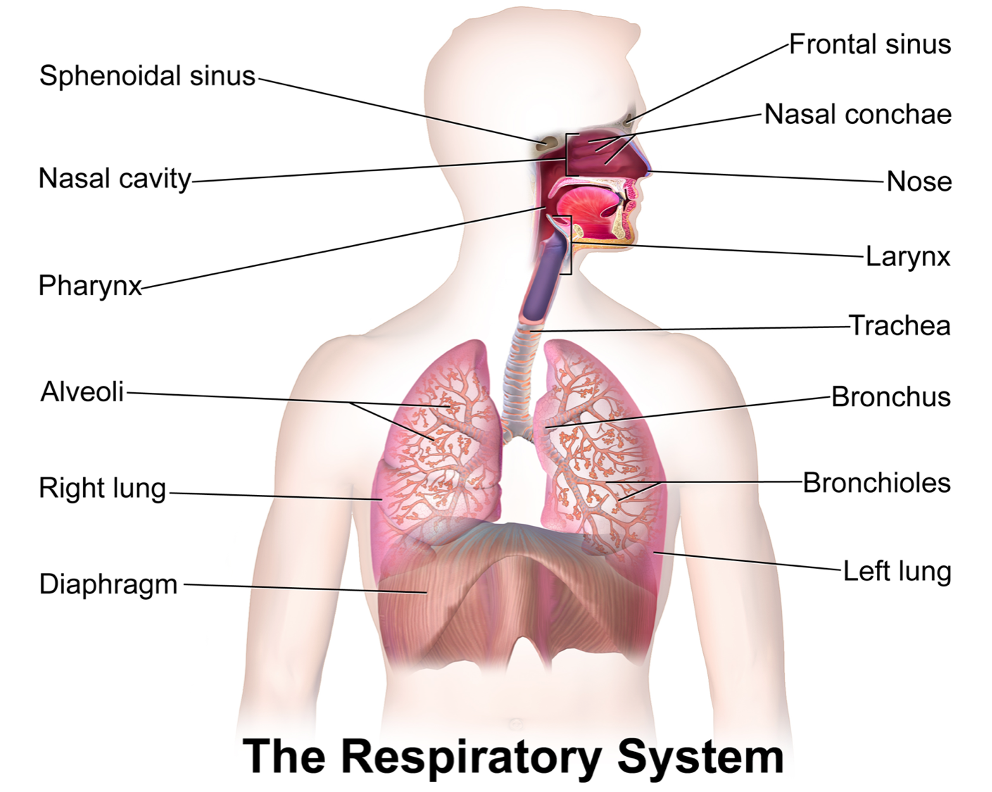
Flow of Air
Air is brought into the alveoli through a system of passages:
Mouth/Nose → pharynx → larynx → trachea → bronchi → bronchioles → alveoli
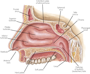
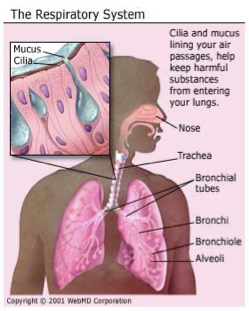
Features of the respiratory system
Nostril – conducts air into hollow nasal cavity.
-best way to intake air as it cleans and purifies air
Nose Hair
Nose hairs help to filter out dust and dirt particles from entering the respiratory tract
Hairs are covered in mucus that trap particles as they are breathed in
Turbinates
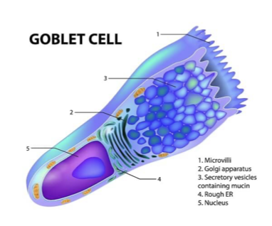
Thin bones found in the nasal cavity that increase surface area and secrete mucous (Goblet cells).
This helps to moisten the air.
The turbinates are lined with capillaries that warm and increase humidity of incoming air.
Ciliated Cells
– These cells secrete mucous.
The mucous traps the foreign particles.
Continual sweeping of the cilia propels these particles back to the nose and throat where they can be expelled.
Main type of ciliated cell = goblet cell
Goblet Cells
Epithelial cell that has a primary function in creating and secreting mucus.
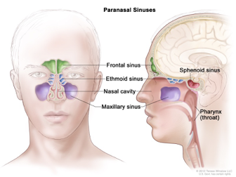
Sinuses
Main function is to produce mucus that moisturize and humidifies the inside of the nose.
The mucus layer protects the nose from pollutants, micro-organisms, dust and dirt
Pharynx
Pharynx- connects the nose and mouth to the throat. This is broken into several different areas.
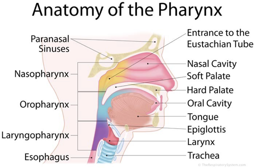
Epiglottis
Glottis – opening to the trachea
Epiglottis – flap of cartilage located behind the tongue that helps prevent food from entering trachea
Snaps shut when swallowing
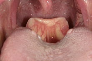
Larynx
Larynx – houses the vocal cords.
There are two folded structures of the vocal cords.
When you breathe normally there is a large gap between the cords.
When you speak, muscles contract bringing cords closer. The air that passes through them vibrates the cords producing sound.
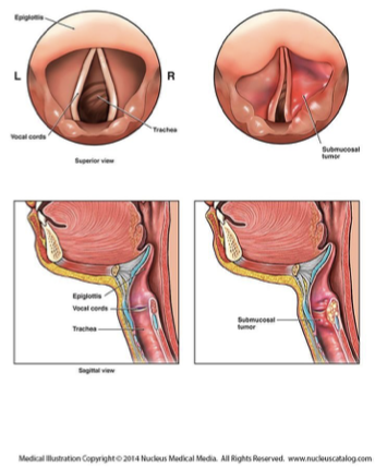
Trachea
Trachea – Flexible tube that is the passageway of air.
Supported by semi-circular cartilage rings to ensure it does not collapse from the passage of food in the esophagus.
lined with ciliated goblet cells that secrete mucous.
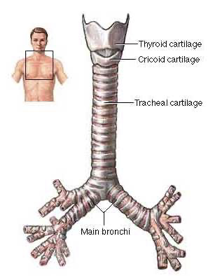
Bronchi
Bronchi – 2 smaller branches of the of the trachea leading to the left and right lung
Bronchioles – tiny subdivisions of the bronchi
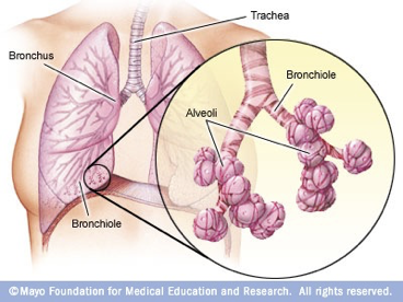
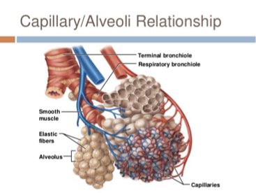
Alveoli
Alveoli – grape like clusters of tiny sacs at the end of the bronchioles.
These sacs are always kept moist.
This is the site of gas exchange.
The sacs provide an abundance of surface area for the exchange of gases.
They are surrounded by capillaries.
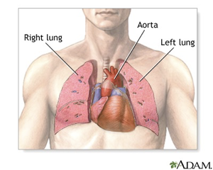
Lungs
Lungs:
Flexible membranes that expand and contract.
Right lung: 3 lobes | Left lung: 2 lobes.
Contain bronchi, bronchioles, and alveoli.
Pleura:
Tissue layer that envelops lungs.
Flexible, allows expansion/contraction.
Each pleura has two layers separated by lubricating fluid.