ANAT 100- Module 6 - The PNS
1/81
There's no tags or description
Looks like no tags are added yet.
Name | Mastery | Learn | Test | Matching | Spaced | Call with Kai |
|---|
No analytics yet
Send a link to your students to track their progress
82 Terms
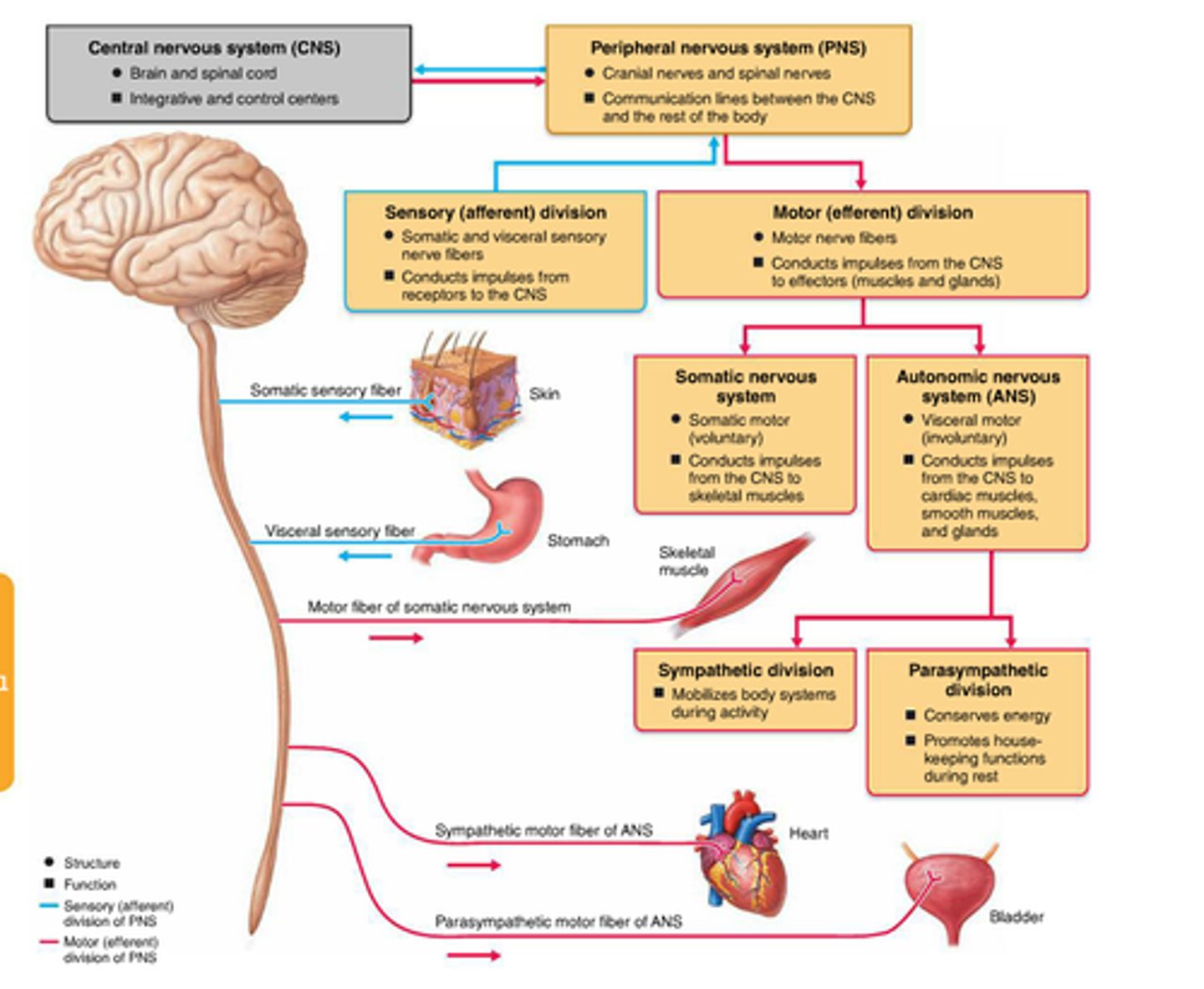
Peripsheral Nervous System
- composed of all the nerves outside the brain and spinal cord
- the PNS contains the spinal and cranial nerves
Divisions of the PNS
Cranial Nerves
- emerge in pairs from the brain and brain stem
- there are 12 pairs
- can carry sensory information, motor information, or both
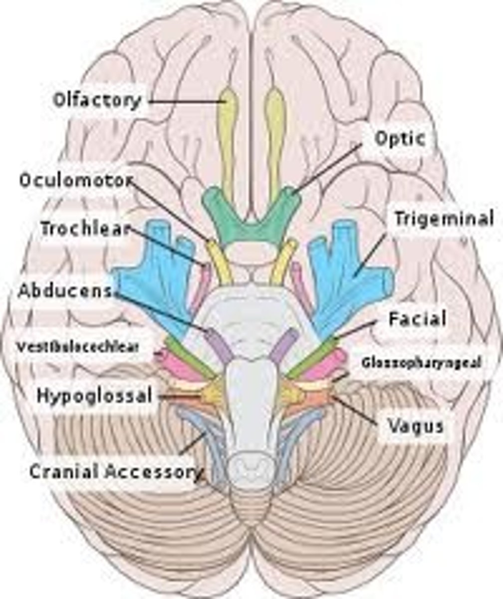
CN I- Olfactory Nerve
- sensory only
Function
- sensory: snell
- motor: none

CN II- Optic Nerve
- sensory only
function
- sensory: vision
- motor: none

CN III- Oculomotor Nerve
- motor only
function
- sensory: none
- motor: movement of the eye
CN III is 1/3 CN that send motor input to the muscles to move the eye*

CN IV- Trochlear Nerve
- motor only
function
- sensory: none
- motor: movement of the eye
*CN IV is the 2nd CN invoked with eye movement*

CN V- Trigeminal Nerve
- sensory and motor
function
- sensory: forehead, eye, upper+lower jaw
- motor: muscles of mastication
(has 3 divisions, that's why tri is in the name)

CN VI - Abducens Nerve
- motor only
function:
- sensory: none
- motor: Movement of the eye
*3/3 cranial nerves involved with eye movement

CN VII - Facial Nerve
- sensory and motor
function
- sensory: taste for the anterior 2/3 of the tongue
- motor: muscles of facial expression, parasympathetic innervation of tear and salivary glands

CN VIII- Vestibulocochlear Nerve
- sensory
function
- sensory: equilibrium and hearing
- motor: none

Nerve IX - Glossopharyngeal Nerve
- sensory and motor
function:
- sensory: sensory innervation of the pharynx, taste for the posterior 1/3 of the tongue
- motor: pharynx muscles, parasympathetic innervation of the salivary glands

CN X - Vagus Nerve
- sensory and motor
function:
- sensory: sensory innervation of the larynx
- motor: pharynx and larynx muscles, parasympathetic innervation of the thoracic and abdominal organs

CN XI - Accessory Nerve
- motor
function
- sensory:none
- motor: trapezius and sternocleidomastoid muscle

Nerve XII- Hypoglossal Nevre
- motor
function:
- sensory: none
- motor: muscles of the tongue
CN involved with eye movement
CN III- Oculomotor Nerve
CN IV- Trochlear Nerve
CN VI - Abducens Nerve
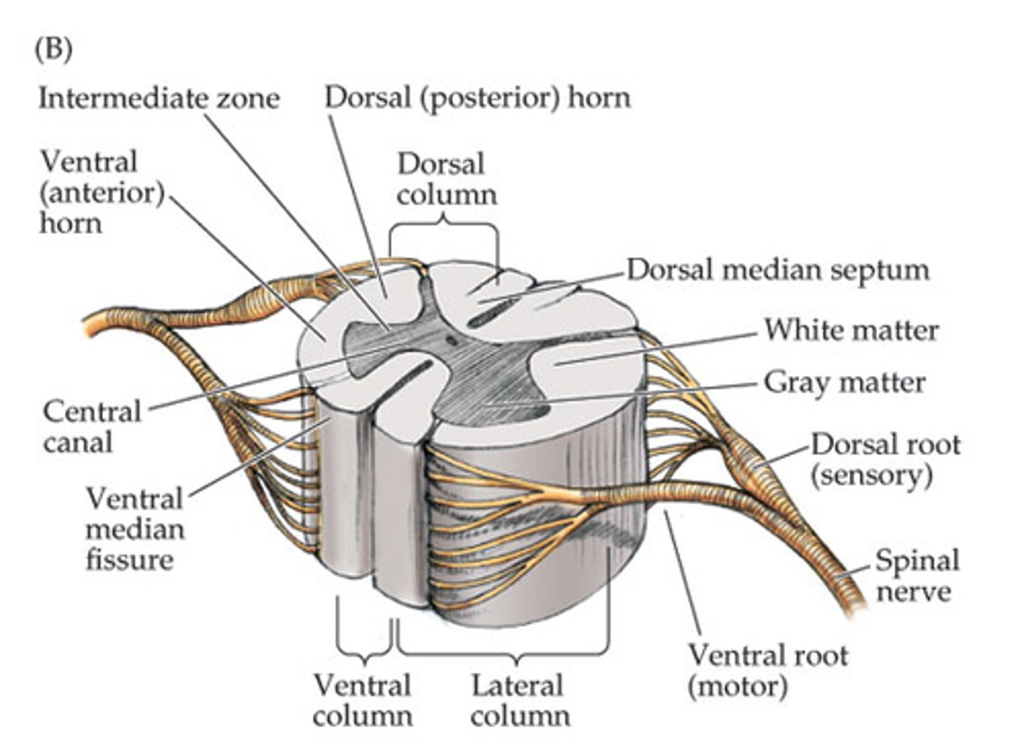
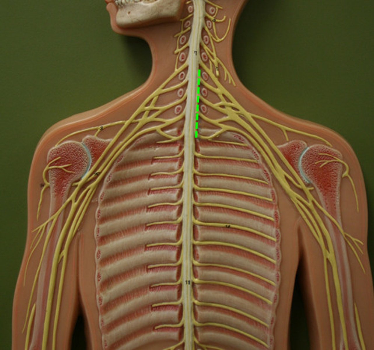
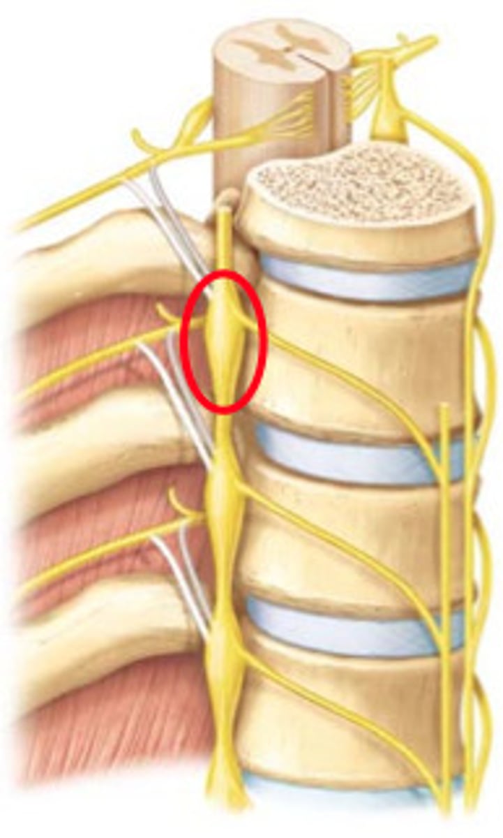
Spinal Nerves
- emerge from the spinal cord
- 31 pairs that carry inffomrtion through the PNS
- a spinal nerve is formed by the union of the posterior (sensory) and anterior (motor) roots of the spinal cord
Classifying Spinal Nerves
- the letter is the region of the spinal cord
- the number is the level of the spinal cord
IN THE CERVICAL REGION (above C8)
- the spinal nerve emerge superior to their corresponding vertebrae
BELOW C8
- all spinal nerves emerge inferior tp their corresponding vertebrae
Nerve Plexuses
- once the spinal nerves exit the vertebral column, fibres from the anterior roots (motor) come together and then redistribute into a network of nerves (plexuses) that mostly innervate muscles of the limb
- each plexus contains fibres (a collection of axons) from a combination of spinal nerves (this is important because in case of injuries, having multiple spinal nerves in the nerve plexus ensures supply to the muscles)
Cervical and Brachial Plexuses/ Lumbar and sacral plexus
- the cervical and brachial plexuses correspond to the cervical enlargement
- the lumbar and sacral plexuses correspond to the lumbosacral enlargement
autonomic nervous system (ANS)
- also known as the visceral motor system
- responsible for transmitting involuntary signals from the CNS to smooth muscle, cardiac muscle, and glands
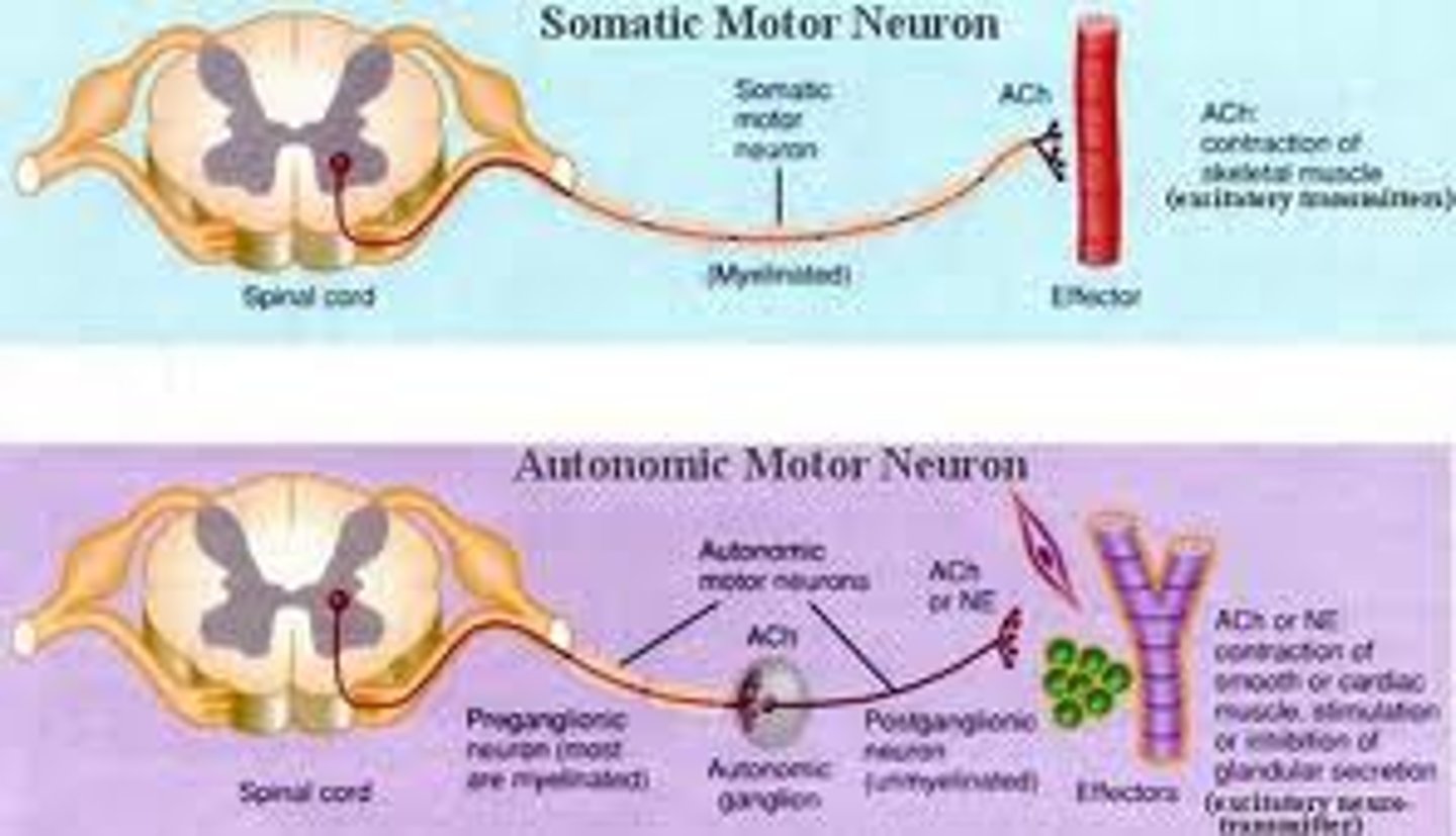
Organization of the ANS
- ANS is a 2 neural pathways (between the CNS and any target organ, there are 2 neurons)
1. Preganglionic Neuron
2. Autonomic Ganglion
3. The Post Ganglion
1. Preganglionic Neuron
- has its cell body within the CNS
- the axon enters into the PNS and travels to the autonomic ganglion
2. Autonomic Ganglion
- is where the preganglionic and postganglionic neurons meet and communicate
- located in the PNS
3. Postganglionic Neuron
- entirely in the PNS
- its cell body Is in the autonomic ganglion, while its axon travels to the target organ
Differences between sympathetic and parasympathetic
\
ORIGIN
- sympathetic: spinal roder segment T1-L2
- parasympathetic: brainstem and spinal cord segment S2-S4
RESPONSE
- sympathetic: fight or flight
- parasympathetic: rest and digest
EFFECT
-sympathetic: widely distributed to many parts of the body
- parasympathetic: more limited distribution to target organs, with localized effects
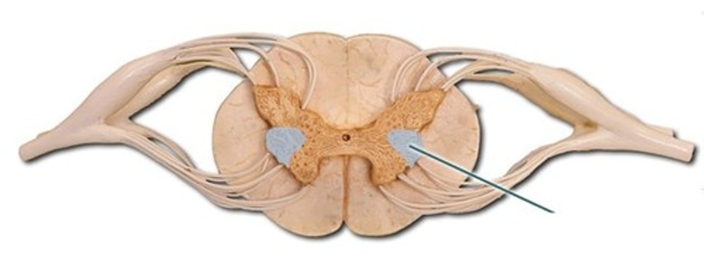
Autonomic Pathways: Sympathetic
- the nerves exit from the spinal cord to supply innervations to the body
Things:
1. lateral horn of the spinal cord
2. Sympathetic Trunk
3. Ganglion
4. Effector Organ
1. Lateral Horn of the Spinal cord
- sympathetic preganglion axons leave the lateral horn of the spinal cord, along with somatic motor axons, to form the anterior root
(Look at pic in notes)
2. Sympathetic Trunk
- the preganglion axons pass through the spinal nerve to the sympathetic trunk (a structure lateral to the spinal cord on either side)
- the sympathetic trunk provides a pathway for the sympathetic fibres to travel through the body, both superiorly and inferiorly
- look at picture in notes
3. Ganglion
- preganglionic neurone synapse with postgangklionic neurons in the ganglia
4. Effector organs
- postganglionic axons leave the sympathetic trunk to enter the spinal nerve and extend to the effector tissue/organ at approximately that vertebral level
Autonomic Pathways: Parasympathetic
- as parasympathetic neurons originate from the brain stem or the sacrum, you can follow the pathway starting at either organ
1. Preganglionic neurons leave the brain or spinal cord at the level of the sacrum
2. Preganglionic neurons meet postganglionic neurons in ganglia close to viscera
3. Postganglionic neuron travels to effector organs (neurons from CN II, VII, IX supply the head, CN X supplies the thorax to the descending colon. S 2,3,4 supply the terminal guy and pelvic viscera-bladder and reproductive organs)
LOOK AT NOTES
Vision
- requires visual receptors called photoreceptors (at the back of the eye)
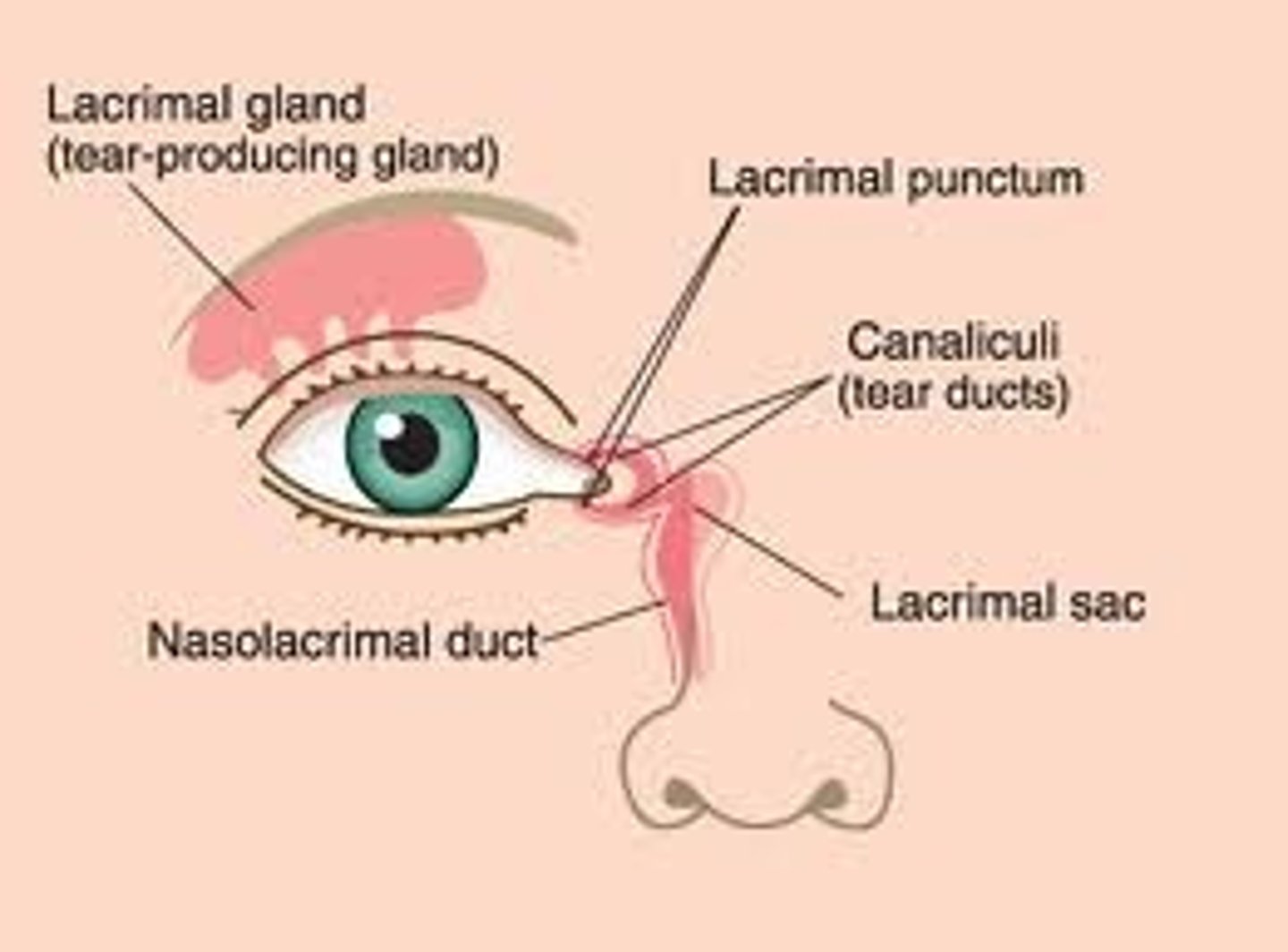
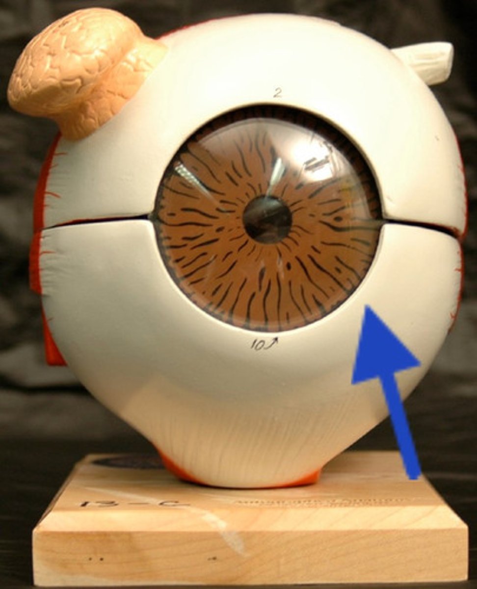
Accessory Structures of the Eyeball
- accessory structures are important structures outside the eyeball that protect and move the eyeball
1. Lacriminal Gland
2. Eyelid
3. Extrinsic Eye Muscles
1. Lacriminal Gland
- produces tears (which keeps the cornea/outside moist and clean
- tear drainage system is also very important and is part of the lacrimal apparatus
2. eyelid
- the upper eyelid contains the levator palpebral superiororis muscles and the orbcularis oculists muscle (which controls the opening and closing of the eyelid)
3. Extrinsic Eye muscles
- there are 6 extrinsic eye muscles that allow you to move your eyes to increase your range of motion
Types of Extrinsic Eye muscles
- medial rectus
- inferior rectus
- superior rectus
- lateral rectus
- inferior oblique
- Superior Oblique
Cavities of the Eye
the eye is separated by 2 fluid filled cavities:
- anterior cavity
- posterior cavity
(the cavities are delineated by the lens)
Anterior Cavity
- composed of the anterior and posterior chambers (both are filled with aqueous humour (watery fluid) )
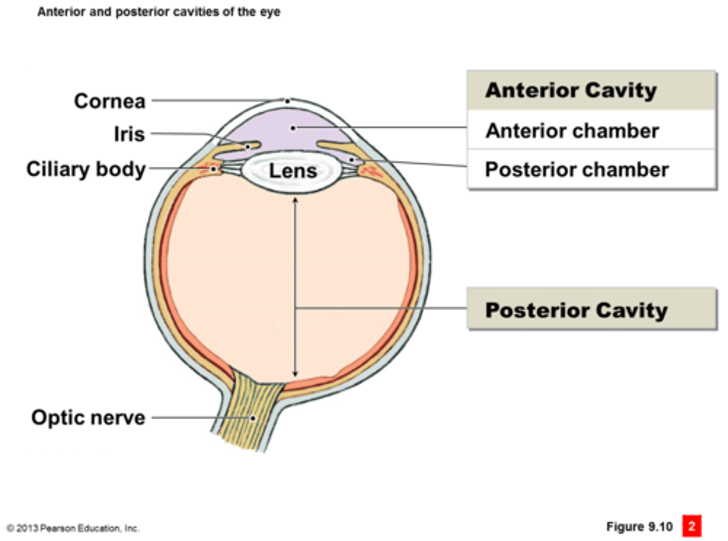
lens
- separates the anterior cavity from the posterior cavity of the eye
Posterior Cavity
- filled with vitreous humour (gelatinous)

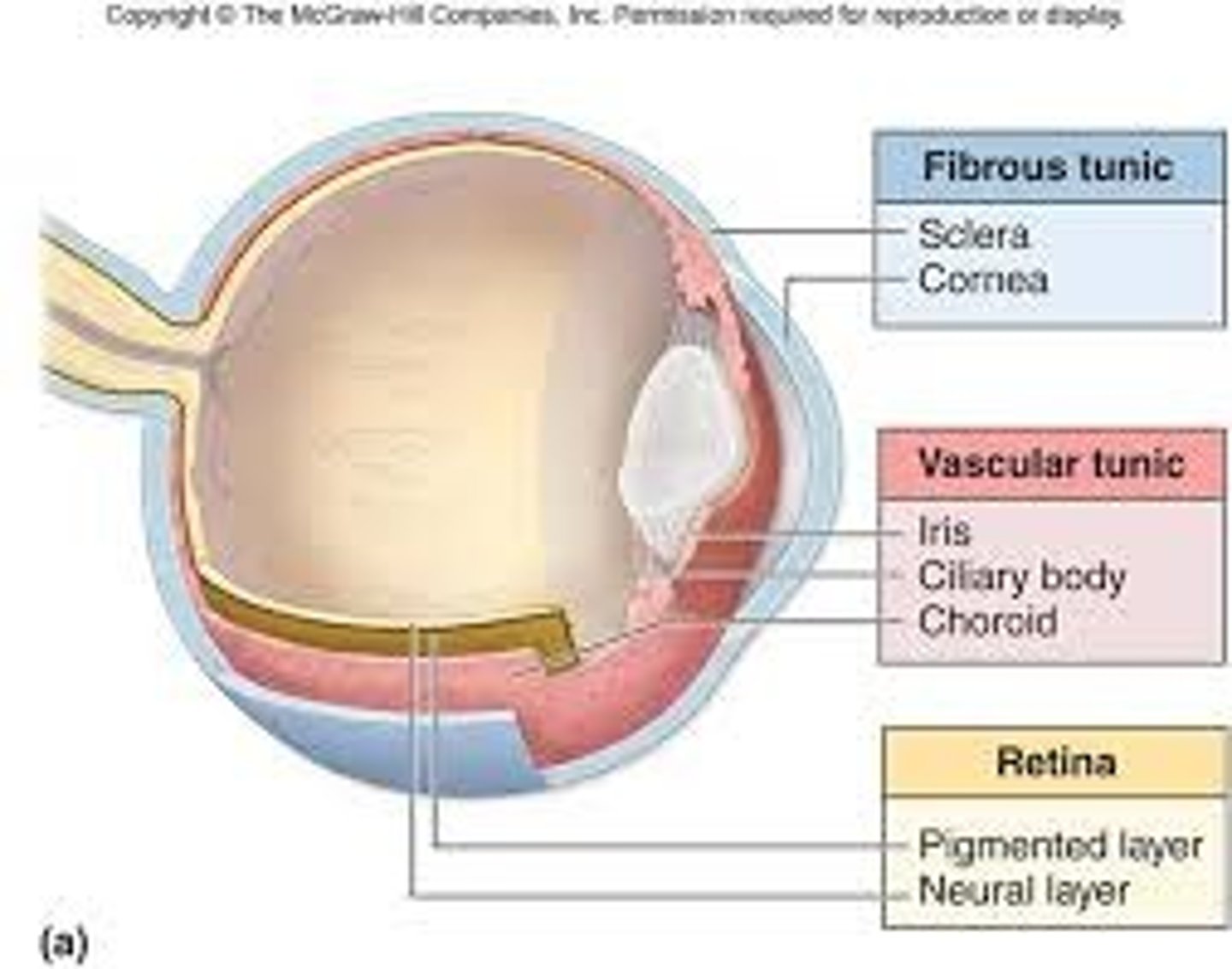
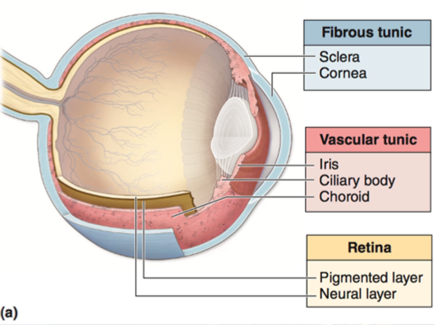
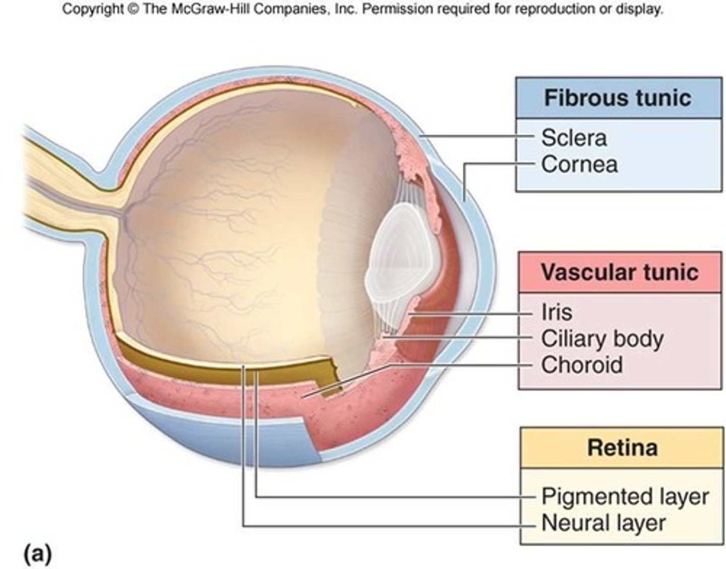
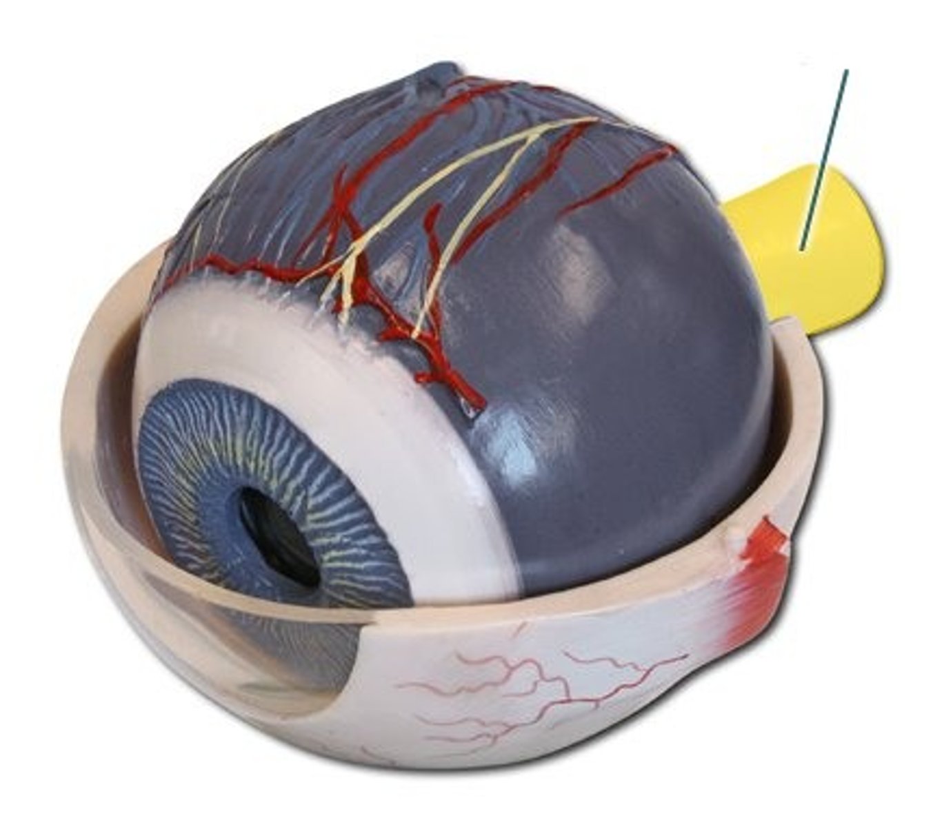
Layers of the eye
- outer layer (fibrous)
- middle layer (Vascular)
- deep layer (retina)
Outer layer of the eye (Fibrous)
- the fibrous tunic is the outermost layer of the eye and has 2 portions located internally and externally to the lens
- these 2 portions are continuous with each other
SCLERA
CORNEA
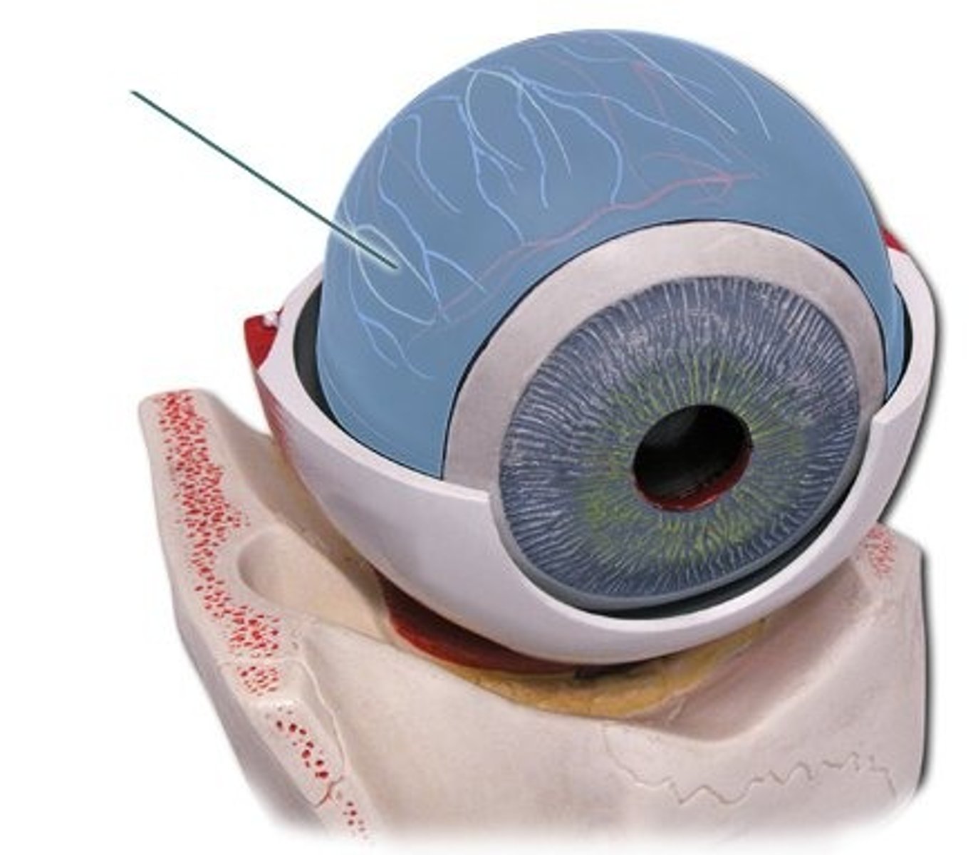
Outer Layer of the eye: sclera
- internal aspect of the fibrous tunic, located posterior to the lens
- the sclera forms the white portion of the eye
- made of dense connective tissue that acts as a point of attachment for the extrinsic eye muscles
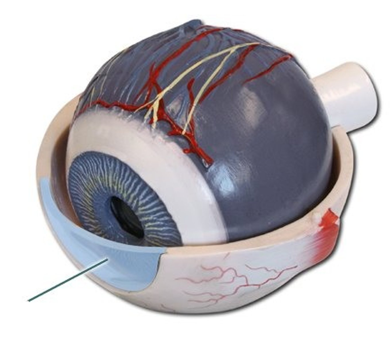
Outer Layer of the eye: cornea
- an external aspect of the fibrous tunic, located anterior to the lens
- transparent (allowing for light transmission)
Middle Layer of the Eye (Vascular)
- the vascular layer is the middle layer of the eye
- made up of:
CHOROID
CILIARY BODY
IRIS
Middle Layer of the Eye: choroid
- contains the blood vessels that supply the eye with oxygen and nourishment
Middle Layer of the Eye: ciliary Body
- produces body aqueous humour and contains the ciliary muscles (intrinsic eye muscles) which control the shape of the lens
LOOK AT PICTURE IN NOTES
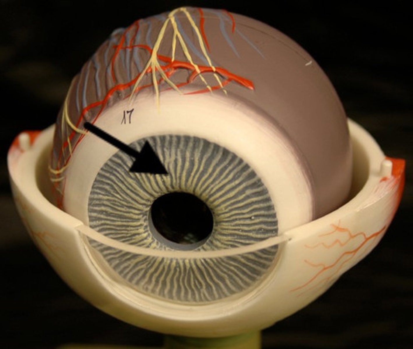
Middle Layer of the Eye: Iris
- the iris contains the sphincter and dilator muscles (intrinsic eye muscles) which controls the narrowing and widening of the pupil
- the iris also contains pigments, which give the eye its colour
Deep Layer of the Eye
- the sensory tunic is the innermost layer of the eye
- this layer is also known as the retina
- contains:
NEURAL LAYER
OPTIC DISC
MACULA
CN II
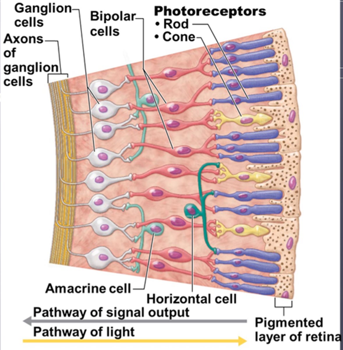
Deep Layer of the eye: Neural Layer
- the outermost region of the neural layer is made up of photoreceptors (rods and cones) which are light sensitive cells
- these receptors send signals through smaller nerves to the axons of the ganglion cells (innermost layer of the retina) exit the posterior aspect of the globe to the brain to form CN II
LOOK AT NOTES
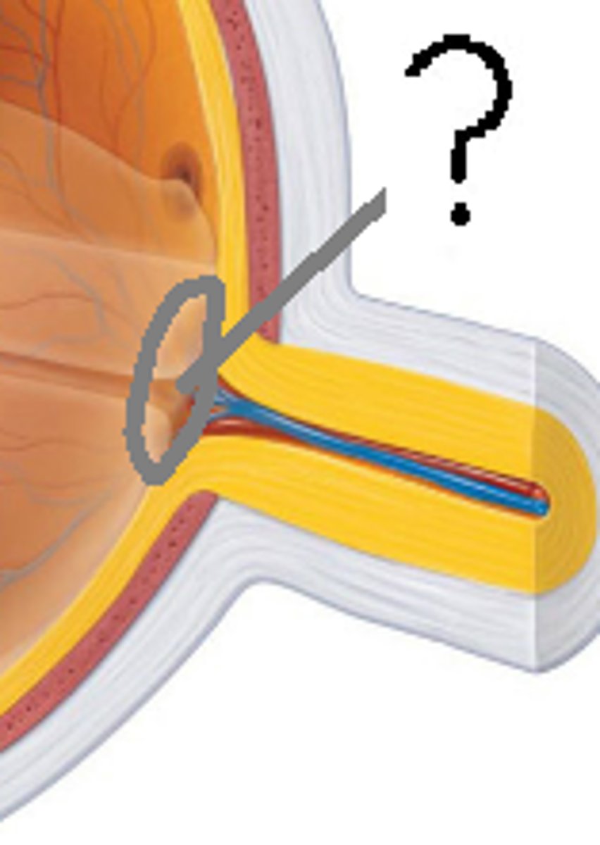
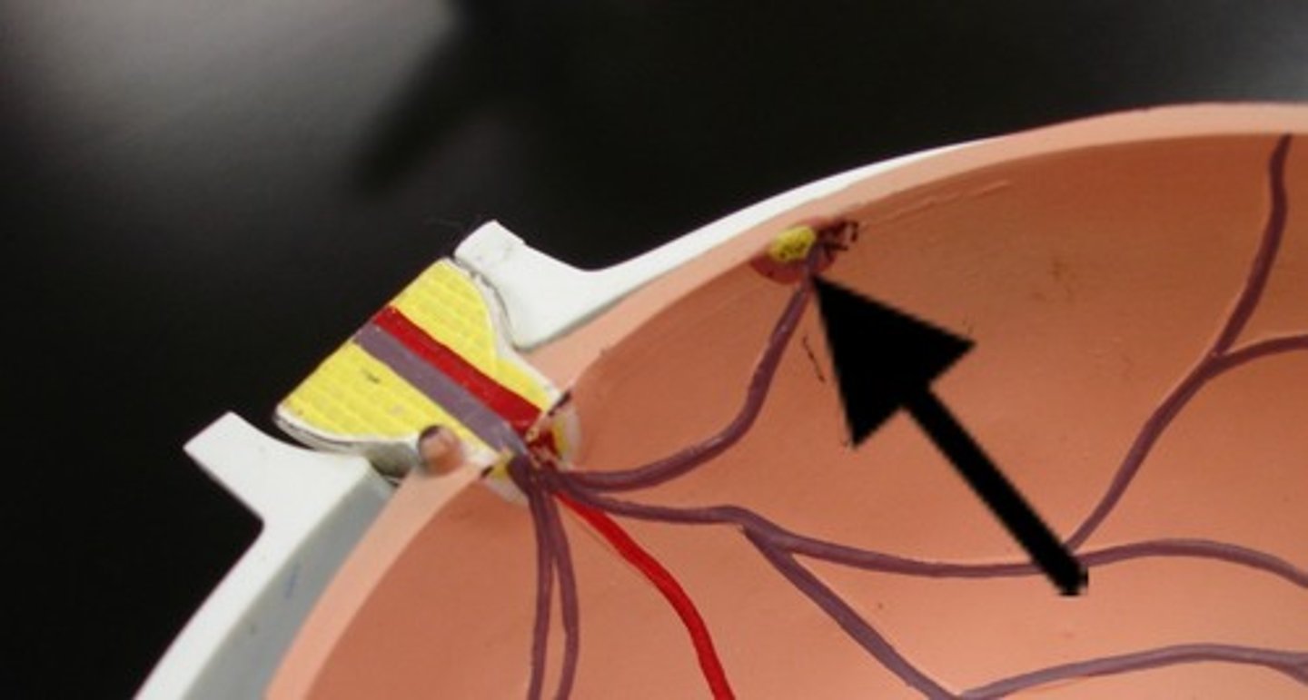
Deep Layer of the eye: optic disc
- the optic disc is the area where smaller nerve cell axons converge
- it is the location we can see when we look into the eye
Deep Layer of the eye: macula
- the central area of the retina (lateral to the optic disc)
- the central depression of the macula (the fovea) is the area where fine detailed vision occurs
Deep Layer of the eye: optic nerve (CN II)
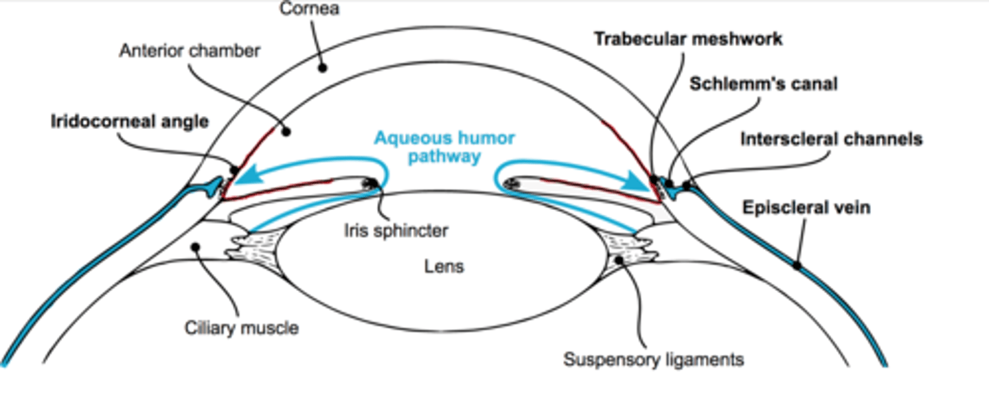
Flow of Aqueous Humour
- it is produced by the ciliary processes in the posterior chamber
- flows through the pupil into the anterior chamber and exits the at anterior chamber angle (junction of crisis, ciliary body, and cornea) through TRABECULAR MESHWORK into Schlemm's canal and into episcleral venous channels which drain into the systematic circulation
LOOK AT PICTURE
Hearing and Equilibrium
- the receptors located in the inner ear are responsible for hearing and equilibrium/ balance and position in space
structure of the ear
- the ear is divided into 3 sections:
external
middle
inner
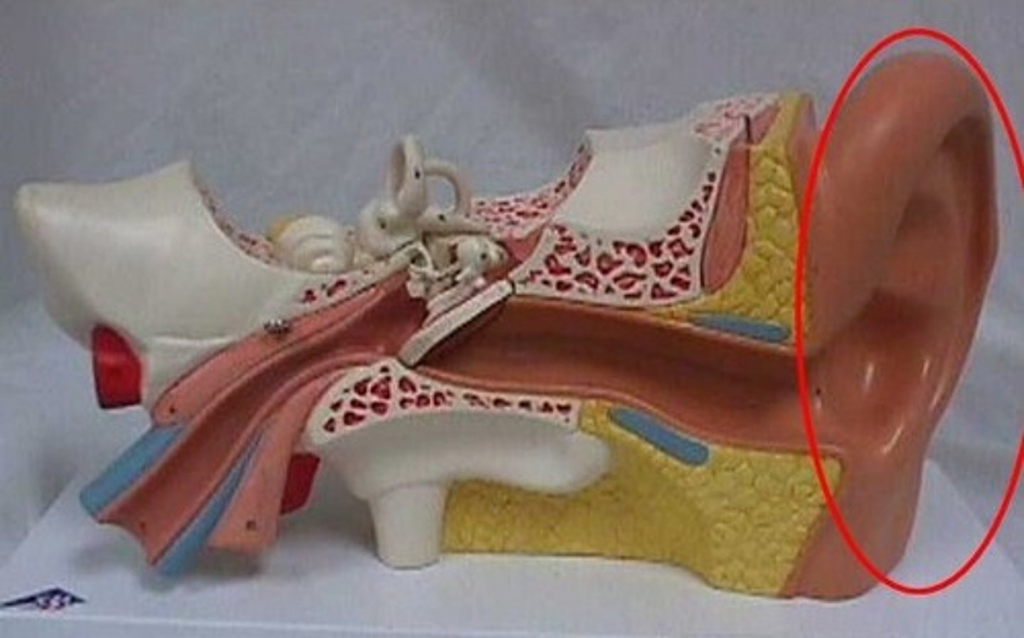
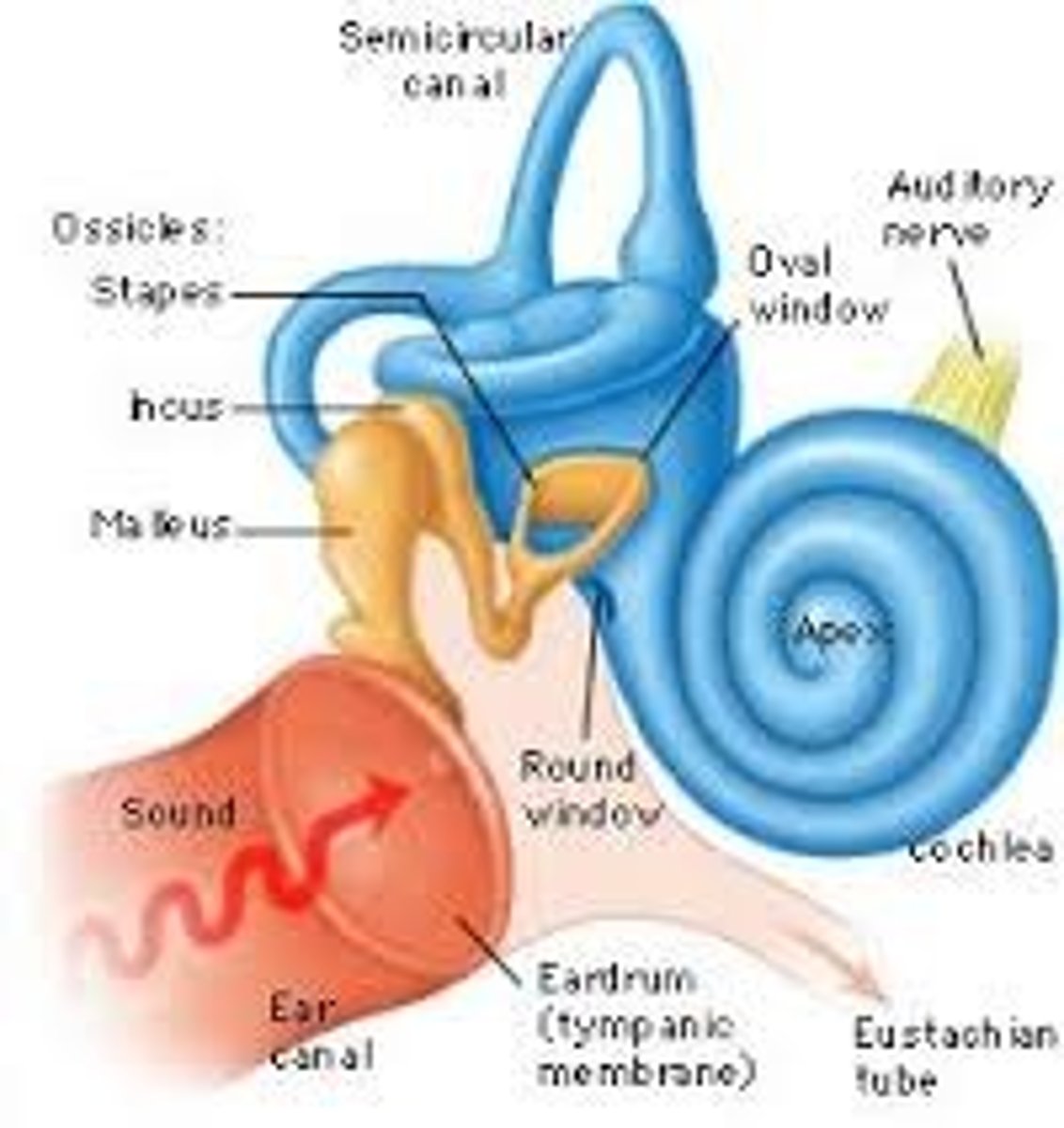
The external ear
- the external ear is the part you can see and touch
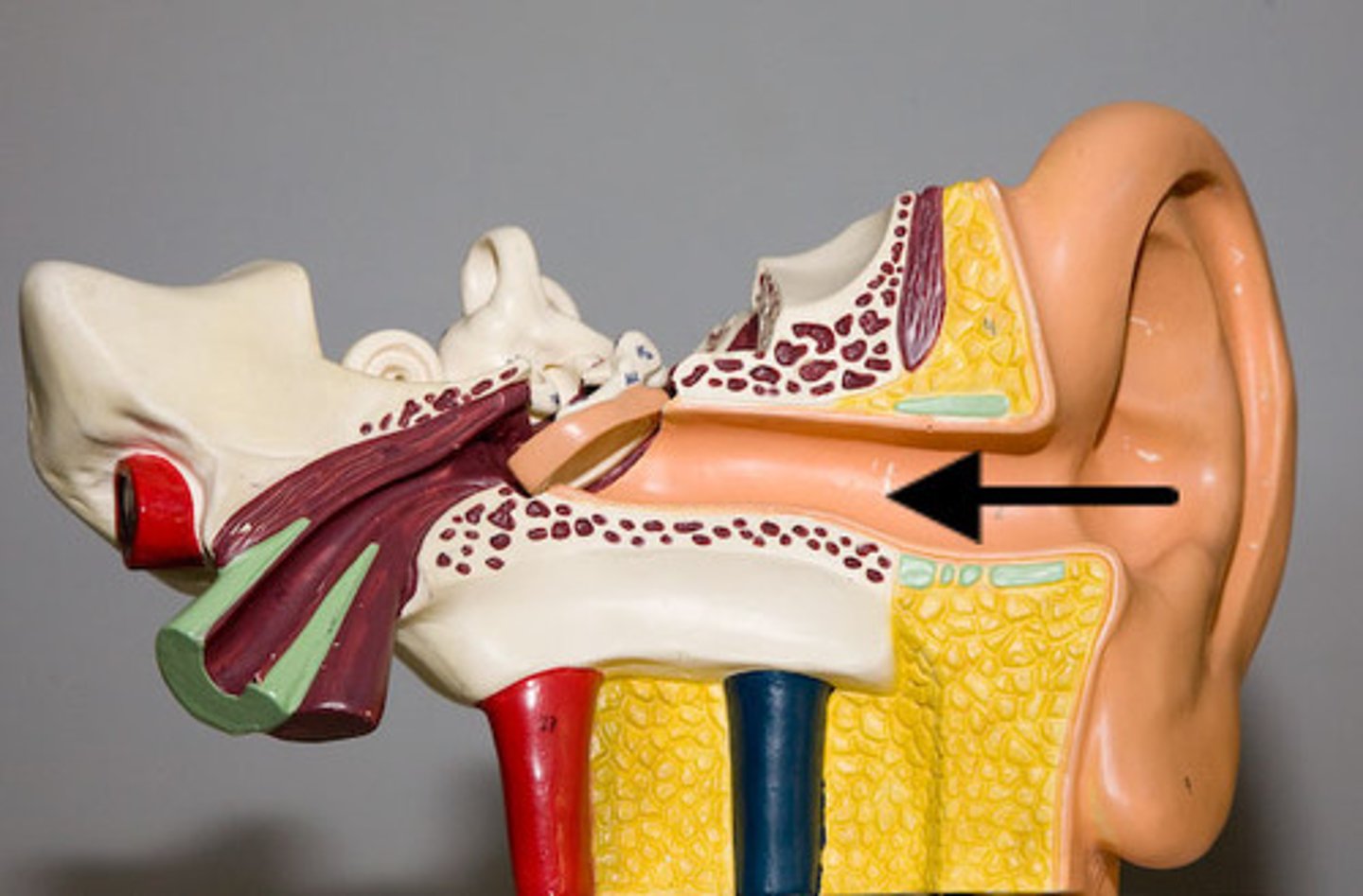
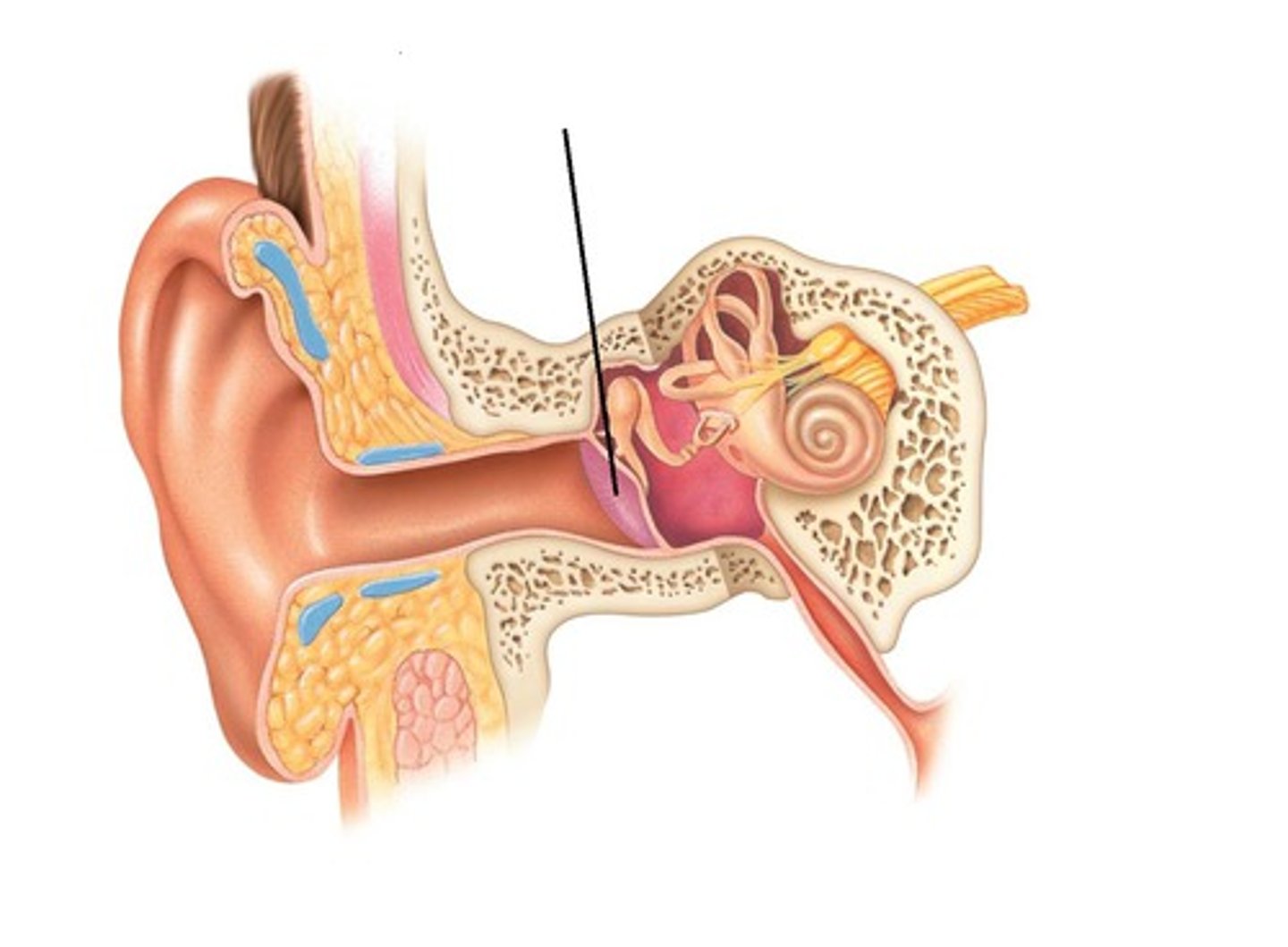
- sound waves from the external enviroment travel through the auditory canal to reach the tympanic membrane, causing it to vibrate
(the tympanic membrane marks the boundary between the external and middle ear)
- cerumonous glands secrete ear wax
Auricle
external auditory canal
tympanic membrane
perforated tympanic membrane
- the tympanic membrane (eardrum) is susceptible to physical damage or injury
- it transmit sound from the external environment to the ossicles of the middle ear
- perforated tympanic membrane is likely to cause disrupted hearing
Middle Ear
- located within the temporal bone
- spans from the tympanic membrane to the oval window
- sound waves involved with hearing are also transmitted through the middle ear
Middle Ear: Ossicles
- small bone in the middle ear
- help transmit sound waves to the receptors in the inner ear
- the muscles attached to the ossicles (the tensor tympani and stapedius) function to dampen vary loud noises
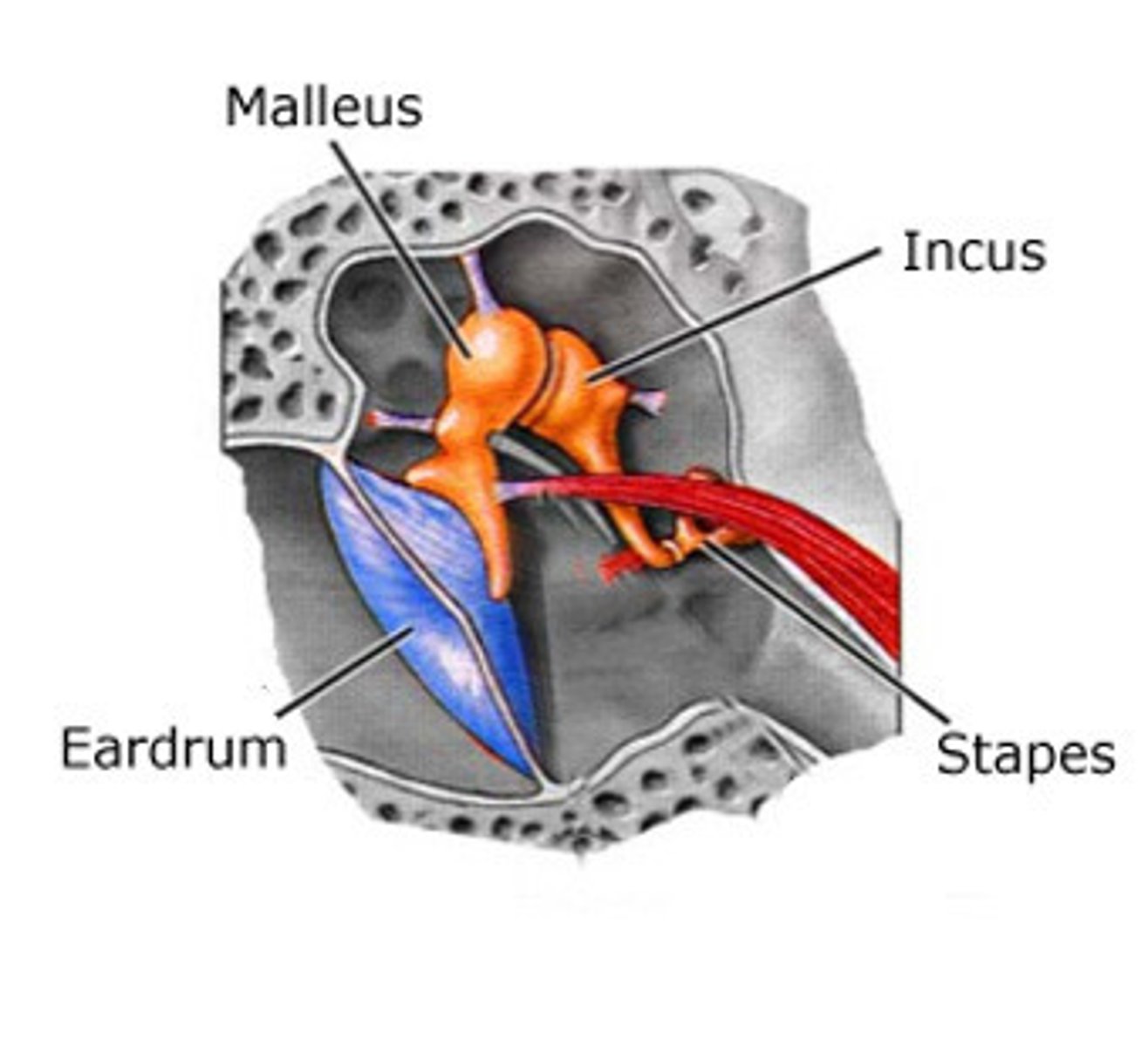
Ossicle- Malleus (Hammer)
- the most lateral ossicle that is attached to and behind the tympanic membrane
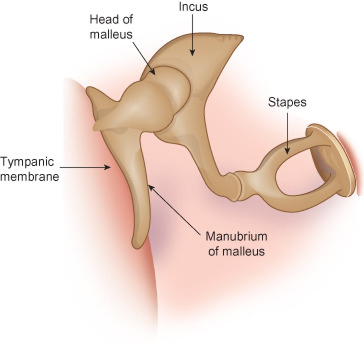
Ossicle- Incus (anvil)
- sits between the malleus and the stapes
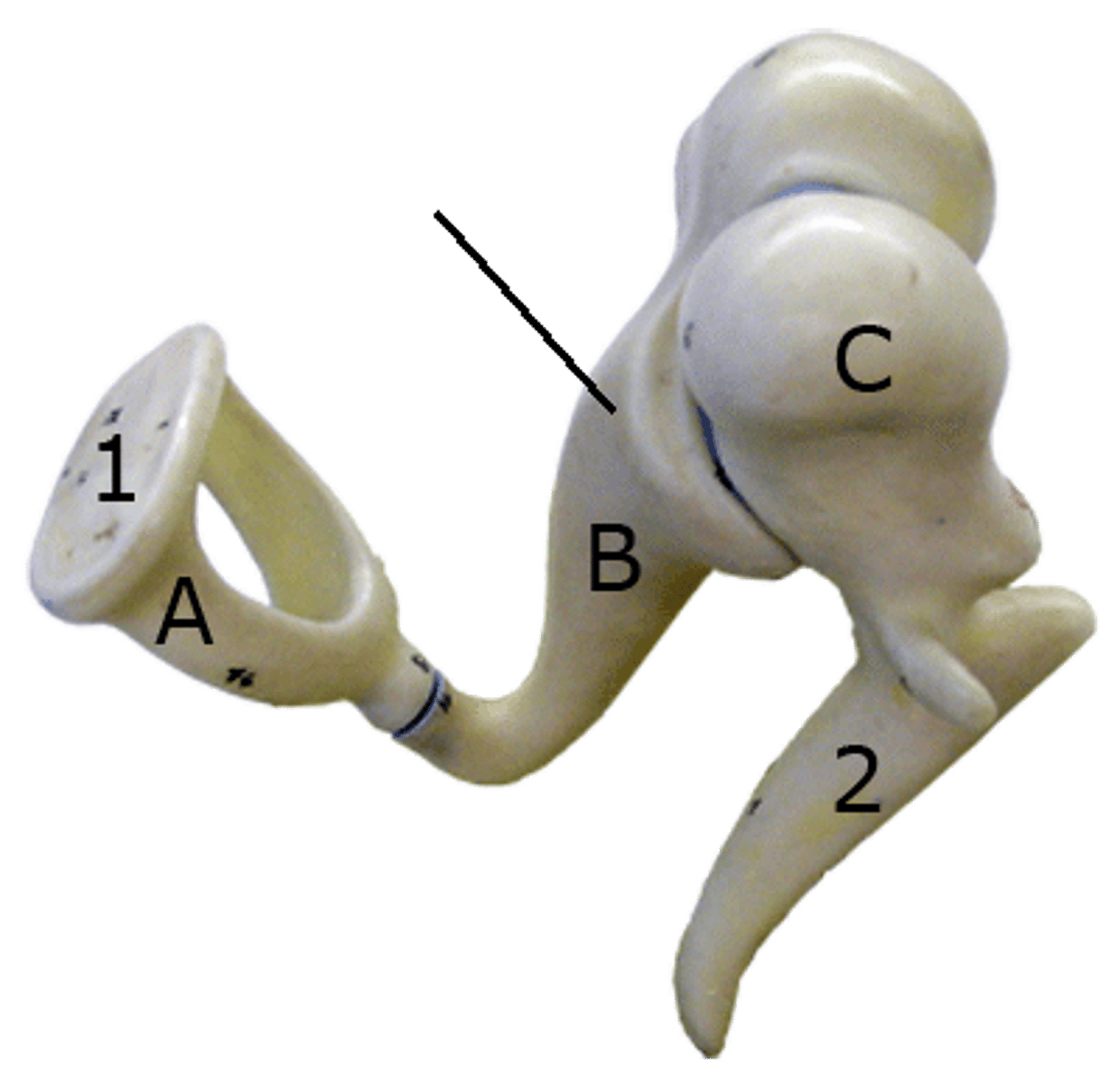
ossicle- stirrup (stapes)
- smallest bone in the oval window
- its about 1/3 the mass of the other ossicles
Middle Ear: tympanic cavity
- chamber in the temporal bone
Middle ear: Round and Oval windows
- areas that connect the middle ear to the inner ear
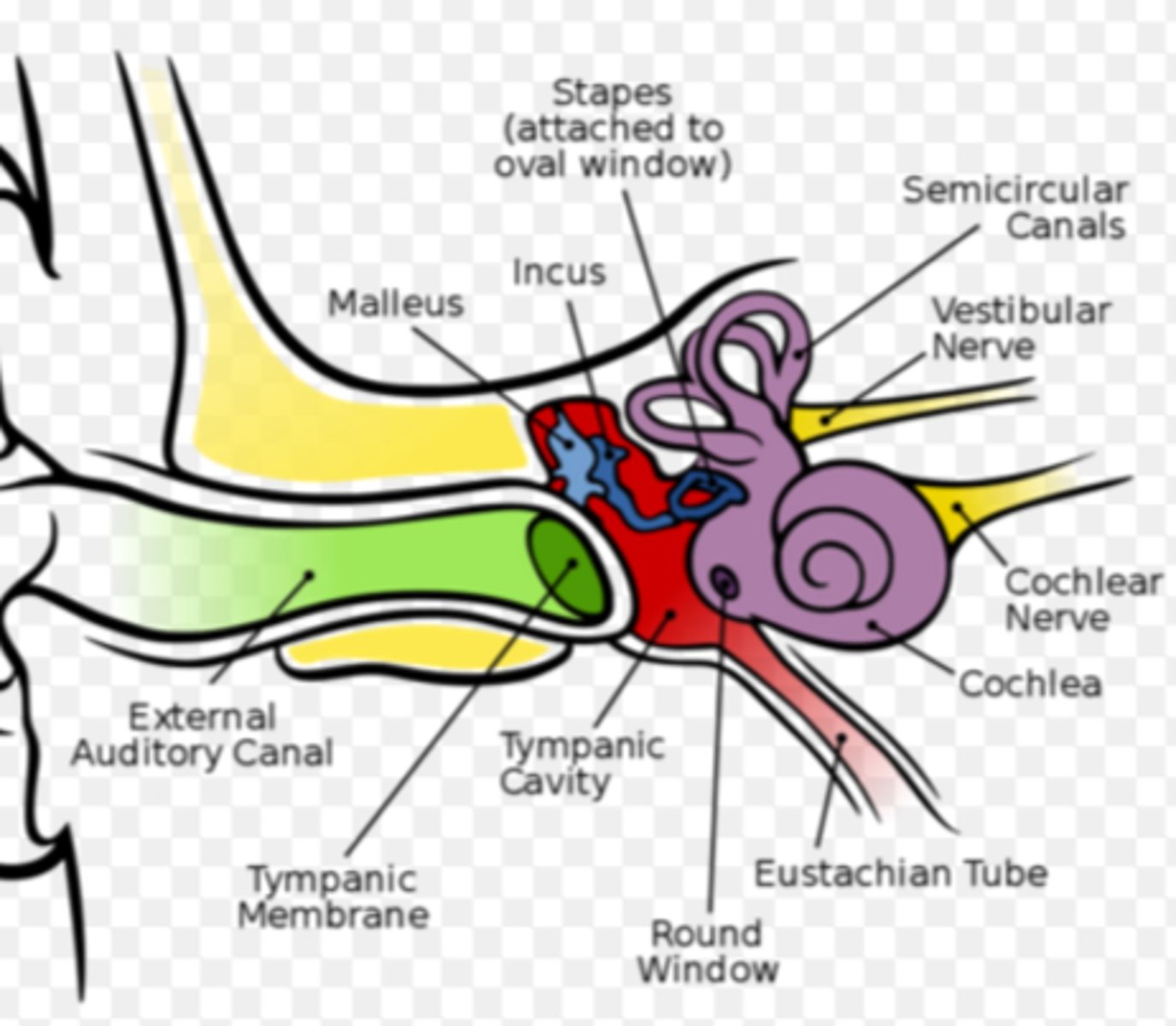
Middle Ear: Eustachian Tube
- connects the middle ear to the nasopharynx

Inner Ear
- located within the temporal lobe.
- houses the receptors for hearing and equilibrium
VESTICULIAR APPARATUS
COCHLEA
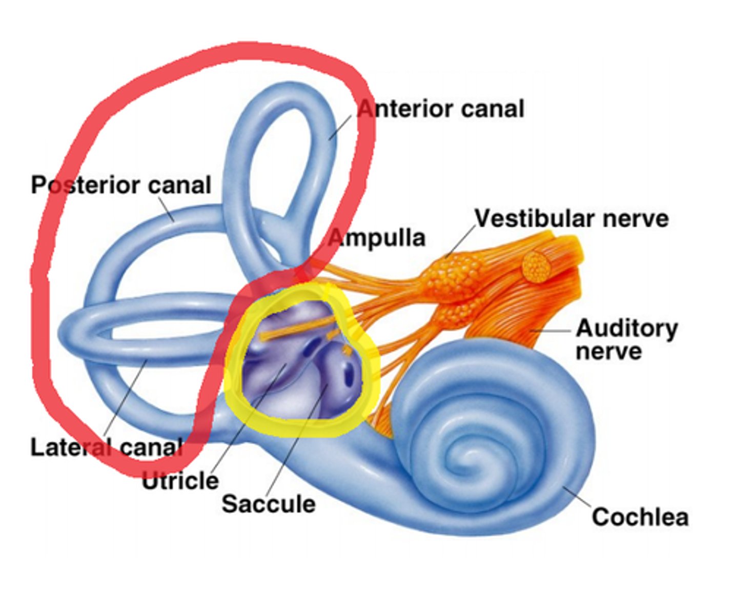
Inner Ear: Vestibular Apparatus
- contains the receptors for equilibrium
- made up of 3 parts
Inner Ear: Cochlea
- contains the receptors for hearing
- it looks like a seashell
Labyrinth
- both the cochlea and vestibular apparatus are structured as 2 channels in the bones, called labyrinths: the MEMRANOUS and BONY LABYRINTH
- the membranous labyrinth is within the bony labyrinth
LOOK AT NOTES
both labyrinths
- are filled with fluids that allow us to hear (cochlea) or become aware of and monitor the position of our head (vestibular apparatus)
membranous and bony labyrinth
- filled with endolymph and the bony labyrinth is filled with perilymph
Vestibular Apparatus
composed of 3 structures:
1. Semicircular Canals
2. Saccule and Utricle
Vestibular Apparatus: Semicircular Canals
- detect rotational movement

Vestibular Apparatus: Saccule and Utricle
- the utricle and saccule are responsible for detecting positional movement
Cochlea
- contains the receptors for hearing
- composed of 3 ducts:
VESTUBULAR DUCT
TYMPANIC DUCT
COCHLEAR DUCT
(the cochlear duct contains the organ of corgi, which is special structure for hearing)
LOOK AT NOTES
Organ of Corti
- made up of specialized cochlear hair cells that rest on basilar membrane
- when the membrane is moved by sound waves, the hair cells fire an impulse, which is sent to the brain via CN VIII for interpretation