Lecture 14: The Auditory System
1/29
There's no tags or description
Looks like no tags are added yet.
Name | Mastery | Learn | Test | Matching | Spaced | Call with Kai |
|---|
No analytics yet
Send a link to your students to track their progress
30 Terms
The Auditory System
sense of hearing
detects and localizes sounds
perceives and interprets its nuances
sound = mechanical energy (pressure waves)
Sound
variations in air pressure
Cycle
distance between successive compressed patches of air
Frequency
number of cycles per second (Hertz-Hz)
Human range of hertz
20-20k
Intensity
Amplitude
The volume of a sound (how loud/soft it is)
How BIG is each wave?
Frequency (Hz)
related to pitch
How MANY waves/sec
Ear Structure
Tympanic Membrane (eardrum) —> Ossicles (little bones) —> Oval Window —> Cochlea (fluid-filled)
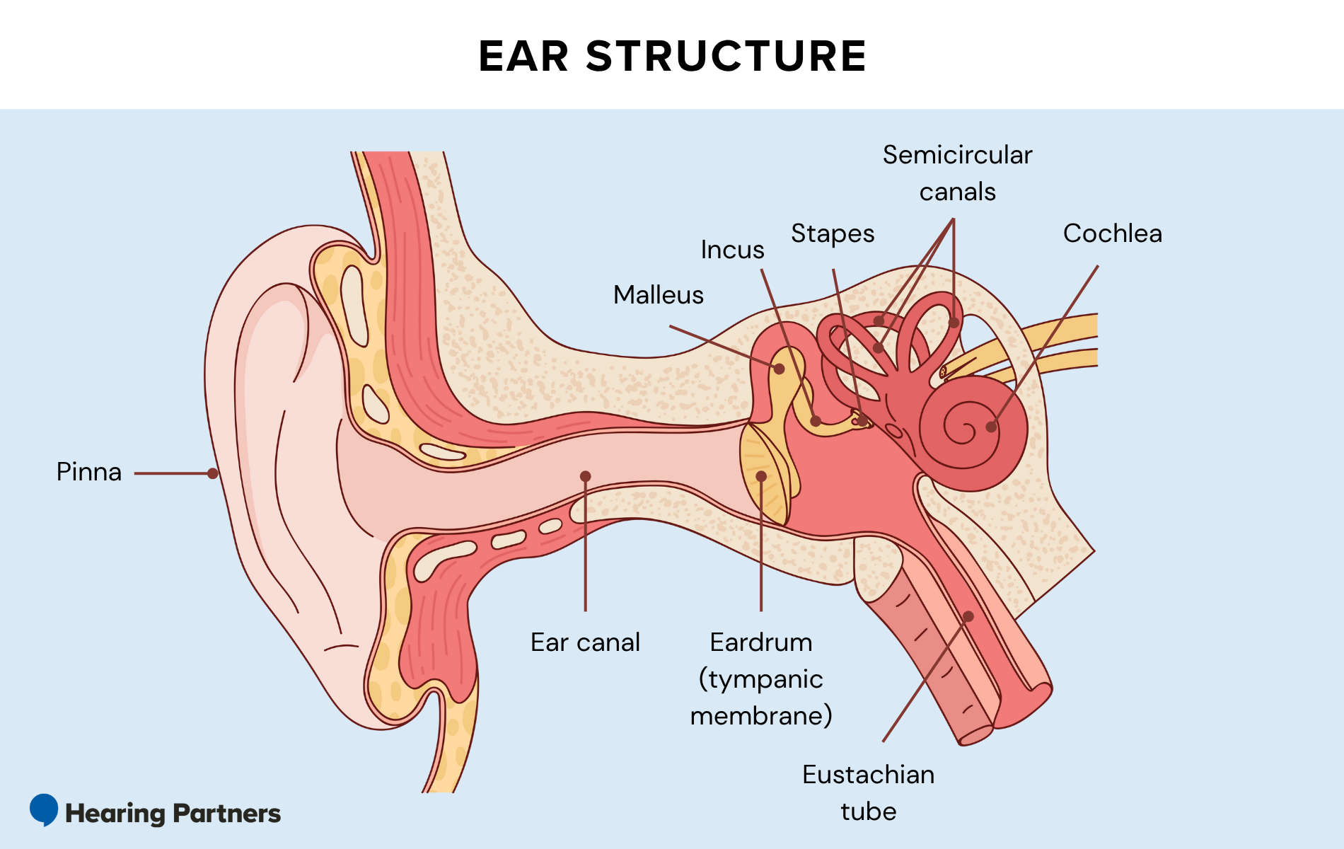
Pinna
visible outer part of our ear; collects sound; many mammals (not humans) can move the pinna to focus their hearing in a certain direction
Auditory Canal
tube that runs thru to the middle ear; funnels sound to middle ear
Amplification in the Middle Ear
Sound waves move the tympanic membrane (eardrum)
Moves the ossicles (small bones)
Malleus (hammer)
Incus (anvil)
Stapes (stirrup)
Ossicles move the oval window (another membrane)
Can air directly move the oval window?
No
cochlea is fluid-filled, so sound waves arriving at the oval window would cause no movement
more pressure is need to vibrate cochlear fluid than provided by air
The Middle Ear: Amplification
middle ear transmits vibrations to fluid-filled cochlea
amplify pressure by increasing force and decreasing surface area
P = F/SA
pressure at the oval window is 20x greater than at the tympanic membrane
Force (F) - ossicle (bones) act like a lever; turning weaker large amplitude waves into stronger smaller amplitude waves
Surface Area (SA) - tympanic membrane —> oval window
oval window is smaller than tympanic membrane so force is concentrated on a smaller surface area.
Inner Ear: Frequency Analyzer
Cochlea
spiral-shaped structure
contains 3 parallel fluid filled chambers (scala vestibuli, scala media, scala tympani)
Basilar Membrane: auditory receptors sit on top of here in the organ of corti
Perilymph: fluid in scala vestibuli and scala tympani
Endolymph: fluid in scala media
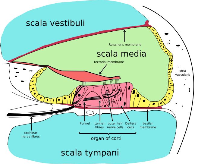
Basilar Membrane and Organ of Corti
Organ of Corti contains the auditory receptor neurons (hair cells)
Organ of corti is covered by the tectorial membranef
fluid in the scala media is endolymph, which has high concentration of K+
Basilar Membrane
pressure at oval window causes movement of the perilymph
makes basilar membrane oscilate like a wave
basilar membrane is flexible (not uniform)
widens towards apex
Base: narrow and stiff
apex: wide and floppy
Frequency Map in the Basilar Membrane
tonotopic map: a place code on the basilar membrane for the frequency that produces maximum amplitude of deflection
diff tones / frequencies will give u bends @ diff spots
waves of diff frequencies travel various lengths down basilar membrane
High frequency: vibrates narrow, stiff base and dissipates
low frequency: propogates to wide, floppy apex
Auditory Receptor Cells: “Hair Cells”
Auditory Receptor Cells = hair cells
inner and outer hair cells
The organ of corti is a collection of hair cells (along w support cells).
Hair cells extend stereocilia from their apical surface into the endolymph
Stereocilia tips end in OHCs or js below IHS the tectorial membrane
The bending of stereocilia is a critical event in tranduction of sound into neural signals
Movement of Hair Cell Stereocilia
Hair cells are responsible for transduction; they convert mechanical energy into a change in membrane potential.
Lifting of the basilar membrane and the organ of corti pushes the sterocilia up against the tectorial membrane. This bends the stereocilia
Stereocilia and K+ Channels
the bending of stereocilia causes mechanically-gated K+ channels to open
tip of each stereocilia has a special type of K+ ion channel that opens/closes as the stereocilia bends
Each channel is covered by a “lid”
Tip Link connects each lid to the neighboring stereocilia
as the stereocilia bends, the tip link pulls the lid open or closed
When the mechanically-gated channel is open, K+ flows INTO the cells and depolarizes it
Mechanism of Stereocilia
at rest (straight cilia) channel partly open (small leak of K+ in)
movement of cilia one direction - tension on tip link opens channels; K+ influx; depolarization
cilia go other direction - close tip links; hyperpolarization
Output
depolarization from K+ influx causes voltage-gated Ca 2+ channels to open, Ca2+ influx and NT release
Glutamate released onto spiral ganglion neurons
axons of spiral ganglion neurons = auditory nerve - wher 1st APs of the auditory pathway occur
most spiral ganglion neurons receive input from INNER hair cells
One inner hair cell —> 10 spiral ganglion cells
Outer hair cells
There are more outer hair cells than inner (3:1), but OHCs only innervate 5% of spiral gangion cells
Several OHCs synapse on 1 spiral ganglion cells*
OHCs function as a cochlear amplifier
Outer hair cells amplify the movement of the basilar membrane during low-intensity sound
motor proteins change the length of the outer hair cells in response to sound
Changes distance between basilar and tectorial membranes
causes the stereocilia of the inner hair cells to bend more - produces a greater response in the auditory nerve
amplifies movement of the basilar membrane 100-fold
Damage to OHCs
medications like antibiotics, chemotherapy drugs, and edema meds can cause ototoxicity
hearing loss or deafness due to damage of the amplifier
Auditory Pathway
Auditory Receptor Cells (hair cells)
Depolarize (+ release NT) in rhythm w sound wave
Spiral Ganglion Cells
Auditory Nerve
Brainstem
(up to 3 synapses); multiple parallel pathways
Thalamus
Auditory Cortex
Encoding the properties of sound
intensity
frequency
location
Sound Intensity
Firing rate of neurons
more movement of basilar membrane
more depolarization
spiral ganglion cells fire APs at greater rates
Number of active neurons
greater movement of basilar membrane activates more hair cells
broadening of frequency response range
Sound Frequency 1
Tonotopy
the frequency of sound that a hair cell responds to is largely determined by its location on the basilar membrane
organizational pattern continues through the brain regions in the auditory pathway, all the way to auditory cortex
Creates a frequency map
Why encoding the frequency of a sound isn’t due to just tonotopy…
no specificity at low frequencies
20hZ and 50hZ have same site of activation
not very specific for sounds w/great intensity (loud)
louder sound will deform greater region of the basilar membrane