AP Unit 4: Cell Communication & Cell Cycle
1/55
There's no tags or description
Looks like no tags are added yet.
Name | Mastery | Learn | Test | Matching | Spaced | Call with Kai |
|---|
No analytics yet
Send a link to your students to track their progress
56 Terms
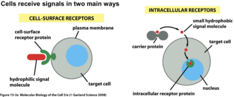
Ligand
A signaling molecule including hormones, small chemicals, or environmental stimuli. Polar (or large) ligands require surface (extracellular) receptors; nonpolar ligands use intracellular receptors.
Receptor
Protein that accepts the ligand and moves the signal forward by changing shape in response to the presence of the ligand. After the ligand binds, the intracellular domain of an extracellular receptor protein changes shape initiating "transduction" of the signal.
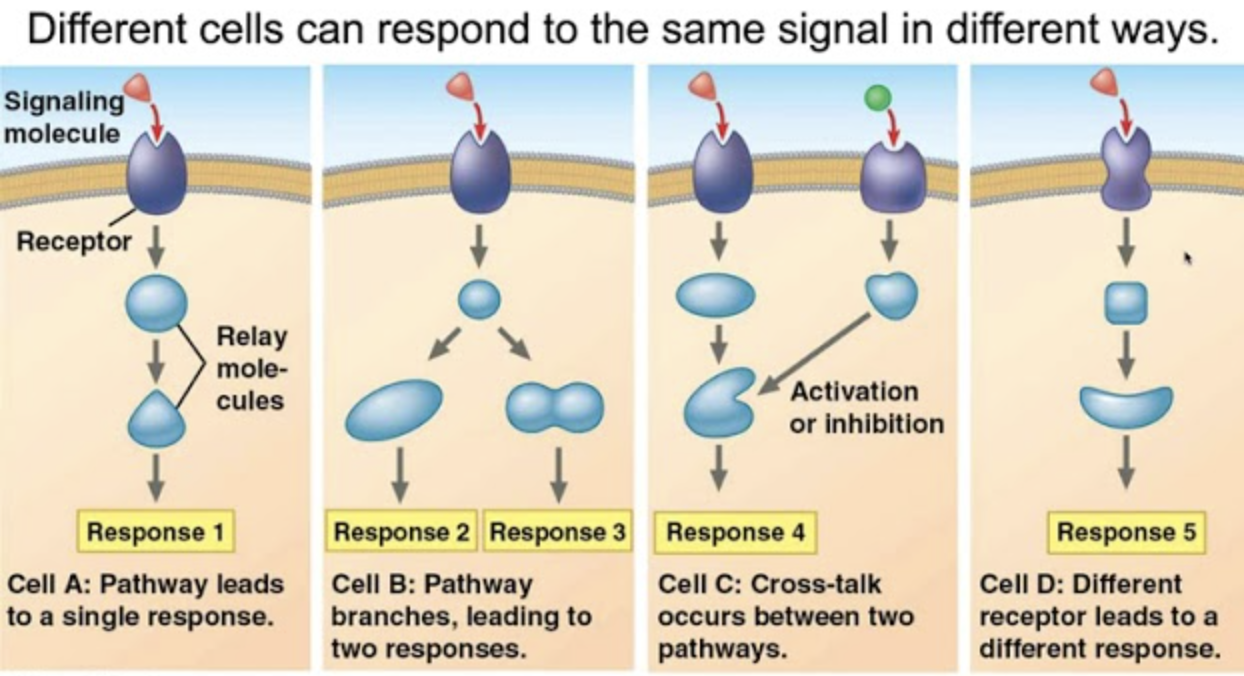
Signal Transduction
The movement of a cell signal from the ligand toward the cell response. May involve the modification of relay proteins, phosphorylation cascades, and 2nd messengers (like Ca2+ and cAMP).

Cellular Response
Includes responses to stimuli like sensory triggers, hormones, and stress. Responses include: activating/deactivating a gene or an enzyme (gene expression), mitosis (cell growth), secretion (releasing cell products), or apoptosis.
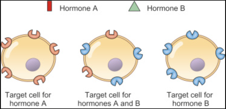
Target Cell
Cells that possess the receptors for a particular ligand and, therefore, respond to the signal.
Local Signaling
Includes paracrine, synaptic, gap junctions (animal), plasmodesmata (plants), autocrine, and cell-to-cell recognition. Ligands come from other cells nearby.
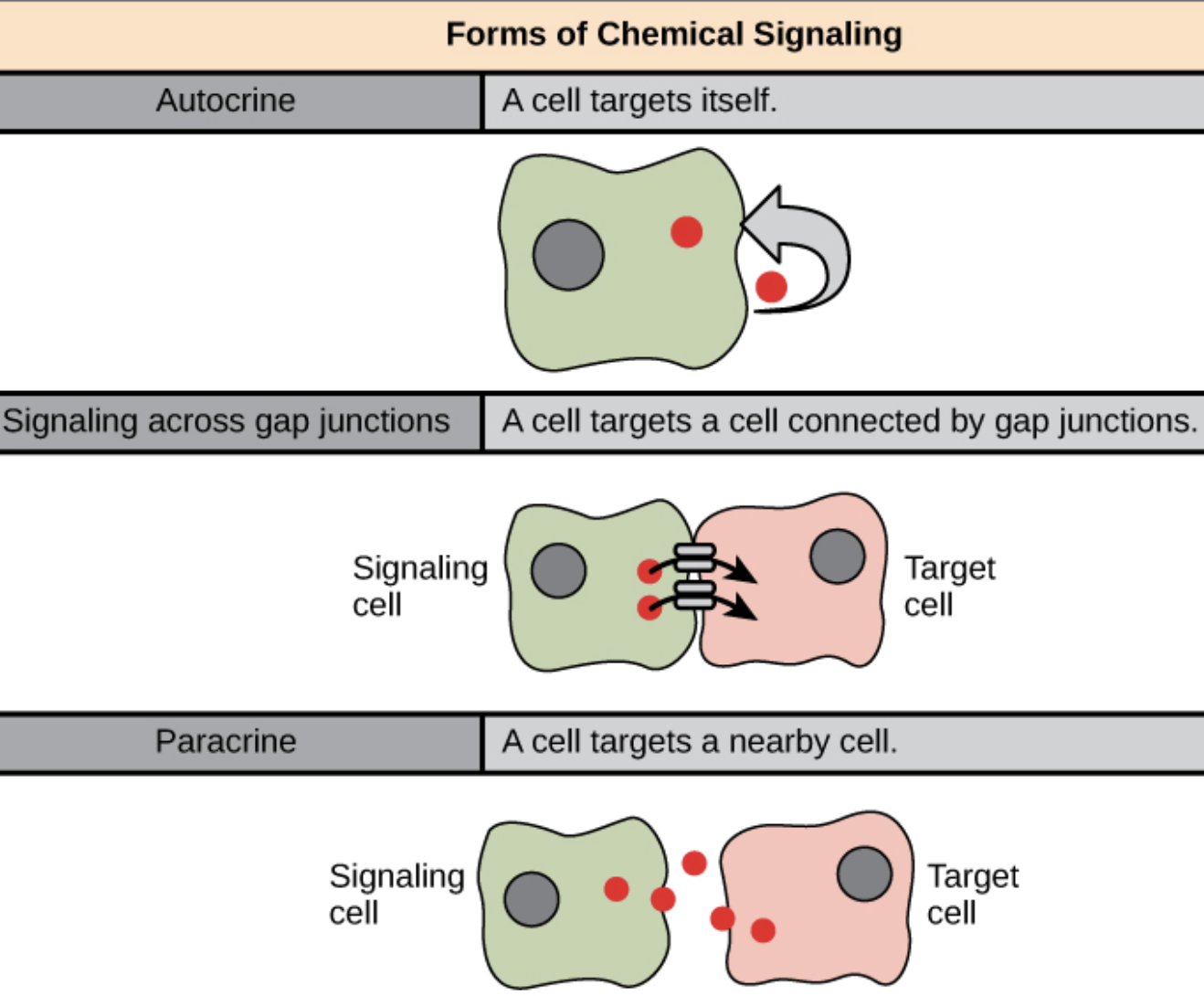
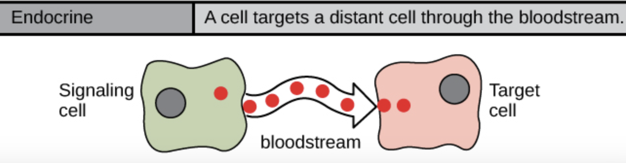
Long-Distance Signaling
Examples include the endocrine system (hormones that travel through the blood stream) and the nervous system
"Widely conserved among all domains"
Means: Almost all organisms share the signaling pathways, an indicator of common ancestry and that these pathways evolved early.
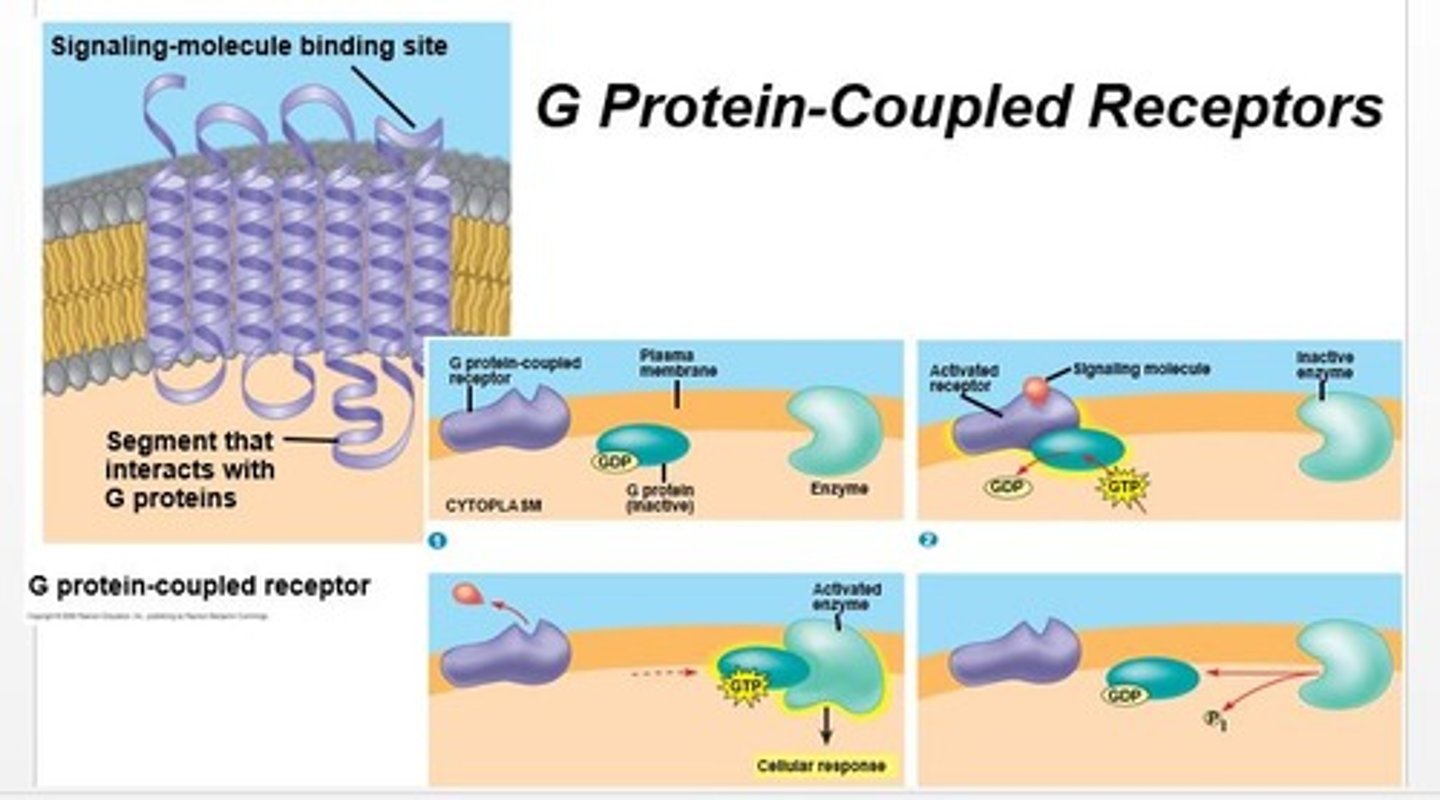
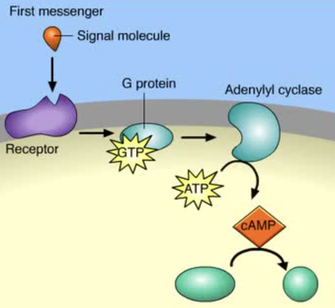
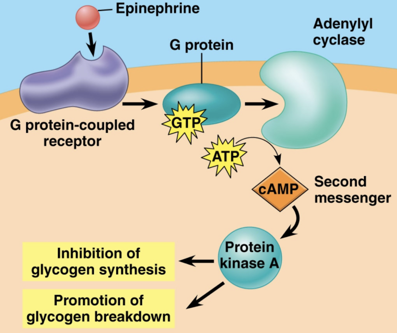
GPCR
G-Protein Coupled Receptors. An example of a receptor in a signaling pathway that involves G-proteins (which use GTP instead of ATP).
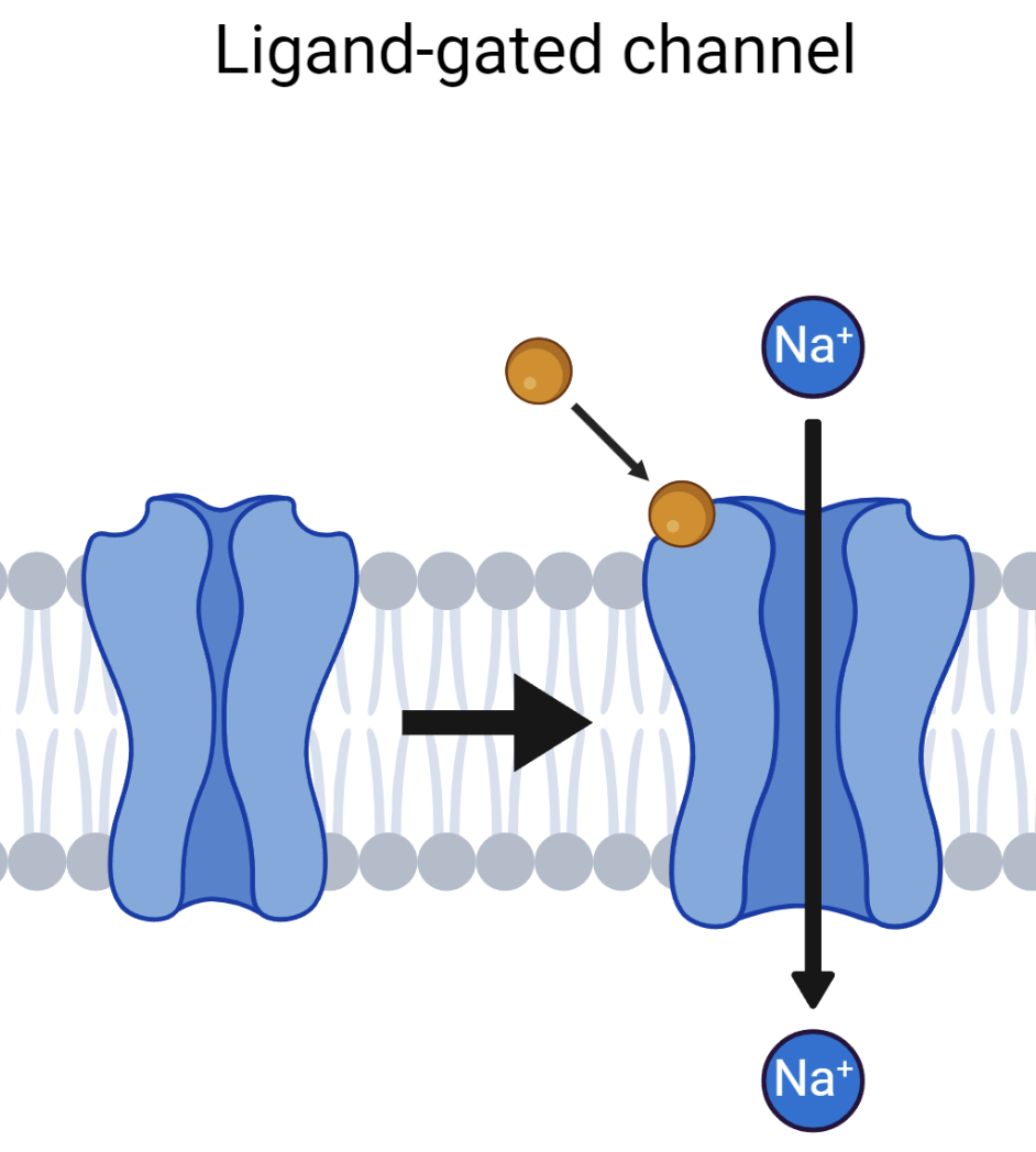
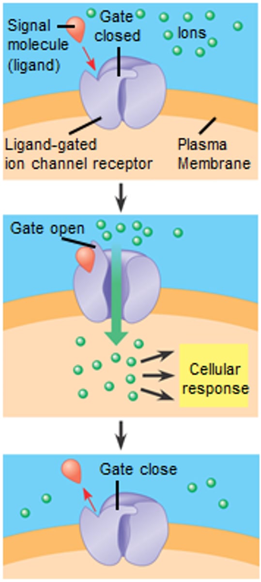
Ligand Gated Channel
A channel protein that opens or closes in response to the presence of a ligand
Second Messenger
An INTERNAL signaling molecule (ex: Ca2+ and cAMP) released after the initial reception of a ligand. 2nd messengers always lead to additional/amplified response pathways.
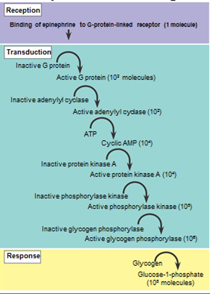
Amplification
The INCREASE in a cellular response by pathways like phosphorylation cascades. A single ligand can then lead to 10^8 responses due to amplification pathways.
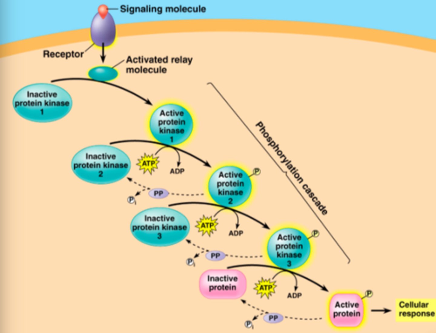
Phosphorylation Cascade
A transduction pathway involving a series of kinases (proteins) that can be activated sequentially when they are phosphorylated by ATP.
Role of the cell's environment
Signal transduction pathways influence how the cell responds to its environment. The cell response should only trigger when the appropriate ligand is present in the environment. Cells respond to changes in: pH, hormones, temp, hydration, new molecules/chemicals
Impact of shape changes in proteins/ligands
If the shapes of any proteins or ligands are changed due to mutations or the presence of chemicals (think toxins), the signaling pathway will be affected downstream (after) the change.
Epinephrine/Adrenaline
An example of a polar hormone (ligand). Uses GPCR pathways to trigger "flight or fight" responses including the inhibition of glycogen synthesis and the promotion of glycogen breakdown to increase glucose levels in the blood
Why do cells need to divide?
To replace damaged cells ("tissue repair"), to grow, for asexual reproduction (cloning of cells like white blood cells)
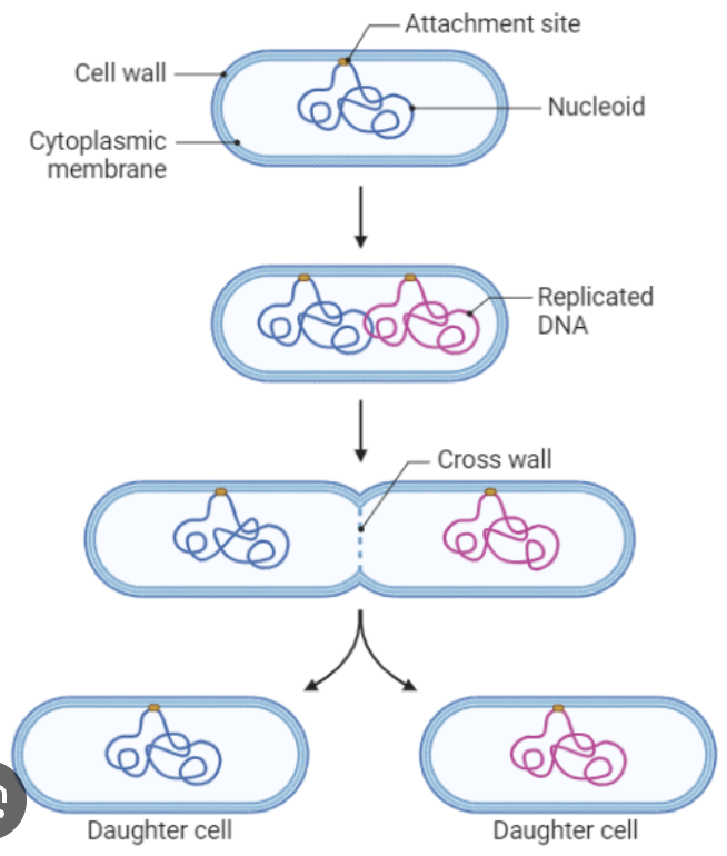
Binary Fission
Mitosis in bacterial cells where the one circular chromosome is replicated and then the bacteria divides.
Cell Cycle
Includes Interphase (G1, S, and G2), Mitosis (P, M, A, T) and Cytokinesis. Some cells leave the cycle and enter a state of non-division called G0
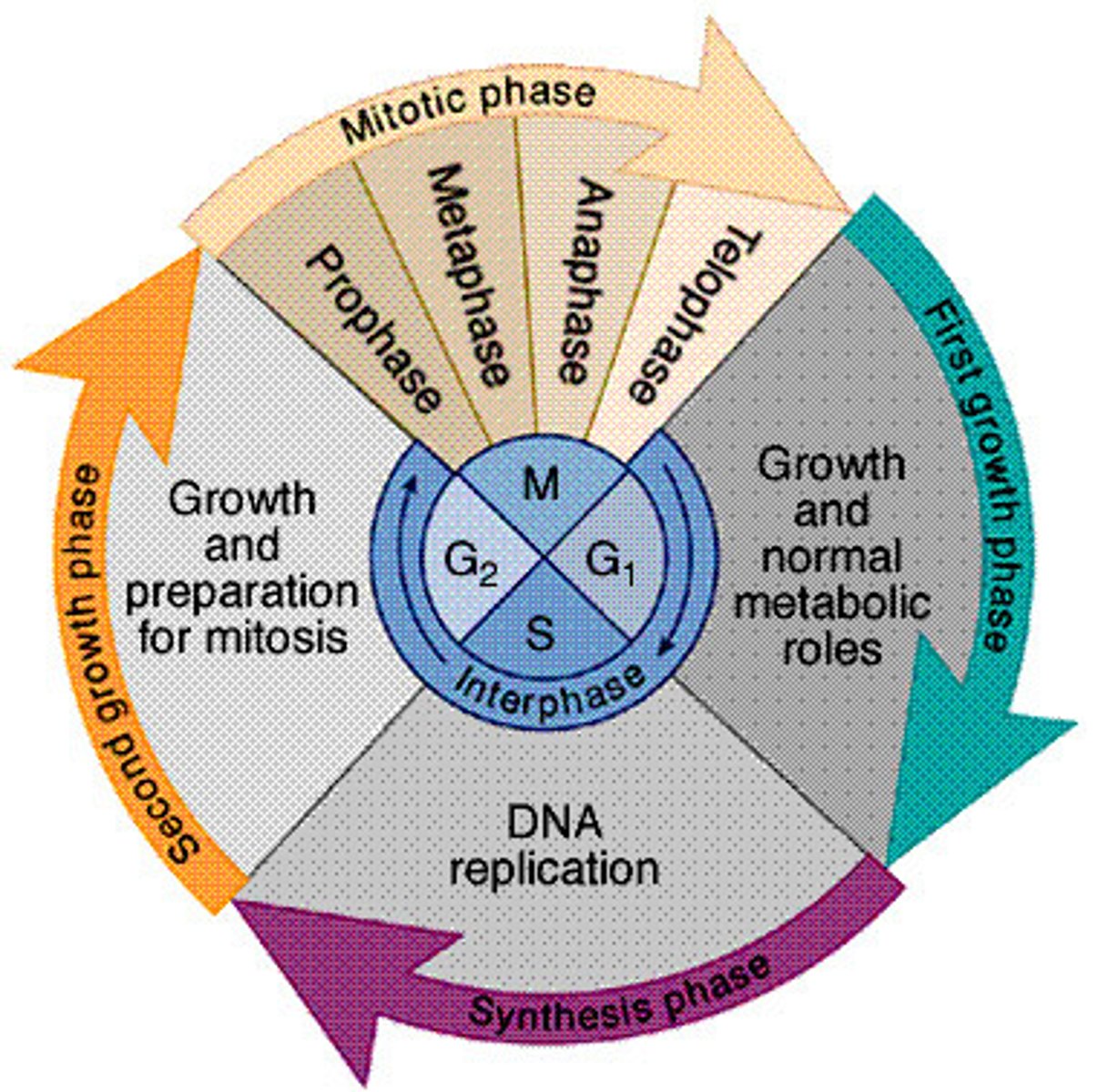
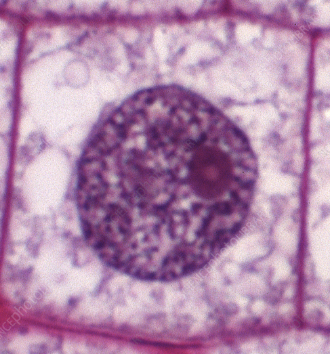
Interphase
The majority of a cell's life; when "normal" cell functions occur. Includes G1 (growth), S (replication) and G2 (prep for mitosis) stages.
G1
Stage of Interphase where the cell is primarily growing.

S
Stage of Interphase where the primary event is the replication of DNA. Mutations MAY occur during the S phase.
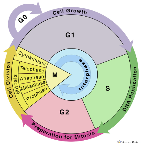
G2
Stage of Interphase where the cell is preparing for mitosis. (Nothing more needed here...)

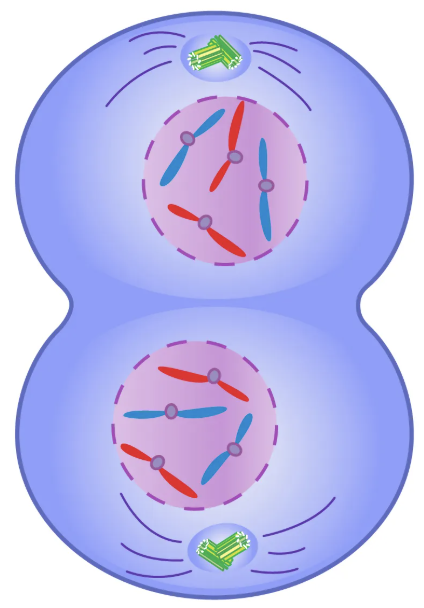
Mitosis
The division of the NUCLEUS of a cell leading to 2 IDENTICAL nuclei. Occurs after Interphase and includes the stages of Prophase, Metaphase, Anaphase, and Telophase. Cytokinesis then divides the two new nuclei into 2 cells.

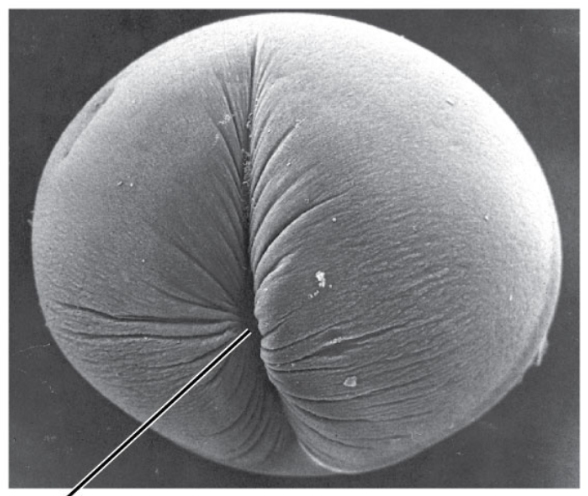
Cytokinesis
The pinching in of the cell membrane in an animal cell after mitosis of the nucleus. Involves the formation of a cell plate in plant cells.
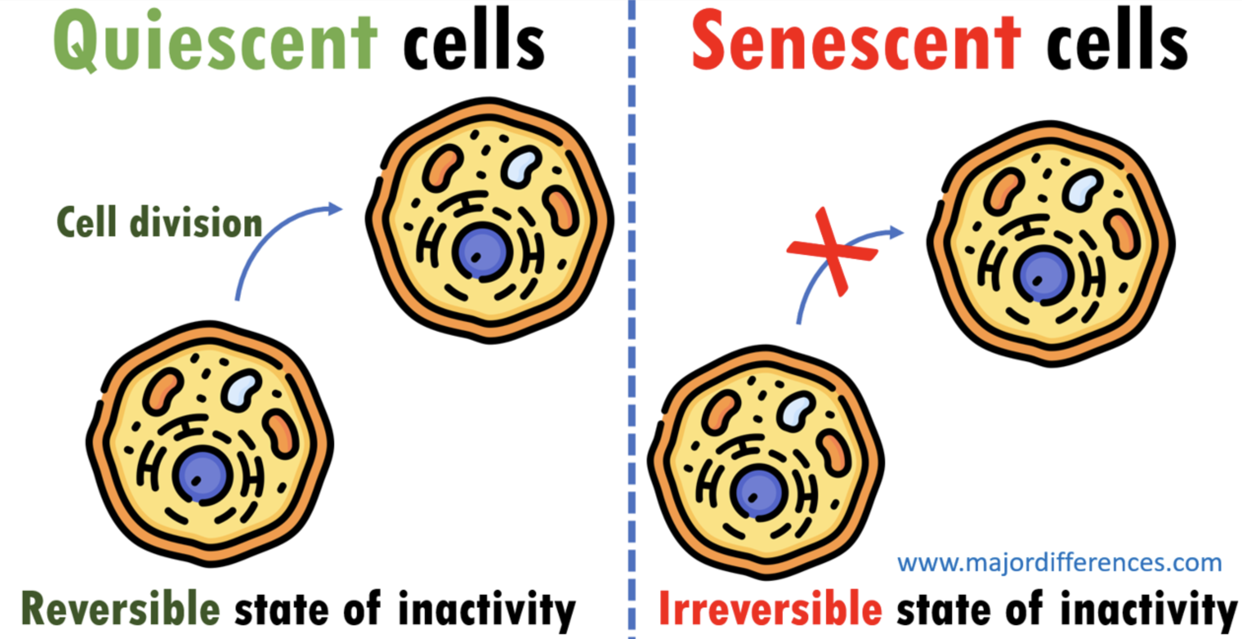
G0
The non-dividing state of cells that are quiescent (waiting), differentiated (functional, but never dividing), or senescent (old).

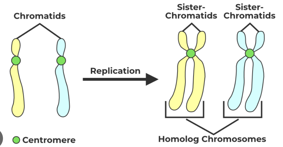
Bivalent Chromosome
Twisted form of DNA ('braids") that includes 2 copies of the original DNA (sister chromatids) attached by a centromere.
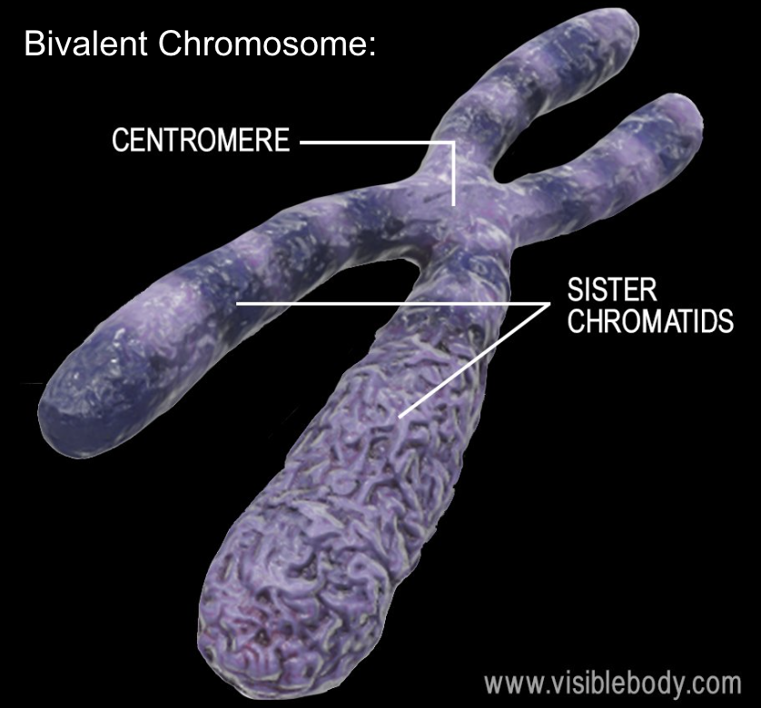
Centromere/Kinetochore
Protein that holds the two identical sister chromatids of a bivalent chromosome together. The kinetochore is attached to the spindle fibers as the chromosomes are moved and separated during mitosis.

Monovalent Chromosome (“Chromatid”)
DNA that has NOT been replicated.
2N
Indicates a cell that has 2 copies of each chromosome (one from bio mom and one from bio dad). All somatic (body) cells in a human body are 2N (diploid w/23 pairs of chromosomes). FYI: 2N/diploid has NOTHING to do with the term bivalent.

Plasmid
Small, circular piece of DNA found in prokaryotic (bacterial) cells that carries a few genes. Genes for antibiotic resistance are found here.
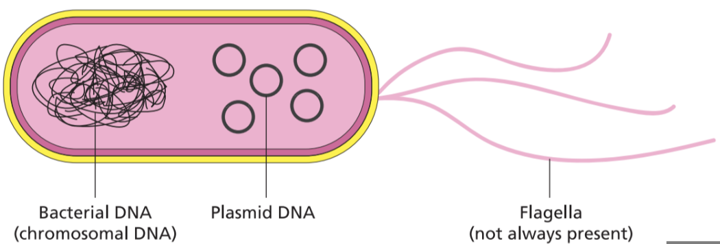
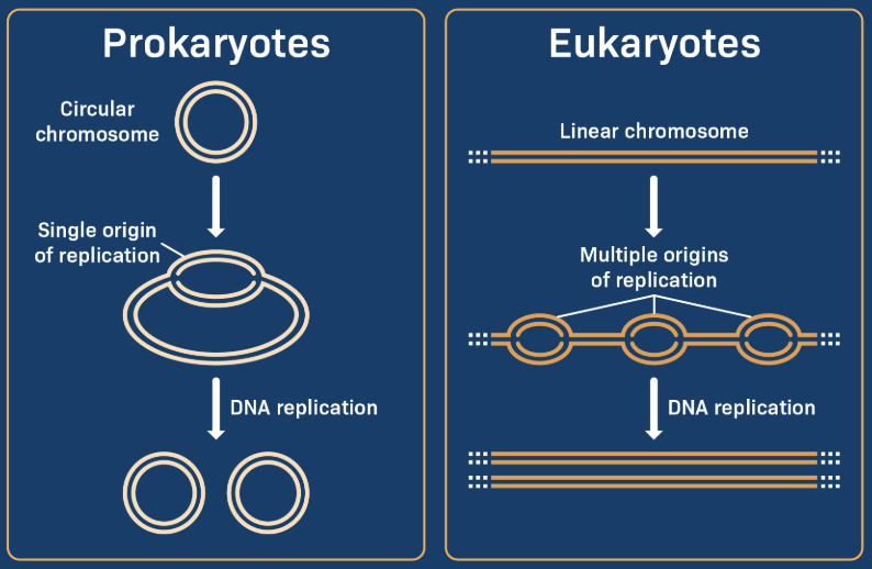
Linear Chromosomes
The shape of eukaryotic chromosomes (like human DNA). Contrasted with the circular chromosome of a prokaryote (bacteria).
Haploid
Cells with HALF of the organism's DNA. These cells are always gametes (sperm, eggs, or pollen). 1N. The haploid number for a human is 23
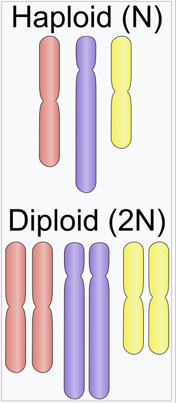
Diploid
Cells with pairs of chromosomes (one from each parent). Include somatic (body) cells. 2N. The diploid number for a human is 46.

Non-Reductive Cell Division
MITOSIS- The number of chromosomes is NOT REDUCED. Ex: A cell with 46 bivalent chromosomes will have 46 monovalent chromosomes after mitosis.
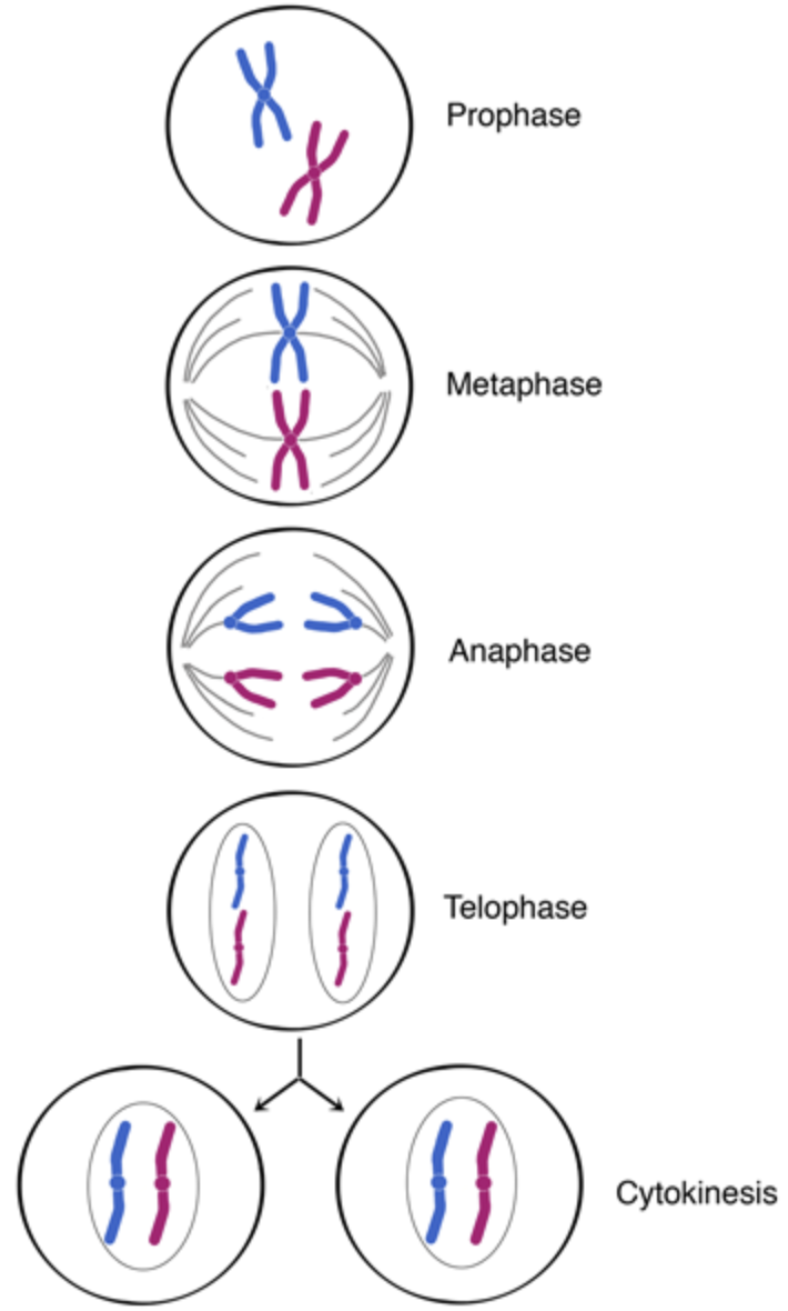
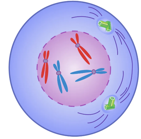
Prophase
1st stage of mitosis. Chromosomes "condense" (wrap up to form bivalent chromosomes). Nucleus disappears. Centrioles move to poles. Spindle begins to form.
Prometaphase
Between prophase and metaphase.
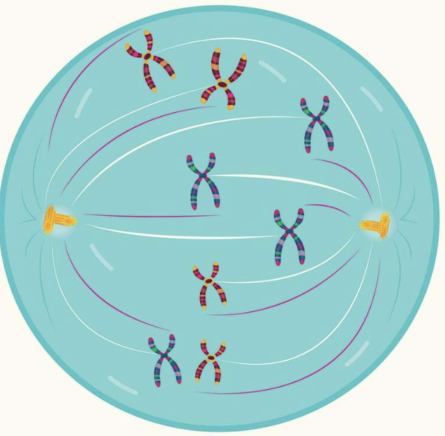
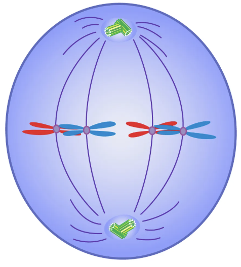
Metaphase
2nd stage of mitosis. Spindle attaches to kinetochore of each chromosome and moves them to the equator (Middle) of the cell.
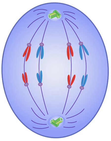
Anaphase
3rd stage of mitosis. Spindle retracts to the centrioles, ripping APART the bivalent chromosomes. This moves IDENTICAL copies (sister chromatids) of the chromosomes to each pole of the cell.
Telophase
Final (4th) stage of mitosis. The nuclei appear, and the chromosomes unwind to return to the chromatin form. Telophase is followed by cytokinesis.
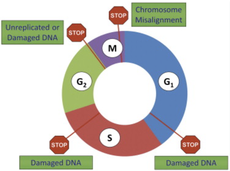
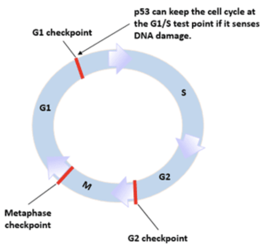
Cell Cycle Checkpoints
G1, G2, and M checkpoints. Failure to "pass" these checkpoints stops cell division, triggers repair enzymes, and leads to apoptosis if repair is not possible. Passing the checkpoints inappropriately could lead to cancer.
Quiescent
Cells in the G0 stage that are "quiet", waiting to be reactivated. Ex: Memory B cells
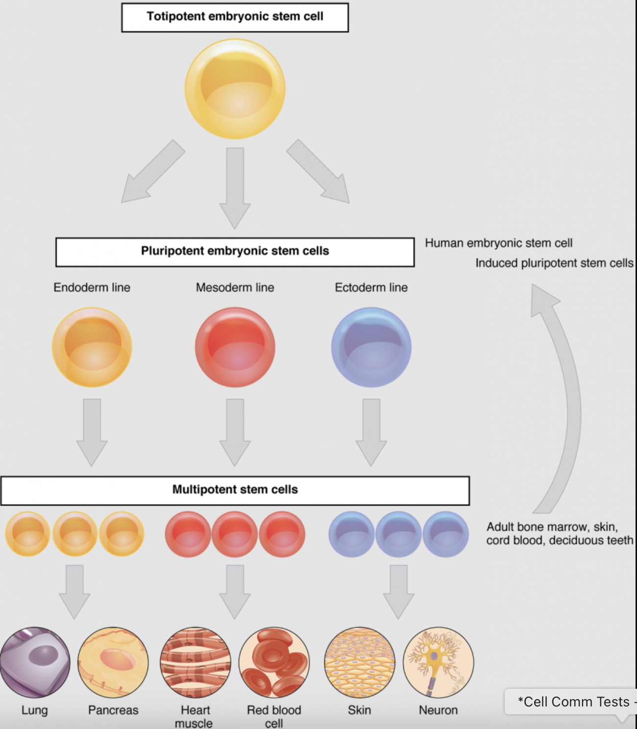
Differentiated
When a less specialized cell develops to acquire a more specialized form and function. Cells in the G0 stage that are functioning, but that will not re-enter the cell cycle and divide again. Ex: Cells of the nervous system
Senescent
Cells in the G0 stage because they are old and may be malfunctioning.

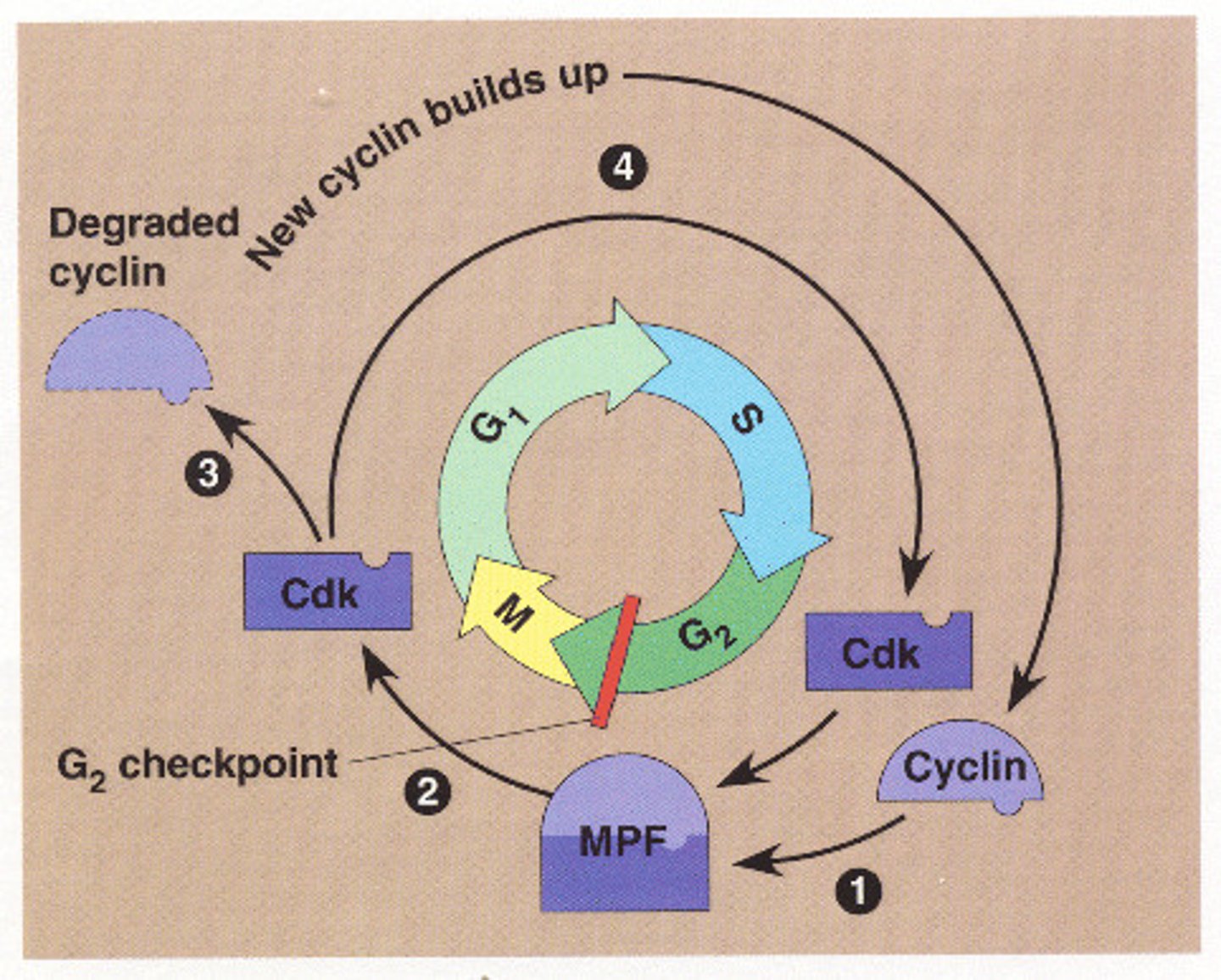
Mitosis Promoting Factor (MPF)
Example of an INTERNAL control of cell division. CdK, present in constant amounts, combines with Cyclin, present in increasing amounts, to form MPF, an enzyme that triggers mitosis.
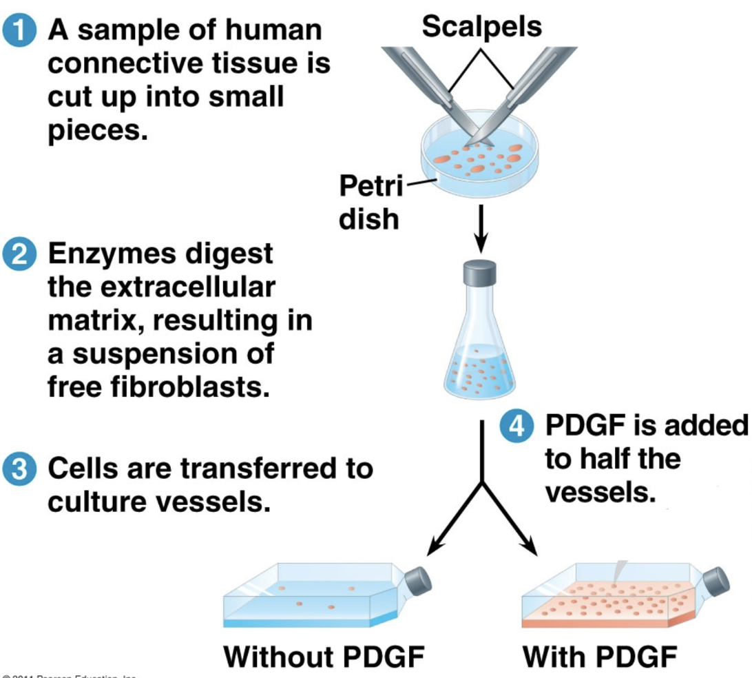
Platelet Derived Growth Factor (PDGF)
Example of an INTERNAL control of cell division. Platelets, released to help clot blood, also release growth factors to increase cell division in the injured tissue.
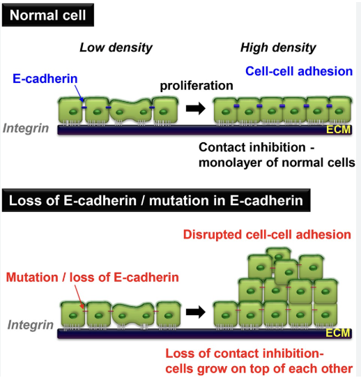
Positional (or "contact") Inhibition
Example of an EXTERNAL control of cell division. Cells that are touching stop dividing, unless they are cancer cells forming a tumor...
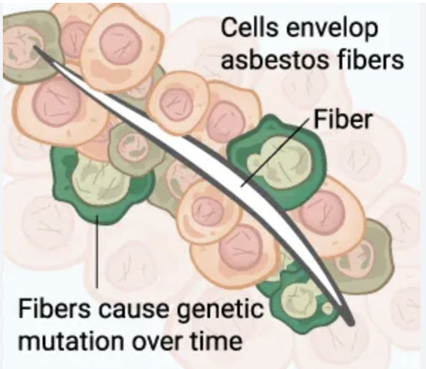
Carcinogens
"Agents" that can cause cancer by leading to mutations in DNA. Ex: asbestos, PFAS, UV light, HPV
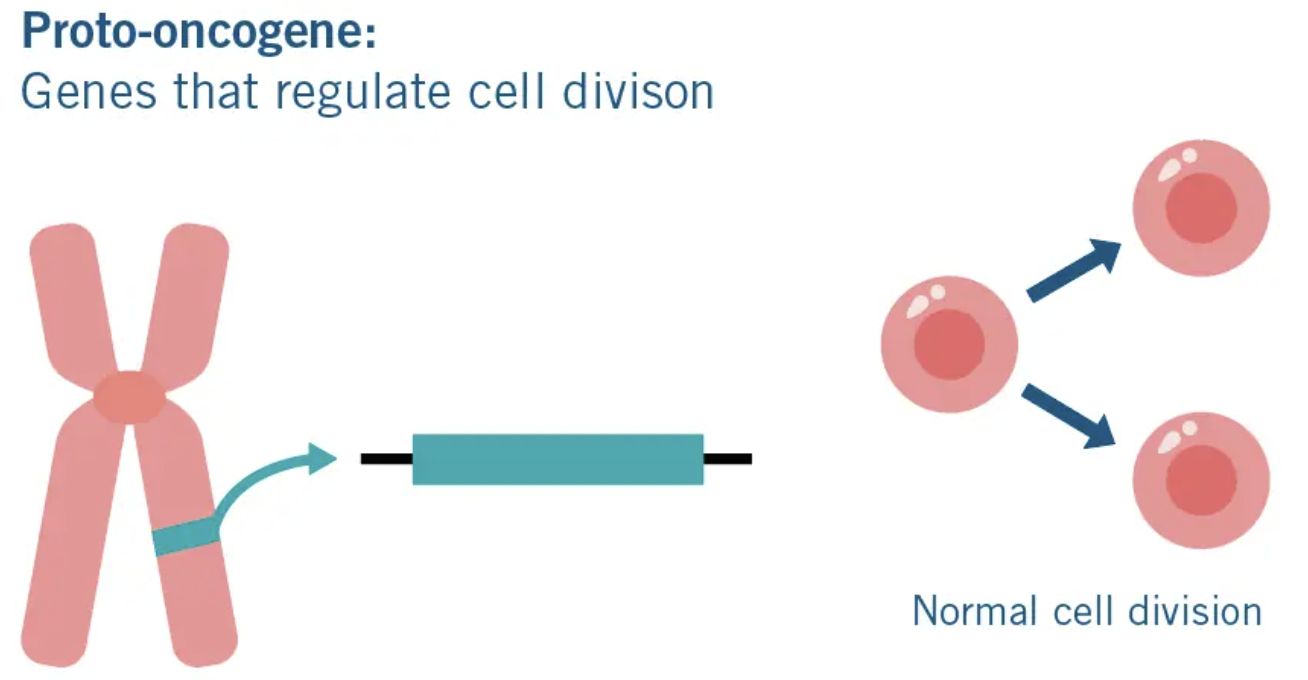
Proto-oncogenes
Normal, healthy genes that start cell division. Mutations in proto-oncogenes may cause them to become "oncogenes" that are always ON.
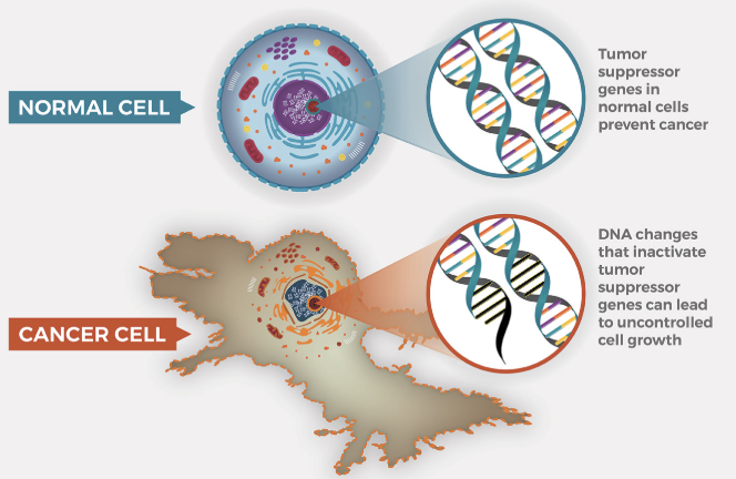
Tumor Suppressor Genes
Normal, healthy genes that stop cell division. Mutations may convert them to "oncogenes" that are always OFF.
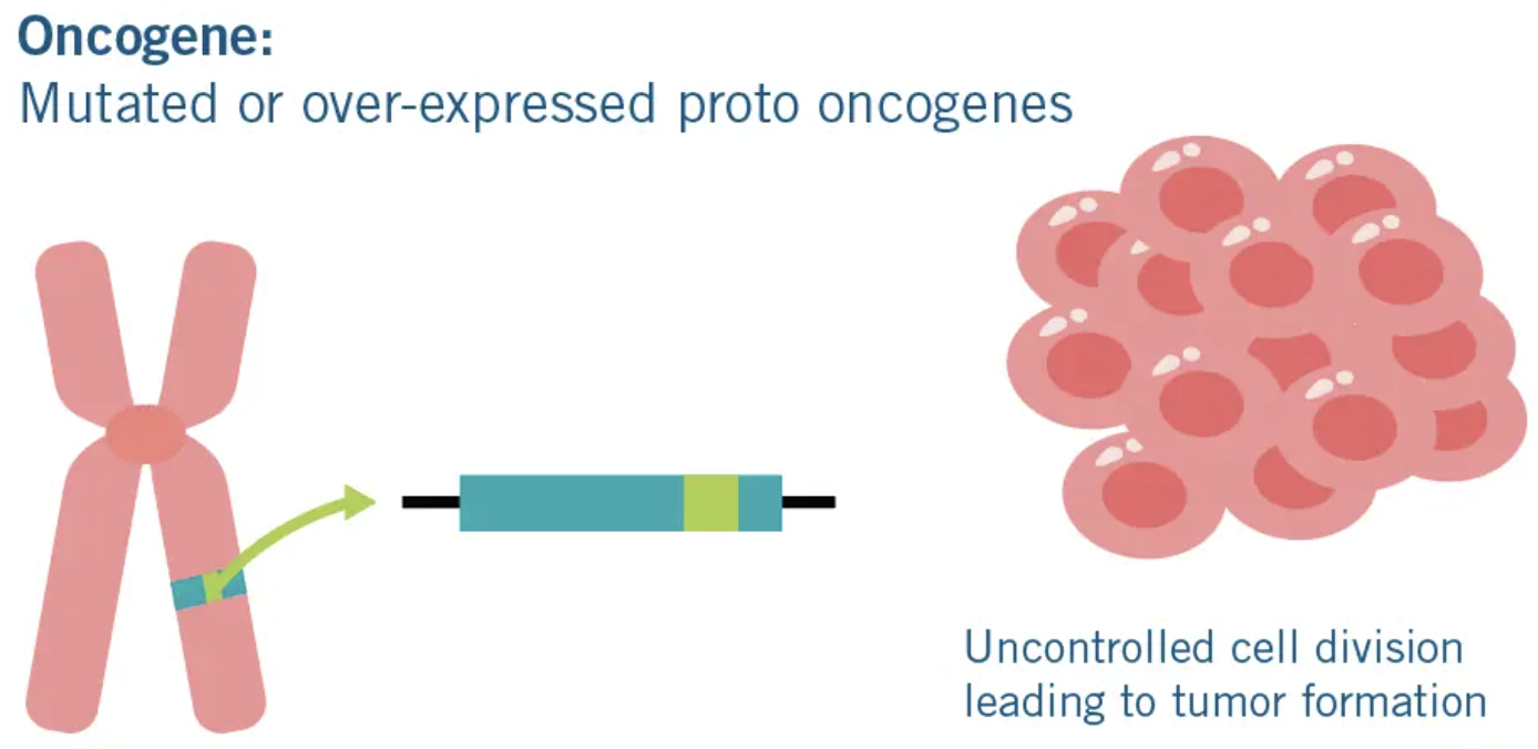
Oncogenes
Genes that lead to excessive mitosis (cancer). Caused by carcinogens, mutations during replication, or inherited.
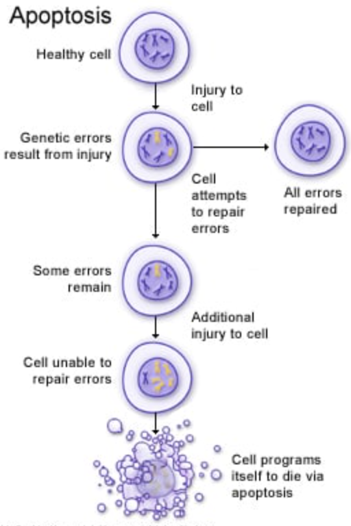
Apoptosis
Programmed cell division. May be used to prevent cancer (cell division of a damaged cell).
p53
An example of a tumor suppressor gene. Mutations in p53 are found in 50% of cancers.
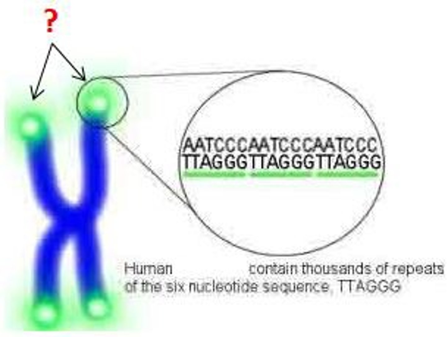
Telomeres
Tips on the ends of chromosomes that shorten with each cell division. Once they are too short to protect the DNA, the cell enters G0 and senescence. Cancer cells have a mutation that protects telomeres so cells can keep dividing indefinitely.
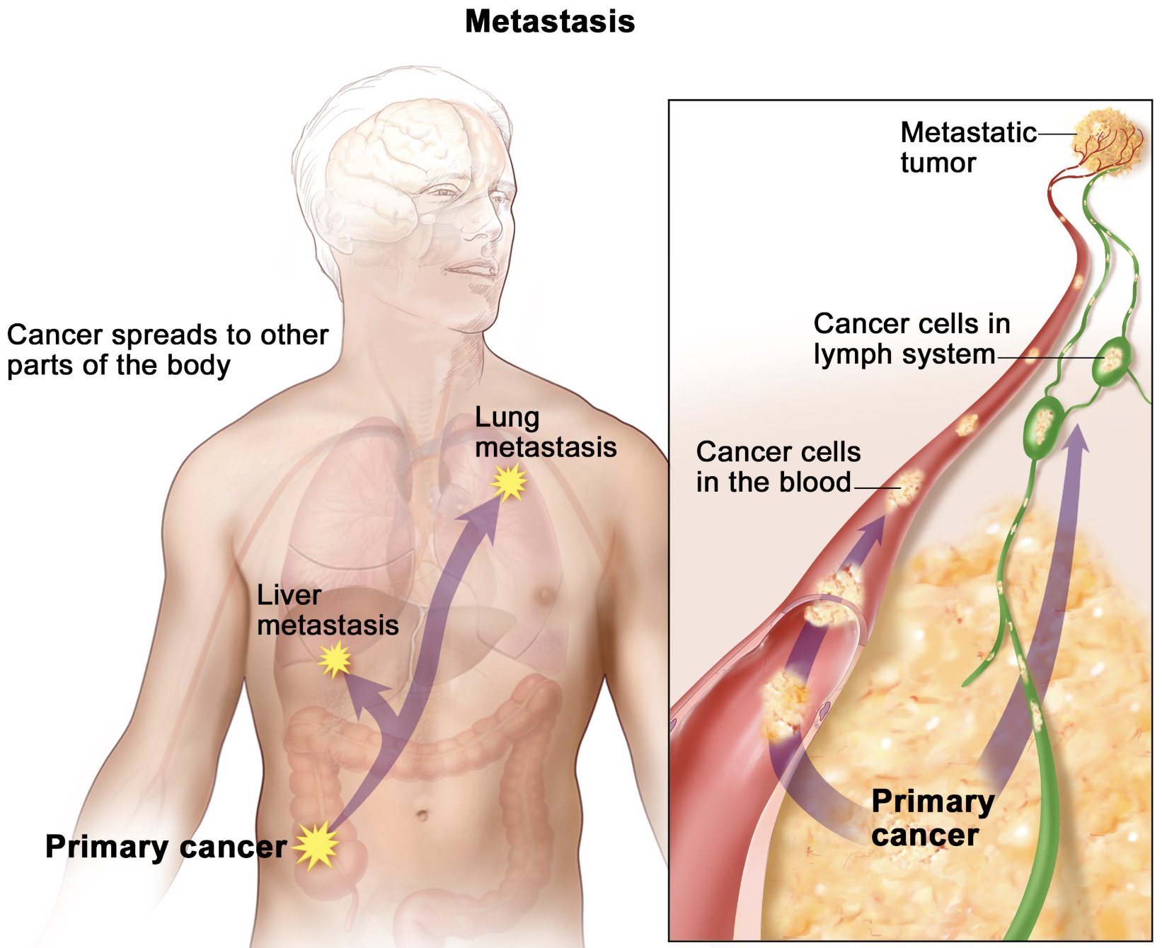
Metastasis
The spreading of cancer cells to other parts of the body.
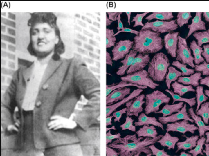
HeLa Cells
"Immortal" cancer cells taken from Henrietta Lacks without her permission. Have been used in countless scientific discoveries, including COVID vaccines.