Exam 1 Learning Objectives
1/129
Earn XP
Description and Tags
Technical Procedures – Administration of Drugs and Other materials, Technical Procedures – Sample Collection, Surgical Techniques, Sutures and Suturing, Anesthesia & Euthanasia, Confounding Factors / Variables Affecting Animal well Being and Research Outcomes, Lab Animal Diseases / Conditions
Name | Mastery | Learn | Test | Matching | Spaced |
|---|
No study sessions yet.
130 Terms
Identify several routes of Enteral administration
In food; in water; by mouth; by oral gavage
Identify several routes of Parenteral administration
Dermal/topical; Inhalation; Injections (IM, SC, IP, IV, intracardiac, intranasal)
IM
Intramuscular
SC
Subcutaneous
IP
Intraperitoneal
IV
Intravenous
Intracardiac
IC
What is the angle for an Intradermal (ID) injection
10-15°
What is the angle for an Subcutaneous (SC or SQ) injection
45°
What is the angle for an Intramuscular (IM) injection
90°
What is the angle for an Intravenous/ intravascular (IV) injection
10-25°
Describe the steps in performing gavage in rodents
Measure from corner of mouth to last rib for proper length/depth of insertion
restrain the animal
using needle, push back on hard palate to rock head back so that passage is straight
ensure that maximum recommended volume does not exceeded
What are the appropriate dose/volume ranges for a gavage in a Mouse
For Average Adult Body weight: Mouse .5-1 mL
What are the appropriate dose/volume ranges for a gavage in a Rat
For Average Adult Body weight: 3 mL
What are the appropriate dose/volume ranges for a gavage in a Hamster
For Average Adult Body weight:
1-1.5 m
Identify variables/concerns related to whole-body inhalation exposure
besides being inhaled, also deposits on fur and skin, and can then be ingested orally– thus greater dose than originally calculated
Identify variables/concerns related to nose-only inhalation exposure
limits to just inhaled dose
Describe why one might choose a luer-lock syringe over a standard syringe
With thick liquids, pressure may build when pushing plunger, and needle may shoot off. With a luer-lock syringe, the needle is screwed on and won’t fly off
For the Diameter (gauge) the lower number means (16 ga,18ga), and who would you use it on?
large gauge, cat/dogs
For the Diameter (gauge) the higher number means (23 ga,27 ga) and who would you use it on?
Smaller gauge: rodents
Standard needle sizes for Rodents?
23-27 gauge
Standard needle sizes for Rabbits?
23 gauge
Provide several reasons why re-use of needles should be discouraged
Tissue trauma; contamination; pain
Discuss why commercially manufactured (USP) drugs are better than non-pharmaceutical grade agents?
Purity, sterility, acid-base balance, longer storage/shelf-life, absence of pyrogens (produced by bacterial contaminants)
IM common injection sites in rodents
(thigh muscles of hind limbs) -
< 0.05 ml mouse, < 0.1 ml hamster, < 0.3 ml rats
IM common injection sites in Rabbits
(thigh muscles of hind limbs; epaxial muscles which parallel the spine)
< 0.5 ml
SC common injection sites in rodents
(over the back)
< 1 ml mice, < 2 ml rats
IP common injection sites in rodents
(ventral abdomen – near midline of upper portion of lower two quadrants)
< 1.5 ml mice, < 3 ml rats
IM common injection sites in Rabbits
(thigh muscles of hind limbs; epaxial muscles which parallel the spine)
< 0.5 ml
IV common injection sites in Rabbits
marginal ear vein
What should always do before injection in order to prevent Hypothermia?
Warm to near body temperature
IV common injection sites in rodents
(lateral tail veins of rats and mice)
< 0.15 mice, < 1 ml rats
safe blood collection volumes for Mice
less than or equal to 0.3 ml
safe blood collection volumes for Rat
less than or equal to 3 ml
safe blood collection volumes for Hamster
less than or equal to 1 ml
safe blood collection volumes for Rabbit
less than or equal to 30 ml
why the use of a vacutainer for blood collection in rodents may not be successful
Collapses vessel, prevents collection of blood
Identify the location of the common vessels used for blood collection: Tail
lateral tail veins (rats and mice)
Identify the location of the common vessels used for blood collection: Ear
marginal ear vein, central artery (rabbit)
Identify the location of the common vessels used for blood collection: Face
submandibular and facial veins (rats and mice)
Identify the location of the common vessels used for blood collection: Eye
retro-orbital sinus (rats, mice, hamsters)
Identify the location of the common vessels used for blood collection: Front Legs
cephalic vein (rabbit)
Identify the location of the common vessels used for blood collection: Hind legs
lateral and medial saphenous veins (rat, mouse, hamster, gerbil); metatarsal vein (gerbils, guinea pigs)
Describe several methods for promoting vasodilation to facilitate blood collection
Warm the whole animal or the area where blood will be collected. Heating pad, heat lamp, warm water in glove, dipping tail in warm water
Why is indwelling catheter maintenance required
To maintain sterility and prevent contamination; to prevent clot (thrombus) formation; to prevent release of emboli.
steps to take to prevent thrombus formation
After administration or blood collection, use a “heparin lock” solution to prevent clot formation
Describe proper “surgical scub” procedures practiced at the site of the skin incision
Three alternating scrubs of Betadyne solution and alcohol. Start at center, and spiral outward with scrub
Describe the approach for the invasive procedure: laparotomy
clip site
surgical scrub
Incise skin with scalpel (NOT scissors – they crush cells and delay healing)
Use rat-tooth forceps to handle skin and abdominal muscles (less trauma, less crushing of tissues)
Elevate abdominal muscles
cut along linea alba to open abdomen
Describe the approach for the invasive procedure: thoracotomy
Insert endotracheal tube.
Clip site
surgical scrub Incise skin with scalpel (NOT scissors – they crush cells and delay healing)
Use rat-tooth forceps to handle skin and intercostal muscles (less trauma, less crushing of tissues).
Cut intercostal muscles (between ribs). NOTE –loss of negative pressure in chest cavity results in collapse of the lungs. Need to manually inflate lungs or use a mechanical ventilator
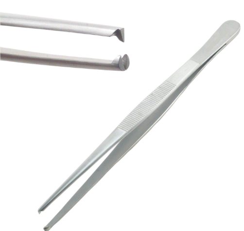
Appropriate use of several surgical instrument, and explain why other instruments may be inappropriate for the same use: rat-tooth forceps
Skin and muscle – to prevent excessive crushing of cells
Appropriate use of several surgical instrument, and explain why other instruments may be inappropriate for the same use: Scalpel
Skin and Muscle-as cut causes less trauma to cells
Appropriate use of several surgical instrument, and explain why other instruments may be inappropriate for the same use: Scissors
Skin and Muscle-crush cells/tissue and delays healing
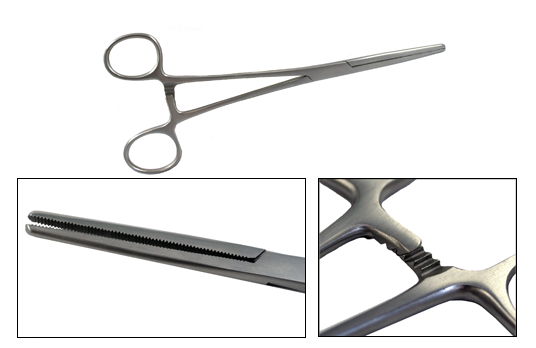
Appropriate use of several surgical instrument, and explain why other instruments may be inappropriate for the same use: Hemostats
Skin and Muscle-are used for blunt dissection of subcutaneous tissues, and for clamping off bleeding vessels
What are swedged needles, why is one better than the other?
have the suture contained within the end of the needle – does not stick out beyond cross-section of needle, less drag, les trauma to tissues
What are non-swedged needles, why is one better than the other?
needles have eyelet, and suture must be looped through/around it, and thus has a wider diameter than the needle itself, allowing dragging/friction/trauma to the tissues, delaying healing
Recognize when taper point should be used
are used for hollow organs, as cutting needles would allow suture to tear through the tissue
Recognize when cutting points should be used
are used for skin, to facilitate passage of the needle through the denser tissue
Why is “perfect” apposition of the wound margin appropriate?
allows faster healing with minimal scar tissue formation
what happens when eversion or inversion of the wound margin occurs?
results in scarring – scar tissue is not as strong as normal tissue
Recognize (in general) which tissues have greater or lesser tensile strength (e.g., fat, muscle, muscle fascia, skin)
Skin > muscle fascia > muscle > subcutaneous fat
proper techniques for suture removal to prevent contamination under the skin
Elevate suture
Elevate suture
cut portion that was underneath the skin, then remove – this prevents entry and tracking of bacteria/contaminants under the skin surface
Recognize the types of anesthetic methods used
Local; Regional; General
Simple Interrupted Suture Pattern (image)
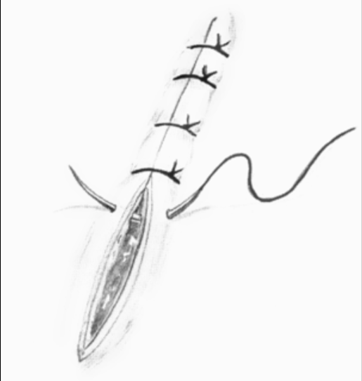
Simple Interrupted Suture Pattern
Advantages:
Good apposition of the margins
Loss of one suture has minimal effect
Disadvantage
Takes a little longer to do than continuous suture patterns
May tear through tissue when tension present
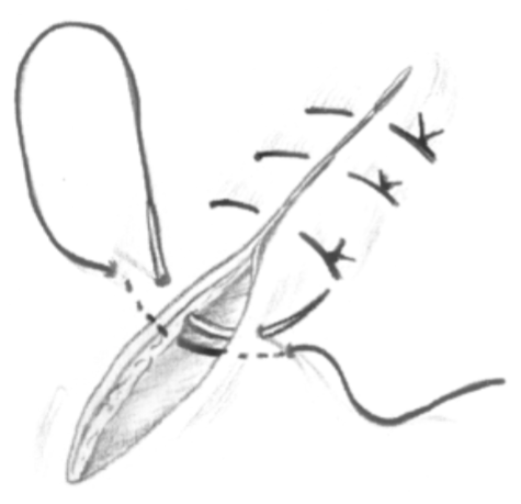
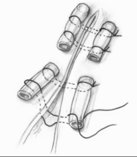
Horizontal Mattress Suture Pattern (image)
Horizontal Mattress Suture Pattern
Advantages:
Good for areas where tension tries to pull the margins apart
Loss of one suture has minimal effect
Disadvantages:
Takes a little longer to do than continuous suture patterns
Causes eversion of the margins
May "strangulate" blood flow and affect healing
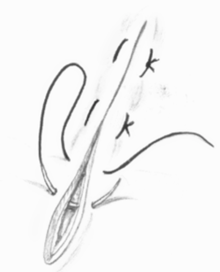
Vertical Mattress Suture Pattern (image)
Vertical Mattress Suture Pattern
Advantages:
Good areas where tension tries to pull the margins apart
Loss of one suture has minimal effect
Not as likely to restrict blood flow
Disadvantages:
Takes a little longer to do than continuous suture patterns
Causes eversion of the margins
Simple Continuous Suture Pattern (image)
Simple Continuous Suture Pattern
Advantages:
Very fast to complete
Fair apposition of the margins
Disadvantages:
May tear through tissue when tension present, or will break
Breaking a suture allows entire wound to gape open
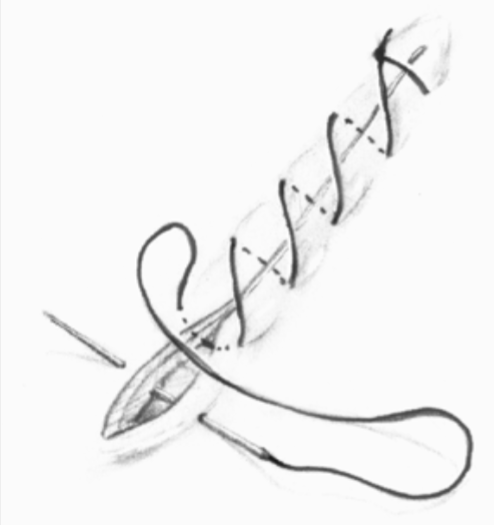
Continuous Interlocking Suture Pattern (image)
Continuous Interlocking Suture Pattern
Advantages:
Very fast to complete
Fair apposition of the margins
Good for tension relief
Disadvantages:
Breaking of suture allows entire wound to gape open
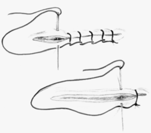
Use of Stents for Wound Closure (image)
Use of Stents for Wound Closure
necessary in areas where significant tension is present
provides a measure of tension relief to prevent sutures from tearing through the skin
rat toothed forceps (image)
Hemostats (image)
factors affecting anesthesia, identifying the different factors which affect metabolic rate
Size, age, gender, recent feeding, fear and activity, tranquilizers, concurrent disease
Differing Metabolic Rates (BMR): Age
neonates have low ___; old & young differ from "adults"
Differing Metabolic Rates (BMR): Sex Male
7% higher than
Differing Metabolic Rates (BMR): Sex female
increases with pregnancy
Differing Metabolic Rates (BMR): Recent feeding
increase, chylomicrons (fat globules in blood to which anesthetic can be bound rendering it ineffective)
Differing Metabolic Rates (BMR): Fear & activity
increase, so sedate initially
Differing Metabolic Rates (BMR): Tranquilizers
Lower
Differing Metabolic Rates (BMR): Fever
increases
Differing Metabolic Rates (BMR): toxemia
decreases
Differing Metabolic Rates (BMR):liver disease
decreases
Differing Metabolic Rates (BMR): shock
decreases
Differing Metabolic Rates (BMR): Vit. C deficiency in guinea pigs
decreases
Differing Metabolic Rates (BMR):hyperthyroidism
increases
Differing Metabolic Rates (BMR): thyroidectomized
decreases
List 4 methods of anesthetic delivery.
Dermal/topical; Injectable (IM, IV, IP, SC); Inhalant
Describe 2 body systems affected by "anesthetic emergencies"
Cardiac; Respiratory
List the 4 stages of anesthesia and the appropriate plane of anesthesia
Stage I – rising pain threshold
Stage II – excitement phase
Stage III – surgical anesthesia – Plane 3
Stage IV – respiratory paralysis
List methods for assessing adequate anesthesia/analgesia
Jaw tone;
swallowing reflex,
withdrawal reflex (“toe pinch”),
palpebral (eyelid) reflex;
corneal reflex
Signs Used to Assess Depth of Anesthesia: Jaw Tone
open the animal’s mouth; if jaw is relaxed and easy to open, then the animal is adequately anesthetized. If there is resistance, then the animal is too light
Signs Used to Assess Depth of Anesthesia: Swallowing Reflex
gently pull on the tongue; if chewing and swallowing can be seen, the animal is too light. When the tongue is not retracted, the animal is adequately anesthetized
Signs Used to Assess Depth of Anesthesia: Withdrawal Reflex (“toe pinch”)
gently extend one leg and pinch the web of skin between the toes or the footpad with your fingernail; a positive reflex is indicated by the flexion (pulling back) of the leg.
Note: this reflex may be preserved in some anesthetized animals
Signs Used to Assess Depth of Anesthesia: Palpebral Reflex
gently touch the medial canthus of the eye (the corner of the eye nearest the nose); the animal will blink its eyelids if not adequately anesthetized
Signs Used to Assess Depth of Anesthesia: Corneal Reflex
gently touch the cornea of the eye with a moistened gauze pad or Q-tip swab; the animal will blink its eyelids if not adequately anesthetized