Biopsychology
0.0(0)
Card Sorting
1/119
Earn XP
Description and Tags
Study Analytics
Name | Mastery | Learn | Test | Matching | Spaced |
|---|
No study sessions yet.
120 Terms
1
New cards
Key features of the nervous system
The nervous system is a specialised network of cells and our primary communication system. It is based on electrical (and chemical) signals whereas the endocrine system is based on the hormones.
The nervous system has two main functions:
1. To collect, process and respond to information in the environment
2. To co-ordinate the working of different organs and cells in the body
The nervous system has two main functions:
1. To collect, process and respond to information in the environment
2. To co-ordinate the working of different organs and cells in the body
2
New cards
The structure of the central nervous system (CNS)
The CNS is made up of the brain and the spinal cord.
The brain is the centre of conscious awareness.
The outer layer of the brain, the cerebral cortex (3mm thick), is highly developed in humans and is what distinguishes our higher mental functions from those of animals.
The brain is divided into two hemispheres.
The brain is the centre of conscious awareness.
The outer layer of the brain, the cerebral cortex (3mm thick), is highly developed in humans and is what distinguishes our higher mental functions from those of animals.
The brain is divided into two hemispheres.
3
New cards
The function of the central nervous system (CNS)
The spinal cord is an extension of the brain and is responsible for reflex actions.
It passes messages to and from the brain and connects nerves to the PNS.
It passes messages to and from the brain and connects nerves to the PNS.
4
New cards
The peripheral nervous system (PNS)
The PNS transmits messages, via millions of neurons, to and from the nervous system.
It’s subdivided into:
* Autonomic nervous system (ANS)
* Somatic nervous system (SNS)
It’s subdivided into:
* Autonomic nervous system (ANS)
* Somatic nervous system (SNS)
5
New cards
\
Autonomic nervous system (ANS)
Autonomic nervous system (ANS)
\
Governs vital functions in the body such as breathing, heart rate, digestion, sexual arousal and stress responses.
Governs vital functions in the body such as breathing, heart rate, digestion, sexual arousal and stress responses.
6
New cards
Somatic nervous system (SNS)
Governs muscle movement and receives information from sensory receptors.
7
New cards
Major subdivisions of the human nervous system
\
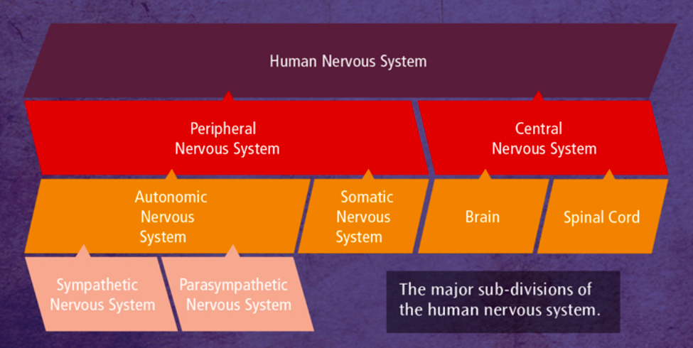
8
New cards
The endocrine system
Works alongside the nervous system to control vital functions in the body through the action of hormones.
It works more slowly than the nervous system but has widespread and powerful effects
It works more slowly than the nervous system but has widespread and powerful effects
9
New cards
Glands
Organs in the body that produce hormones.
The key endocrine gland is the pituitary gland, located in the brain. It is called the ‘master gland’ because it controls the release of hormones from all the other endocrine glands in the body.
The key endocrine gland is the pituitary gland, located in the brain. It is called the ‘master gland’ because it controls the release of hormones from all the other endocrine glands in the body.
10
New cards
Hormones
They are secreted in the bloodstream and affect any cell in the body that has a receptor for that particular hormone.
For example, thyroxine produced by the thyroid gland affects cells in the heart and also cells throughout the body which increase metabolic rates. This in turn affects growth rates.
For example, thyroxine produced by the thyroid gland affects cells in the heart and also cells throughout the body which increase metabolic rates. This in turn affects growth rates.
11
New cards
Fight or flight response
The endocrine system and the ANS work together, for instance during a stressful event
1. Stressor perceived hypothalamus which activates the pituitary.
2. The sympathetic nervous system is now aroused.
3. Adrenaline (the stress hormone) is released from the adrenal medulla into the bloodstream. This delivers the aroused state causing changes in target organs in the body e.g. increased heart rate, dilation of pupils, decreased population of saliva. This is called the fight or flight response.
4. Immediate and automatic - this response happens the instant a threat is perceived.
5. Parasympathetic nervous system (rest and digest) takes over once the threat has passed. This returns the body to its resting state. This acts as a ‘brake’ and reduces the activities of the body that were increased by the actions of the sympathetic branch (rest and digest).
12
New cards
Types of neurons
There are 100 billion nerve cells (neurons) in the human nervous system, 80% of which are located in the brain.
By transmitting signals electrically and chemically, these provide the nervous system with its primary means of communication.
There are three types of neuron:
1. Sensory neurons
2. Relay neurons
3. Motor neurons
By transmitting signals electrically and chemically, these provide the nervous system with its primary means of communication.
There are three types of neuron:
1. Sensory neurons
2. Relay neurons
3. Motor neurons
13
New cards
Sensory neurons
Carry messages from the PNS to the CNS. They have long dendrites and short axons. Located in the PNS in clusters called ganglias.
14
New cards
Relay neuron
Connect sensory neurons to motor or relay neurons. They have short dendrites and short axons. Of all neurons, 97% are relay neurons and most are in the brain and visual system.
15
New cards
Motor neuron
Connect the CNS to effectors such as muscles and glands. They have short dendrites and long axons. Cell bodies may be in the CNS but long axons form part of the PNS.
16
New cards
Structure of cell body
Includes a nucleus which contains the genetic material of the cell
17
New cards
Structure of dendrites
Branchlike structures that protrude from the cell body. These carry nerve impulses from neighbouring neurons towards the cell body.
18
New cards
Structure of axon
Carries the electrical impulse away from the cell body down the length of the neuron.
* It is covered in a fatty layer of myelin sheath that protects the axon
* Gaps in the axon called nodes of Ranvier speed up the transmission of the impulse
* It is covered in a fatty layer of myelin sheath that protects the axon
* Gaps in the axon called nodes of Ranvier speed up the transmission of the impulse
19
New cards
Structure of terminal buttons
At the end of the axon communicate with the next neuron in the chain across a gap called the synapse.
20
New cards
Electrical transmission
When a neuron is in a resting state the inside of the cell is negatively charged compared to the outside.
When a neuron is activated, the inside of the cell becomes positively charged for a split second causing an action potential to occur.
This creates an electrical impulse that travels down the axon towards the end of the neuron.
When a neuron is activated, the inside of the cell becomes positively charged for a split second causing an action potential to occur.
This creates an electrical impulse that travels down the axon towards the end of the neuron.
21
New cards
A synapse
Each neuron is separated from the next by an extremely tiny gap called the synapse.
22
New cards
Chemical transmission
The event that occurs at the synapse - Signals within neurons are transmitted electrically, but signals between neurons are transmitted chemically across the synapse.
When the electrical impulse reaches the end of the neuron (the presynaptic terminal) it triggers the release of neurotransmitter from tiny sacs called synaptic vesicles.
Once a neurotransmitter crosses the gap, it is taken up by a postsynaptic receptor site on the next neuron, so the impulse only ever travels in one direction.
The chemical message is converted back into an electrical impulse and the process of electrical transmission begins.
23
New cards
Neurotransmitters
Chemicals that diffuse across the synapse to the next neuron in the chain.
Many neurotransmitters have been identified. Each has its own specific molecular structure that fits perfectly into a postsynaptic receptor site, like a lock and key.
Each has specific functions
Many neurotransmitters have been identified. Each has its own specific molecular structure that fits perfectly into a postsynaptic receptor site, like a lock and key.
Each has specific functions
24
New cards
Function of acetylcholine
Found where a motor neuron meets a muscle, causing muscles to contract.
25
New cards
Function of serotonin
Affects mood and social behaviour (among other things) which is why it has been implicated as a cause of depression.
26
New cards
Adrenaline effect on neighbouring neuron
Generally excitatory, increasing the positive charge of the postsynaptic neuron, making it more likely the postsynaptic neuron will fire.
27
New cards
Serotonin effect on neighbouring neuron
Generally inhibitory, increasing the negative charge of the postsynaptic neuron, making it less likely the postsynaptic neuron will fire.
28
New cards
Dopamine effect on neighbouring neuron
It’s an unusual neurotransmitter as it is equally likely to have excitatory or inhibitory effects on the postsynaptic neuron.
29
New cards
Summation
Excitatory and inhibitory influences are summed and must reach a certain threshold in order for the action potential of the postsynaptic neuron to be triggered.
If the net effect of the neurotransmitters in inhibitory then the postsynaptic neuron is less likely to fire (i.e. no electrical signal is transmitted). It is more likely to fire id the net effect is excitatory.
If the net effect of the neurotransmitters in inhibitory then the postsynaptic neuron is less likely to fire (i.e. no electrical signal is transmitted). It is more likely to fire id the net effect is excitatory.
30
New cards
Holistic theory
In the early 19th century holistic theory suggested tha all parts of the brain were involved in processing thought and action.
31
New cards
Localisation theory
Later, after the holistic theory specific areas of the brain were linked with specific physical and psychological functions which is the localisation theory.
If an area of the brain is damaged through illness or injury, the function associated with that area is also affected.
If an area of the brain is damaged through illness or injury, the function associated with that area is also affected.
32
New cards
Lateralisation
Some physical and psychological functions are controlled by a particular hemisphere.
Generally, the left side of the body is controlled by the right hemisphere, the right side of the body by the left hemisphere.
Generally, the left side of the body is controlled by the right hemisphere, the right side of the body by the left hemisphere.
33
New cards
Cerebral cortex
The outer layer of the brain.
It’s like a ‘tea cosy’ that covers the inner part of the brain. It’s about 3mm thick and is what separates us from lower animals as it is highly developed
The cortex appears grey due to the location of cell bodies - hence the phrase ‘grey matter’
It’s like a ‘tea cosy’ that covers the inner part of the brain. It’s about 3mm thick and is what separates us from lower animals as it is highly developed
The cortex appears grey due to the location of cell bodies - hence the phrase ‘grey matter’
34
New cards
Four lobes of the cerebral cortex
frontal, parietal, occipital and temporal
35
New cards
Motor area
At the back of the frontal lobe (both hemispheres).
It controls voluntary movement.
Damage may result in loss of control over fine movements.
It controls voluntary movement.
Damage may result in loss of control over fine movements.
36
New cards
Somatosensory area
At the front of the parietal lobes.
Processes sensory information from the skin (touch, heat, pressure, etc).
The amount of somatosensory area devoted to a particular body part denotes its sensitivity.
Processes sensory information from the skin (touch, heat, pressure, etc).
The amount of somatosensory area devoted to a particular body part denotes its sensitivity.
37
New cards
Visual area
In the occipital lobe at the back of the brain.
Each eye sends information from the right visual field to the left visual cortex, and from the left visual field to the right visual cortex.
Each eye sends information from the right visual field to the left visual cortex, and from the left visual field to the right visual cortex.
38
New cards
Auditory area
In the temporal lobe.
Analyses speech-based information.
Damage may produce partial hearing loss - the more extensive the damage, the more serious the loss.
Analyses speech-based information.
Damage may produce partial hearing loss - the more extensive the damage, the more serious the loss.
39
New cards
Broca’s area
Speech production
Identified by Broca in the 1880s, in the left frontal lobe.
Damage to this area causes Broca’s aphasia which is characterised by speech that is slow, laborious and lacking in fluency. Broca’s patients may have difficulty finding words and naming certain objects.
People with Broca’s aphasia have difficulty with prepositions and conjunctions (e.g. ‘a’, ‘the’, ‘and’)
40
New cards
Wernicke’s area
Language understanding
Identified by Wernicke in the 1880s, in the left temporal lobe.
People with Wernicke’s aphasia produce language but have problems understanding it, so they produce fluent but meaningless speech.
They will often produce nonsense words (neologisms as part of the content of their speech).
Identified by Wernicke in the 1880s, in the left temporal lobe.
People with Wernicke’s aphasia produce language but have problems understanding it, so they produce fluent but meaningless speech.
They will often produce nonsense words (neologisms as part of the content of their speech).
41
New cards
AO3 of localisation theory (neurosurgery)
Strength of localisation theory - Neurosurgery is used to treat mental disorders e.g. cingulotomy involves isolating the cingulate gyrus - dysfunction of this area may be a cause of OCD.
Dougherty et al. (2002) studied 44 people with OCD who has a cingulotomy. At follow up, 30% met the criteria for successful response and 14% for partial response.
The success of such procedures strongly suggests that behaviours associated with serious mental disorders may be localised.
Dougherty et al. (2002) studied 44 people with OCD who has a cingulotomy. At follow up, 30% met the criteria for successful response and 14% for partial response.
The success of such procedures strongly suggests that behaviours associated with serious mental disorders may be localised.
42
New cards
AO3 of the localisation theory (brain scan)
Strength of localisation theory - Petersen et al. (1988) used brain scans to show activity in Wernicke’s area during a listening task and in Broca’s area during a reading task.
Also, a study of long-term memory by Tulving et al. (1994) revealed semantic and episodic memories are located in different parts of the prefrontal cortex.
There now exists a number of sophisticated and objective methods for measuring activity in the brain, providing sound scientific evidence of localisation of function.
43
New cards
Counterpoint to AO3 of localisation theory (brain scan)
Lashley removed areas of the cortex (up to 50%) in rats learning the route through a maze. Learning required all of the cortex rather than being confined to a particular area.
This suggests that higher cognitive processes (e.g. learning) are not localised but distributed in a more holistic way in the brain.
This suggests that higher cognitive processes (e.g. learning) are not localised but distributed in a more holistic way in the brain.
44
New cards
AO3 of localisation theory (language model)
Dick and Tremblay (2016) found that very few researchers still believed language is only in Broca’s and Wernicke’s area.
Advanced techniques (e.g. fMRI) have identified regions in the right hemisphere and the thalamus.
This suggests that, rather than being confined to a couple of key areas, language may be organised more holistically in the brain, which contradicts localisation theory.
Advanced techniques (e.g. fMRI) have identified regions in the right hemisphere and the thalamus.
This suggests that, rather than being confined to a couple of key areas, language may be organised more holistically in the brain, which contradicts localisation theory.
45
New cards
AO3 of localisation theory (case study)
Unique cases of neurological damage suppport localisation theory, e.g. Phineas Gage who lost some of his brain in an explosion and his personality changed.
However, it is difficult to make meaningful generalisations based on a single individual and conclusions may depend on the subjective interpretation of the researcher.
This suggests that some evidence supporting localisation may lack validity, oversimplifying brain processes and undermining the theory.
46
New cards
Plasticity
This describes the brain’s tendency to change and adapt (functionally and physically) as a result of experiences and new learning.
During infancy, the brain experiences a rapid growth in synaptic connections, peaking at about 15,000 at age 2-3 years. (Gopnik et al 1999)
During infancy, the brain experiences a rapid growth in synaptic connections, peaking at about 15,000 at age 2-3 years. (Gopnik et al 1999)
47
New cards
Synaptic pruning
As we age, rarely-used connections are deleted and frequently-used connections are strengthened.
It was once thought these changes were limited to childhood. But recent research suggests neural connections can change or be formed at any time, due to learning and experience.
It was once thought these changes were limited to childhood. But recent research suggests neural connections can change or be formed at any time, due to learning and experience.
48
New cards
Maguire et al (2000)
Found significantly more volume of grey matter in the posterior hippocampus in London taxi drivers than in a matched control group. This part of the brain is linked with the development of spatial and navigational skills.
As part of their training, London cabbies take a complex test called ‘The Knowledge’ to assess their recall of city streets and possible routes. This learning experience appears to alter the structure of the taxi drivers’ brains. The longer they had been in the job, the more pronounced was the structural difference.
As part of their training, London cabbies take a complex test called ‘The Knowledge’ to assess their recall of city streets and possible routes. This learning experience appears to alter the structure of the taxi drivers’ brains. The longer they had been in the job, the more pronounced was the structural difference.
49
New cards
Draganski et al. (2006)
Imagined the brains of medical students three months before and after final exams. Learning-induced chnages were seen in the posterior hippocampus and the parietal cortex, presumably as a result of learning for the exam.
50
New cards
Functional recovery
A form of plasticity. Following damage through trauma, the brain’s ability to redistribute or transfer functions usually performed by a damaged area(s) to other, undamaged area(s).
Neuroscientists suggest this process occurs quickly after trauma (spontaneous recovery) and then slows down - at which point the person may require rehabilitative therapy.
Neuroscientists suggest this process occurs quickly after trauma (spontaneous recovery) and then slows down - at which point the person may require rehabilitative therapy.
51
New cards
Synaptic connections
The brain is able to rewire and reorganise itself by forming new synaptic connections close to the area of damage.
52
New cards
Secondary neural pathways
Ones that would not typically be used to carry out certain functions are activated or ‘unmasked’ to enable functioning to continue.
53
New cards
Axonal sprouting
A structural change in the brain - Growth of new nerve endings which connect with other undamaged cells to form new neuronal pathways.
54
New cards
Denervation supersensitivity
A structural change in the brain - Axons that do a similar job became aroused to a higher level to compensate for the ones that are lost.
55
New cards
Recruitment of homologous (similar) areas
The opposite side of the brain takes over specific tasks e.g. language production.
56
New cards
AO3 of plasticity (consequences)
Limitation of plasticity - The brain’s adaptation to prolonged drug use leads to poorer cognitive functioning in later life, as well as an increased risk of dementia (Medina et al. 2007).
60-80% of amputees have phantom limb syndrome (experience sensations in missing limb due to changes in somatosensory cortex).
This suggests that the brain’s ability to adapt to damage is not always beneficial and may lead to physical and psychological problems.
60-80% of amputees have phantom limb syndrome (experience sensations in missing limb due to changes in somatosensory cortex).
This suggests that the brain’s ability to adapt to damage is not always beneficial and may lead to physical and psychological problems.
57
New cards
AO3 of plasticity (lifespan)
Strength of plasticity - Ladina Bezzola et al. (2012) demonstrated how 40 hours of golf training produced changes in the neural representation in Ps aged 40-60.
Using fMRI, motor cortex activity in the novice golfers reduced compared to a control group, suggesting greater efficiency after training.
This shows that neural plasticity can continue throughout the lifespan.
Using fMRI, motor cortex activity in the novice golfers reduced compared to a control group, suggesting greater efficiency after training.
This shows that neural plasticity can continue throughout the lifespan.
58
New cards
AO3 of functional recovery
Strength of functional recovery - Understanding plasticity had led to neurorehabilitation. Understanding axonal growth encourages new therapies.
For example, constraint-induced movement therapy involves massed practice with an affected arm while unaffected arm is restrained.
This shows that research into functional recoverey helps medical professionals known when interventions can be made.
For example, constraint-induced movement therapy involves massed practice with an affected arm while unaffected arm is restrained.
This shows that research into functional recoverey helps medical professionals known when interventions can be made.
59
New cards
AO3 of neural plasticity
Limitation of neural plasticity - Schneider et al. (2014) looked at the time brain injury patients had spent in education (indicated their cognitive reserve) and their chances of a disability-free recovery (DFR).
40% of patients who achieved DFR gad more than 16 years’ education compared to about 10% of patients who had less than 12 years’ education.
This suggests that cognitive reserve is a crucial factor in determining how well the brain adapts after trauma.
40% of patients who achieved DFR gad more than 16 years’ education compared to about 10% of patients who had less than 12 years’ education.
This suggests that cognitive reserve is a crucial factor in determining how well the brain adapts after trauma.
60
New cards
Hemispheric laterlisation
The idea that the two halves (hemispheres) of the brain are functionally different and that certain mental processes and behaviours are mainly controlled by one hemisphere rather than the other, as in the example fo language (which localised as well as lateralised).
61
New cards
Localised
Some functions are localised and appear in both left and right hemispheres (LH and RH).
e.g auditory, visual, motor, somatosensory areas
e.g auditory, visual, motor, somatosensory areas
62
New cards
Localised and lateralised
Two main language centres are in the LH (for most people) - Broca’s area (left frontal lobe), Wernicke’s area (left temporal lobe).
RH produces rudimentary words but provide emotional context. LH may be the analyser, RH the synthesiser.
RH produces rudimentary words but provide emotional context. LH may be the analyser, RH the synthesiser.
63
New cards
Contralateral
In the motor area, the right hemisphere controls the left side of the body and vice versa (cross-wired).
64
New cards
Contralateral and ipsilateral
Left visual field (LVF) of both eyes is connected to the RH and right visual field (RVF) of both eyes is connected to the LH.
Enables the visual ares to compare slighty different perspective from each eye and aids depth perception.
Same arrangement for auditory areas.
Enables the visual ares to compare slighty different perspective from each eye and aids depth perception.
Same arrangement for auditory areas.
65
New cards
Sperry (1968) Procedure
Split-brain research
‘Split brain’ = two hemispheres surgically separated by cutting the connection e.g corpus callosum.
Used to treat severe epilepsy to reduce the ‘electrical storm’ across hemispheres.
Eleven split-brian Ps were studied using the set-up shown on the left. Image or word projected to RVF (processed by LH), and same, or different, image could be projected to the LVF (processed by RH).
Presenting the image to one hemishere meant that the information could not be conveyed from that hemisphere to the other.
66
New cards
Sperry (1968) Findings and Conclusions
Object shown to RVF:
* Participant can describe what is seen (language centres in LH)
Object shown to LVF:
* Cannot name object (no language centres in RH)
* Can select matching object behind screen using left hand.
* Can select object closely associated with picture (e.g ashtray if picture of cigarette).
* Pinup picture shown to LVF, participant giggled but reported seeing nothing.
Demonstrates how certain functions are lateralised in the brain, shows that LH is verbal and the RH is ‘silent’ but emotional.
67
New cards
AO3 of lateralised brain functions (evidence)
Strength of lateralised brain functions - PET scans show when ‘normal’ Ps attend to global elements of an image, the RH is more active.
When required to focus on finer detail the specific areas of the LH tend to dominate (Fink et al. 1996).
This suggets that hemispheric lateralisation is a feature of the normal brain as well as the split-brain.
When required to focus on finer detail the specific areas of the LH tend to dominate (Fink et al. 1996).
This suggets that hemispheric lateralisation is a feature of the normal brain as well as the split-brain.
68
New cards
AO3 of lateralised brain functions (differences between hemispheres)
Limitation of lateralised brain functions - There may be different functions in the RH and LH but research suggests people do not have a dominant side, creating a different personality.
Nielson et al. (2013) analysed 1000 brain scans, finding people did use certain hemispheres for certain tasks but no dominance.
This suggests that the notion of right - or left - brained people in wrong (e.g. ‘artist’ brain).
Nielson et al. (2013) analysed 1000 brain scans, finding people did use certain hemispheres for certain tasks but no dominance.
This suggests that the notion of right - or left - brained people in wrong (e.g. ‘artist’ brain).
69
New cards
AO3 on lateralisation versus plasticity
Lateralisation is adaptive, enabling two simultaneous tasks with greater efficiency, e.g. only lateralised chickens better at finding food while watching for predators (Rogers et al. 2004).
On the other hand, neural plasticity is also adaptive. After damage to brain, language function can ‘switch sides; (Holland et al. 1996).
This seems to suggest that lateralisation is first preference but ultimately plasticity is more important because it deals with loss of lateralisation.
On the other hand, neural plasticity is also adaptive. After damage to brain, language function can ‘switch sides; (Holland et al. 1996).
This seems to suggest that lateralisation is first preference but ultimately plasticity is more important because it deals with loss of lateralisation.
70
New cards
AO3 of split-brain research (evidence)
Strength of split-brain research - Luck et al. (1989) showed that split-brain Ps are better than normal controls e.f. twice as fast at identifying the odd one out in an array of similar objects.
In the normal brain, the LH’s superior processing abilities are ‘watered down’ by the inferior right hemisphere (Kingstone et al. 1995).
This supports Sperry’s earlier findings that the ‘left brain’ and ‘right brain’ are distinct in terms of functions and abilities.
In the normal brain, the LH’s superior processing abilities are ‘watered down’ by the inferior right hemisphere (Kingstone et al. 1995).
This supports Sperry’s earlier findings that the ‘left brain’ and ‘right brain’ are distinct in terms of functions and abilities.
71
New cards
AO3 of split-brain research (causal relationships)
Limitation of split-brain research - In Sperry’s research the behaviour of the split-brain Ps was compared to a neurotypical control group.
However, none of the control group has epilepsy. Any differences between the groups may be due to epilepsy not the split-brain (a confounding variable).
This means that some of the unique features of the split-brain Ps’ cognitive abilities might have been due to their epilepsy.
However, none of the control group has epilepsy. Any differences between the groups may be due to epilepsy not the split-brain (a confounding variable).
This means that some of the unique features of the split-brain Ps’ cognitive abilities might have been due to their epilepsy.
72
New cards
AO3 of split-brain research (ethics)
Limitation of split-brain research - Sperry’s Ps were not deliberately harmed and procedures were explained in advance to gain informed consent.
However, Ps may not have understood they would be tested for many years, and participation was stressful.
This suggests that there was no deliberate harm but the negative consequences make the study unethical.
However, Ps may not have understood they would be tested for many years, and participation was stressful.
This suggests that there was no deliberate harm but the negative consequences make the study unethical.
73
New cards
Investigating brain localisation
Techniques for investigating the brain are often used for medical purposes in the diagnosis of illness.
The purpose of scanning in psychological research is often to investigate localisation - to determine which parts of the brain do what.
The purpose of scanning in psychological research is often to investigate localisation - to determine which parts of the brain do what.
74
New cards
fMRI
Highlights active areas of the brain - Functional magnetic resonance imaging (fMRI) detects changes in both blood oxygenation and flow that occur due to neural activity in specific brain areas.
When a brain area is more active it consumes more oxygen and blood flow is directed to the active area (haemodynamic response).
fMRI produces a 3D image showing which parts of the brain are active and therefore must be involved in particular mental processes.
When a brain area is more active it consumes more oxygen and blood flow is directed to the active area (haemodynamic response).
fMRI produces a 3D image showing which parts of the brain are active and therefore must be involved in particular mental processes.
75
New cards
EEG
Shows overall electrical activity - Electroencephalogram (EEG) measures electrical activity within the brain via electrodes using a skull cap (looks a bit like a swimming cap with the electrodes attached).
The scan recording represents the brainwave patterns generated from thousands of neurons. This shows overall brain activity.
EEG is often used as a diagnostic tool. For example unusual arrhythmic patterns of brain activity may indicate abnormalities such as epilepsy, tumours or sleep disorders.
The scan recording represents the brainwave patterns generated from thousands of neurons. This shows overall brain activity.
EEG is often used as a diagnostic tool. For example unusual arrhythmic patterns of brain activity may indicate abnormalities such as epilepsy, tumours or sleep disorders.
76
New cards
ERPs
Brainwaves related to particular events
Event-related potentials (ERPs) are what is left when all extraneous brain activity from an EEG recording is filtered out.
This is done by using a statistical technique, leaving only those response that relate to the presentation of a specific stimulus (for example).
ERPs are types of brainwave that are triggered by particular events.
Research has revealed many different forms of ERP and how these are linked to cognitive processes (e.g. perception and attention).
77
New cards
Post-mortem examinations
A technique involving the analysis of a person’s brain following their death.
Areas of the brain are examined to establish the likely cause of a deficit or disorder that the person experienced in life.
This may also involve comparison with a neurotypical brain in order to assess the extent of the difference.
Areas of the brain are examined to establish the likely cause of a deficit or disorder that the person experienced in life.
This may also involve comparison with a neurotypical brain in order to assess the extent of the difference.
78
New cards
Strengths of fMRI
Unlike other scanning techniques (e.g PET), fMRI does not rely on the use of radiation and is safe.
It also produces images with high spatial resolution, showing detail by the millimetre.
This means fMRI can safely provide a clear picture of how brain activity is localised.
It also produces images with high spatial resolution, showing detail by the millimetre.
This means fMRI can safely provide a clear picture of how brain activity is localised.
79
New cards
Limitations of fMRI
fMRI is expensive compared to other techniques.
It has poor temporal resolution because of 5-second lag between initial neural activity and image.
This means fMRI may not truly represent moment-to-moment brain activity.
It has poor temporal resolution because of 5-second lag between initial neural activity and image.
This means fMRI may not truly represent moment-to-moment brain activity.
80
New cards
Strengths of EEG
EEG has contributed to our understanding of the stages of sleep.
It has high temporal resolution - brain activity in one millisecond.
This shows the real-world usefulness of the technique.
It has high temporal resolution - brain activity in one millisecond.
This shows the real-world usefulness of the technique.
81
New cards
Limitations of EEG
The EEG produces a generalised signal from thousands of neurons
It is difficult to know the exact source of neural activity.
Therefore EEG can’t distinguish the activity of different but adjacent neurons.
It is difficult to know the exact source of neural activity.
Therefore EEG can’t distinguish the activity of different but adjacent neurons.
82
New cards
Strengths of ERP
Measures of neural processes more specific with ERPs than EEGs.
ERPs have excelled temporal resolution, better than fMRI.
This means that ERPs are frequently used in cognitive research.
ERPs have excelled temporal resolution, better than fMRI.
This means that ERPs are frequently used in cognitive research.
83
New cards
Limitations of ERP
Lack of standardisation makes it difficult to confirm findings in studies involving ERPs.
Background ‘noise’ and extraneous material must be completely eliminated.
These issues are a problem because they may not always be easy to achieve.
Background ‘noise’ and extraneous material must be completely eliminated.
These issues are a problem because they may not always be easy to achieve.
84
New cards
Strengths of post-mortems
Broca and Wernicke both relied on post-mortem studies.
Used to link HM’s memory deficits to damage in his brain.
This means they continue to provide useful information.
Used to link HM’s memory deficits to damage in his brain.
This means they continue to provide useful information.
85
New cards
Limitations of post-mortems
Observed damage in the brain may not be linked to the deficits under review.
Post-mortem studies raise ethical issues of consent after death (e.g. HM).
This challenges their usefulness in psychological research.
Post-mortem studies raise ethical issues of consent after death (e.g. HM).
This challenges their usefulness in psychological research.
86
New cards
Biological rhythms
They are periodic activity, governed by:
1. Internal biological ‘clocks’ (endogenous pacemakers
2. External changes in the environment (exogenous zeitgebers)
Some of these rhythms occur many times a day (ultradian rhythms). Others take more than a day to complete (infradian rhythms).
1. Internal biological ‘clocks’ (endogenous pacemakers
2. External changes in the environment (exogenous zeitgebers)
Some of these rhythms occur many times a day (ultradian rhythms). Others take more than a day to complete (infradian rhythms).
87
New cards
Circadian rhythm
Circa meaning ‘about’ and diem meaning ‘day’.
There are several important types of circadian rhythms such as the sleep/wake cycle.
There are several important types of circadian rhythms such as the sleep/wake cycle.
88
New cards
Sleep/wake cycle
Exogenous zeitgebers - the fact we feel drowsy when it’s night-time and alert during the day shows the effect of daylight.
Endogenous pacemakers - a biological clock ‘left to its own devices’ without the influence of external stimuli (e.g light) is called ‘free-running’.
The basic rhythm is governed by the suprachiasmatic nucleus (SCN). The SCN lies just above the optic chiasm which provides information from the eyes about light.
Exogenous zeitgebers (light) can reset the SCN.
89
New cards
Siffre (1962 and 1970s)
French caver Siffre spent long periods in dark caves to examine the effects of free-running biological rhythms - two months (in 1962) and six months (in the 1970s).
In each case study, Siffre’s free-running circadian rhythm settles down to about 25 hours.
Importantly, he did have a regular sleep/wake cycle.
In each case study, Siffre’s free-running circadian rhythm settles down to about 25 hours.
Importantly, he did have a regular sleep/wake cycle.
90
New cards
Aschoff and Wever (1976)
A group of Ps spent four weeks in a World War 2 bunker deprived of natural light.
All but one (whose sleep/wake cycle extended to 29 hours) displayed a circdian rhythm between 24 and 25 hours.
Siffre’s experience and the bunker study suggest that the ‘natural’ sleep/wake cycle may be slightly longer than 24 hours but is entrained by exogenous zeitgebers associated with our 24-hour day (e.g. daylight hours, mealtimes, etc).
91
New cards
Folkard et al. (1985)
They studied a group of 12 people who lived in a dark cave for three week, going to bed when the clock said 11.45 pm and waking when it said 7.45 am.
The researchers gradually speeded up the clock (unbekown to the participants) to a 22-hour day.
Only one participant comfortably adjusted to the new regime. This suggests the existence of a strong free-running circadian rhythm not controlled by exogenous zeitgebers.
The researchers gradually speeded up the clock (unbekown to the participants) to a 22-hour day.
Only one participant comfortably adjusted to the new regime. This suggests the existence of a strong free-running circadian rhythm not controlled by exogenous zeitgebers.
92
New cards
AO3 of circadian rhythm research
Strength of circadian rhythm research - Shift work creates desynchronisation of biological rhythms. Boivin et al. (1996) found shift workers experience a lapse of concentration around 6am (a circadian trough) so accidents are more likely.
Research also suggests a link between shift work and poor health, with shift workers three times more likely to develop heart disease (Knutsson 2003).
Thus, research into the sleep/wake cycle may have economic implications in terms of how best to manage shift work.
Research also suggests a link between shift work and poor health, with shift workers three times more likely to develop heart disease (Knutsson 2003).
Thus, research into the sleep/wake cycle may have economic implications in terms of how best to manage shift work.
93
New cards
Counterpoint to circadian rhythm research having application
The research is correlational, therefore desynchronisation may not be the cause of observed difficulties. For example, Solomon (1993) concluded that high divorce rates in shift workers might be due to missing out on important family events.
This suggests that it may not be biological factors that create the adverse consequences associated with shiftwork.
This suggests that it may not be biological factors that create the adverse consequences associated with shiftwork.
94
New cards
AO3 of circdian rhythms (medical treatment)
Strength of circadian rhythms - Circadian rhythms co-ordinate the body’s basic processes (e.g. heart rate, hormone levels) with implications for chronotherapeutics (timing medication to maximise effects on the body).
Aspirin reduces heart attacks, which are most likely int he morning. Bonten et al. (2015) found taking aspirin is most effective last thing at night.
This shows that circadian rhythm research can help increase the effectiveness of drug treatments.
Aspirin reduces heart attacks, which are most likely int he morning. Bonten et al. (2015) found taking aspirin is most effective last thing at night.
This shows that circadian rhythm research can help increase the effectiveness of drug treatments.
95
New cards
AO3 of circadian rhythms (generalisations)
Studies of the sleep.wake cycle often use small groups of Ps (e.g Aschoff and Wever), or even single individuals (Siffre).
Ps may not be representative of the wider population and this limits making meaningful generalisations. Siffre observed that his internal clock ticked much more slowly at 60 than when he was younger.
This suggest that, even when the same person is involved, there are factors that may prevent general conclusions being drawn.
96
New cards
AO3 of circadian rhythms (school)
Research shows benefits fro teenagers academic and behavioural performance when lessons start later in the day (Adolescent Sleep Working Group 2014).
However, a later start is disruptive for parents and teachers, and teenagers may simply stay up later and still be exhausted.
This suggests changing the school day may not be practical even though it may be desirable.
However, a later start is disruptive for parents and teachers, and teenagers may simply stay up later and still be exhausted.
This suggests changing the school day may not be practical even though it may be desirable.
97
New cards
The female menstrual cycle
The human female menstrual cycle is about 28 days (i.e. less than one cycle in 24 hours - infra diem meaning ‘below’ a day).
Rising levels of oestrogen casue the ovary to release an egg (ovulation). Then progesterone helps the womb lining to thicken, readying the womb for pregnancy. If pregnancy does not occur, the egg is absorbed and the womb lining comes away (menstrual flow).
Rising levels of oestrogen casue the ovary to release an egg (ovulation). Then progesterone helps the womb lining to thicken, readying the womb for pregnancy. If pregnancy does not occur, the egg is absorbed and the womb lining comes away (menstrual flow).
98
New cards
Stern and McClintock (1998)
They studied 29 women with irregular periods. Pheromones were taken from some at different stages of their cycles, via a cotton pad under the armpits. These pads were cleaned with alcohol and later rubbed on the upper lips of the other participants.
68% of women experienced changes to their cycle which brought them closer to the cycle of their ‘odour donor’.
68% of women experienced changes to their cycle which brought them closer to the cycle of their ‘odour donor’.
99
New cards
SAD (Seasonal Affective Disorder)
A depressive disorder (low mood, lack of activity) with a seasonal pattern.
Often called the ‘winter blues’ because the symptoms are triggered during the winter months when the number of daylight hours becomes shorter.
Often called the ‘winter blues’ because the symptoms are triggered during the winter months when the number of daylight hours becomes shorter.
100
New cards
SAD caused by melatonin
During the night, the pineal gland secretes melatonin until dawn when there is an increase in light.
In winter, less light means secretion goes on for longer.
This has a knock-on effect on the production of serotonin in the brain (low serotonin is linked to depressive symptoms.
In winter, less light means secretion goes on for longer.
This has a knock-on effect on the production of serotonin in the brain (low serotonin is linked to depressive symptoms.