Looks like no one added any tags here yet for you.
what comprises the cardiovascular system?
the heart and blood vessels
what is the difference between systemic and pulmonary circulation?
systemic pumps oxygenated blood out of the left ventricle, carrying waste to kidneys, liver, and skin
pulmonary pumps deoxygenated blood out of the heart to lungs
arteries do what?
high pressure with thick walls for oxygenated blood to be carried away
veins do what?
lower pressure with thinner walls for deoxygenated blood from blood to heart
capillaries do what?
they are tiny vessels connecting arteries to veins for gas exchange, only one cell thick
pathway of blood through the heart
sup. inf. vena cava —> right atrium —> tricuspid valve —> right ventricle —> pulmonary valve and artery —> lungs for O2 —> pulmonary vein —> left atria —> mitral valve —> left ventricle —> aorta —> arteries —> capillaries —> veins —> sup. inf. vena cava
layers of the heart
pericardium, myocardium, endocardium
what is the pericardium of the heart?
the outer wall, providing protection and support, 50 ml of fluid between parietal and visceral layers
what is the myocardium of the heart?
the middle layer, muscular and thick in ventricles
what is the endocardium of the heart?
lines the inner epithelial layer, including cardiac valves
T/F the cardiovascular system is an open circuit
F, the cardiac flow is a closed circuit to always move bloodd forward
systole in the cardiac cycle
the heart contraction: ventricles push blood out of the heart, so the aortic valve and pulmonic valve are open, BUT the mitral and tricuspid valve are closed
diastole part of the cardiac cycle
when the heart relaxes: ventricles fill, aortic and pulmonic valve are closed, BUT the mitral and tricuspid valve are closed
Cardiac conduction, 3 components
conductivity - can conduct impulses
excitability - respond to impulses
automaticity - generate impulses
what happens in the heart at 90 milivolts?
nothing, the heart is at rest: K+ is leaking, Na+ and Ca+ channels are closed
what happens in the heart at 40 miliviolts?
the heart is repolarizing, so K+ is moving out of the cell
what happens in the heart when the impulse causes depolarization?
a contraction/systole
T/F all healthy heart cardiac cells should generate an impulse
F, healthy heart cardiac cells should not generate an impulse, the Sinoatrial Node (SA) should generate the impulse
Path of impulses in the heart
SA node (right atrium)—> AV node (right atrium)—> bundle of HIS (interventriclar septum) —> purkinje fibers (inside ventricles)
what is another name for the Sinoatrial Node?
pacemaker
what is the normal BPM range for the SA node in the right atrium of the heart?
60-100 BPM, spreads impulse to cardiac cells around it/ excitable
where does the impulse travel to from the SA node?
the Atrioventricular Node, which generates impulse of 40-60 BPM
why does the AV node generate a lower BPM than the SA node?
it is delayed so ventricle can fill up, moves through the atria of the heart to cause a contraction
what is the bundle of HIS?
branch of nerve cells extending from the AV node that stretches into the right and left septum of the heart
where do the bundle of HIS travel to?
the Purkinje fibers/ network of fibers that spread the impulse quickly to cause ventricle contraction
what machine reads the electrical conduction of the heart over a period of time?
Cardiac Telemetry Monitoring
electrical impulse moves to skin and electrodes can sense them
what does the electrocardiodiagram/EKG do?
it is a snapshot of the heart rhythm
6 lead is most common
12 lead is a snapshot
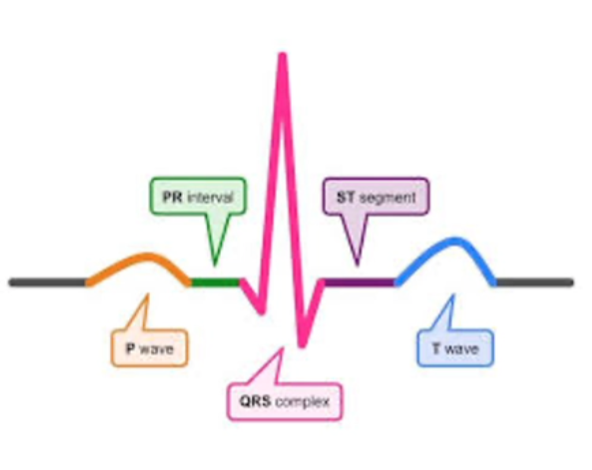
what is the P-wave?
orange: contraction of the R and L atria
what is the PR segment?
green: pause at the AV node so ventricles can fill up/ isoelectric
what is the QRS segment?
pink: largest wave, ventricles contract
what is the ST segment?
purple: when the ventricles repolarize and depolarize
what is the T-wave?
blue: ventricles go back to resting/repolarization
what nervous system largely controls the heart rate?
ANS: sympathetic and parasympathetic
what are chemoreceptors?
detect chemical changes in the blood
what are baroreceptors and where are they found?
found in carotid arteries, detecting pressure changes in heart and arteries
what are adrenocgenic receptors and where are they found?
structures on effector cells that respond to catecholamines, essentially let sympathetic and parasympathetic act on heart and vessels
what are the types of adrenogenic receptors
vascular smooth muscle (A1) | vasculature smooth muscle, vasoconstriction
central nervous system (A2), decrease smooth muscle of GI tract with SNS
(B1), increased Heart, contractility
(B2), contractility in lungs, increased HR
blood pressure def
force of blood on walls of blood vessels haw
what are the two numbers for blood pressure?e
systolic pressure is the larger number / 90-100mmHg
diastolic pressure is smaller / 60-80 mmHg
how do you find the pulse pressure?
Systolic - diastolic = difference
how do you find the mean arterial pressure (MAP)
systolic + diastolic + diastolic / 3 = 70-100 mmHg
what does it mean when the minimum MAP is below 65?
you have poor perfusion
cardiac output def
blood from heart in 1 minute | stroke volume x HR
what is stroke volume (SV)?
how much blood is ejected from the heart with each beat
what is the heart rate?
number of times a heart beats per minute
what is ejection fraction/EF
the percent of blood ejected from the left ventricle
blood ejected/total amount of blood in L.V.
usually 50-70% of blood is still in there
what is peripheral vascular resistance?
the force opposing blood in peripheral circulation
constriction, increased
dilation, decreased = maybe sepsis because not enough blood flow to cells
factors influencing BP
hormones
renin-angiotensin-aldosterone system
viscosity of blood
blood volume
cardiac contractility