biol 223 muscles 1
1/56
There's no tags or description
Looks like no tags are added yet.
Name | Mastery | Learn | Test | Matching | Spaced | Call with Kai |
|---|
No analytics yet
Send a link to your students to track their progress
57 Terms
muscle tissue
specialized for contraction
three types:
skeletal - attached to bone
striated, voluntary
cardiac - found in the heart
striated, involuntary
smooth - lines hollow organs
nonstriated, involuntary
If binding Acetylcholine to a chemically gated sodium ion channel opens the channel
A. sodium ions will move into the cell causing a hyperpolarization
B. sodium ions will move out of the cell causing a hyperpolarization
C. sodium ions will move into the cell causing a depolarization
D. sodium ions will move out of the cell causing a depolarization
C. sodium ions will move into the cell causing a depolarization
skeletal muscle functions
produce skeletal movement
maintain posture and body position (always contracting muscle to maintain posture)
support soft tissue
guard entrances and exits
maintain body temperature
nutrient reserves
gross anatomy of skeletal muscle
attached to bone by tendon
origin - attached to bone that remains relatively stationary during movement
insertion - attached to the bone that moves
synergist muscles - muscles that work together for a common goal; ex. muscles working to create a fit
antagonistic muscles
flexors and extensors
exceptions: circular sphincter muscles
endomysium
covers individual muscle fibers (1 fiber = 1 cell)
contains blood vessels and nerves
perimysium
sheathes bundles of muscle fibers(muscle fascicles)
contains blood vessels and nerves
epimysium
surrounds a muscle
deep fascia
wrap groups of cooperating muscles together
skeletal muscle cells (myo- & sarco-)
muscle cell = muscle fiber
multinucleate, very long cell
each muscle cell is as long as the muscle
like osteoclasts, multinucleate for transcription and translation
formed during embryogenesis by end-to-end fusion of uni-nucleate myoblasts
adult muscle repair is limited
new skeletal muscle cells come from stem cells called satellite cells
structure of a skeletal muscle cell (muscle fiber)
contains large quantities of protein filaments = myofilaments (strands of certain proteins
actin and myosin
myofibrils
sarcoplasm
sarcolemma
sarcoplasmic reticulum
actin
thin filaments
myosin
thick filaments
sarcoplasm
muscle cell cytoplasm
sarcolemma
muscle cell membrane
excitable membrane
conducts action potentials
narrow tubes of sarcolemma extend into cell at right angles to cell surface
transverse tubules (t-tubules)
conduct action potential deep into cell
comes in close contact with sarcoplasmic reticulum
sarcoplasmic reticulum
modified ER
similar to smooth ER
forms a tubular network around each myofibril
terminal cisternae form triads with t tubules
stores high concentration of Ca+2 ions needed for muscle contraction
myofilaments
thick (myosin)
thin (actin)
myofibril
bundles of myofilaments
anchored to inner surface of sarcolemma at either end of cell
sarcomeres
repeating units of myofilaments in myofibrils
can actively shorten
striated sarcomeres
differences in distribution of thick and thin myofilaments gives banded appearance
I bands
A bands
Z disk (line)
I bands
LIght band
contains only thin filaments
A bands
dArk band
contains thick filaments, and some overlap with thin filaments
H band contains only thick filaments
Z disk (line)
border between sarcomeres
transition
Muscles shorten during contraction because
A. the sarcoplamic reticulum pulls sarcomeres out of the myofilaments,
shortening them
B. the myosin and actin filaments can fold up like an accordion
C. the myosin and actin filaments are coiled like a spring and can recoil
after being stretched
D. the myosin and actin filaments slide between each other to shorten
each sarcomere
D. the myosin and actin filaments slide between each other to shorten
each sarcomere
skeletal muscle (levels of functional organization in skeletal muscle fiber)
surrounded by: epimysium
contains: muscle fascicles
muscle fascicle (levels of functional organization in skeletal muscle fiber)
surrounded by: perimysium
contains: muscle fibers
muscle fiber (levels of functional organization in skeletal muscle fiber)
surrounded by: endomysium
contains: myofibrils
myofibril (levels of functional organization in skeletal muscle fiber)
surrounded by: sarcoplasmic reticulum
consists of: sarcomeres (z line to z line)
sarcomere (levels of functional organization in skeletal muscle fiber)
contains: thick and thin filaments
sarcomere structure and function
myofibril in muscle cell consists of thousands of sarcomeres end to end
interactions between thin filaments and thick filaments are responsible for muscle contraction
thin filaments slide over thick filaments, shortening the sarcomere
shortening occurs in every sarcomere in the myofibril, thus shortens the myofibril
sliding filament model of muscle contraction
thin actin filaments - attached to Z disk
as thin filaments move toward center of sarcomere
thin filaments slide ver thick filaments
Z lines are puller closer together
I bands and H band narrow
A band stays the same width
sarcomere is at maximum shortening when it is the width of the A band, no I band or H band are visible
relaxed sarcomere
a relaxed sarcomere showing location of the A band, Z lines, and I band
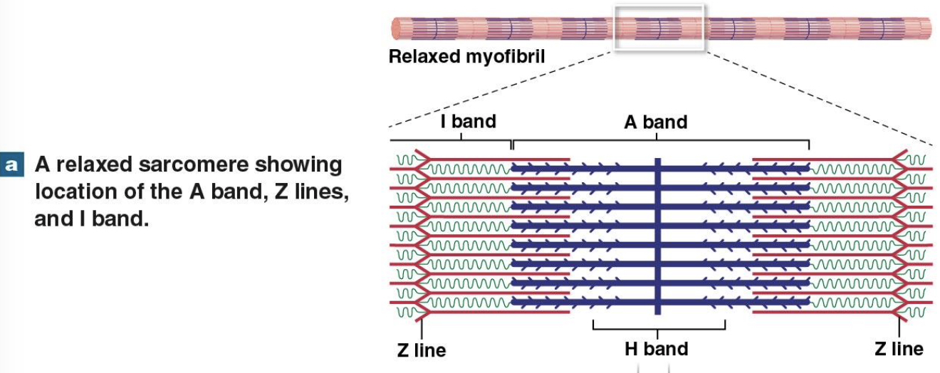
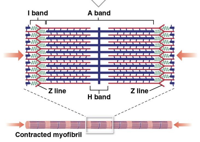
contracted sarcomere
during a contraction, the A band stays the same width, but the Z lines move closer together and the I band gets smaller. when the ends of a myofibril are free to move, the sarcomeres shorten simultaneously and the ends of the myofibril are pulled toward its center
what causes thin and thick myofilaments to slide across each other
myosin filaments have many short projections extending out from the filament
these projections can bind to sites on actin filaments, forming cross bridges
cross bridges, once formed, change shape pulling the actin past the myosin
cross bridges use energy of ATP to change shape and pull the actin - convert chemical energy to mechanical energy
molecular anatomy of thick (myosin) myofilaments
composed of many (~100) identical myosin molecules bundled side by side, in staggered bipolar array
myosin molecules have elongate tail, globular head - golf club shape
arrayed with half facing each end, center is just tails
heads form cross bridges during contraction
interactions between myosin head and actin prevented by tropomyosin during rest
molecular anatomy of thin (actin) myofilaments
composed of multiple actin molecules
twisted strand composed of two rows of individual globular actin molecules
each actin molecule in twisted strand has active site to which a myosin head can attach
strands of tropomyosin cover the actin active sites during rest
tropomyosin strands attached to actin by troponin
sliding filament theory
explains the relationship between thick and thin filaments as contraction proceeds
cyclic process beginning with calcium release from SR
calcium binds to troponin
troponin moves, moving tropomyosin and exposing actin active site
myosin head forms cross bridge to actin, bends toward center of sarcomere, pulling the actin
ATP allows release of cross bridge
role of ATP in molecular mechanism of contraction
ATP supplies the energy for the movement of the myosin head
converting chemical energy to mechanical energy of movement
myosin head in energized position binds to actin active site
releases ADP and P
pivots, pulling on actin and moving to un-energized state
ATP binds to un-energized myosin head
detaching myosin from action
ATP is split and head is energized
Active transport of ions
role of calcium ions in molecular mechanism of contraction
concentration of Ca+2 around sarcomere controls sarcomere contraction
Ca+2 is low around sarcomere at rest
action potential in sarcolemma and t tubules results in contractions
causes voltage-gated Ca+2 channels of SR to open
releases Ca+2 into sarcoplasm around sarcomere
Ca+2 binds to troponin
causes troponin to change shape and pull tropomyosin off of actin active sites
myosin heads bind to available actin sites over and over until Ca+2 level falls
When Ca+2 level falls
tropomyosin covers actin active sites, ending contraction
motor neuron
nerve cell that controls muscle contraction
neuromuscular junction
synapse between motor neuron and muscle cell
where we have chemically gated ion channels
control of skeletal muscle activity occurs at the neuromuscular junction
motor neuron
neuromuscular junction
action potential initiated in motor neuron in response to central nervous system commands
travels through motor neuron and arrives at synaptic neuron
AP in motor neuron causes the neurotransmitter Acetylcholine (ACh) to be released from motor neuron terminal
ACh diffuses across synaptic gap
ACh binds to receptors on chemically-gated sodium channels in muscle membrane
sodium ions flow into the muscle cell
depolarization the muscle cell membrane and starts an action potential in the muscle cell
chemically regulated gates stay open as long as ACh is present
Acetylcholine Esterase (AChE)
excitation of muscle cell
acetylcholine esterase (ache) (Control of skeletal muscle activity occurs at the neuromuscular junction)
located in synaptic gap
rapidly breaks down acetylcholine
excitation of muscle cell (Control of skeletal muscle activity occurs at the neuromuscular junction)
action potential is initiated which spreads across the entire muscle cell membrane including the t tubules
activity at the neuromuscular junction 1
the cytoplasm of the axon terminal contains vesicles filled with molecules of acetylcholine, or ACh. acetylcholine is a neurotransmitter, a chemical released by a neuron to change the permeability or other properties of another cells’s plasma membrane. the synaptic cleft and the motor end plate contain molecules of the enzyme acetylcholinesterase (AChE), which breaks down ACh
the synaptic cleft is a narrow space that separates the axon terminal of the neuron from the opposing motor end plate
activity at the neuromuscular junction 2
the stimulus for ACh release is the arrival of an electrical impulse, or action potential, at the axon terminal. an action potential is a sudden change in the membrane potential that travels along the length of the axon
activity at the neuromuscular junction 3
when the action potential reaches the neuron’s axon terminal, permeability changes in its membrane trigger the exocytosis of ACh into the synaptic cleft. exocytosis occurs as vesicles fuse with the neuron’s plasma membrane
activity at the neuromuscular junction 4
ACh molecules diffuse across the synaptic cleft and bind to ACh receptor membrane channels. ACh binding opens the membrane channel on the surface on the motor end plate. because the extracellular fluid contains a high concentration of sodium ions, and sodium ion concentration inside the cell is very low, sodium ions rush into the cytosol
activity at the neuromuscular junction 5
the sudden inrush of sodium ions results in the generation of an action potential in the sarcolemma. ACh is removed from the synaptic cleft in two ways. ACh either diffuses away from the synapse, or it is broken down by AChE into acetic acid and choline. this removal closes the ACh receptor membrane channels. the muscle fiber pictured above indicates the propagation of the action potential along the sarcolemma
excitation/contraction coupling
action potential along t tubule causes release of calcium from cisternae of SR
initiates contraction cycle
cycle repeats over and over until calcium ion concentration falls to resting level
contraction cycle (excitation/contraction coupling)
Ca+2 binds to troponin, moving tropomyosin
attachment of myosin head to actin
pivot of myosin head pulls on actin
detachment of myosin head with binding of ATP
Action potentials in the muscle cell membrane are conducted into the interior of the muscle via _ and cause Ca2+ ions to be released from the _
A. sarcoplasmic reticulum; myofibril
B. myofibril; mitochondria
C. myosin thick filaments; actin thin filaments
D. transverse tubules; sarcoplasmic reticulum
D. transverse tubules; sarcoplasmic reticulum
how does calcium ion concentration return to resting level
AP depolarization ends, voltage-gated Ca+2 channels in SR close
calcium ion diffusion into sarcoplasm stops
Ca+2 is actively transported out of sarcoplasm
across sarcolemma to outside of cell
across sarcoplasmic reticulum membrane into SR
requires ATP for active transport protein to function
duration of contraction depends on
duration of stimulation at nerve-muscle synapse (neuromuscular junction)
multiple action potentials in motor neuron cause continued release of ACh and multiple AP in muscle fiber
presence of calcium ions in sarcoplasm
contraction cycle continues until calcium ion concentration returns to resting level
availability of ATP
if no ATP is available, contraction cycle stops even if action potential and calcium ions are present
contraction ends and relaxation occurs when
action potentials stop in motor neuron
acetylcholinesterase breaks down ACh in the neuromuscular synaptic gap
ACh gated channels close
sodium ion influx stops
action potentials stop occurring in sarcolemma and t tubules
calcium ion levels in sarcoplasm return to resting levels
tropomyosin covers actin sites and no new myosin cross bridges can form
relaxation requires ATP
ATP needed to pump Ca+2 into the SR
ATP needed to disconnect myosin heads from actin
rigor mortis - lack of ATP after death
steps that initiate a muscle contraction
ACh released
ACh is released at the neuromuscular junction and binds to ACh receptors on the sarcolemma
action potential reaches t tubule
an action potential is generated and spreads across the membrane surface of the muscle fiber and along the t tubules
sarcoplasmic reticulum releases Ca+2
the sarcoplasmic reticulum releases stored calcium ions
active sites exposed and cross-bridges form
calcium ions bind to troponin, exposing the active sites on the thin filaments. cross-bridges form when myosin heads bind to those active sites
contraction cycle begins as repeated cycles of cross-bridge binding, pivoting, and detachment occur—all powered by ATP
steps that end a muscle contraction
ACh is broken down
ACh is broken down by acetylcholinesterase (AChE), ending action potential generation
sarcoplasmic reticulum reabsorbs Ca2+
as the calcium ions are reabsorbed, their concentration in the cytosol decreases
active sites covered, and cross-bridge formation ends
without calcium ions, the tropomyosin returns to its normal position and the active sites are covered again
contraction ends
without cross-bridge formation, contraction ends
muscle relaxation occurs
the muscle returns passively to its resting length
muscular system disorders
nervous system disorders that affect the coordination or control of muscle contraction
blockage of release of ACh (e.g. botulism)
interference with binding o ACh to receptors (e.g. myasthenia graves, autoimmune, can’t contract muscle)
interference with ACh esterase activity (blocks enzyme, continuous muscle contraction, spasm)
loss of motor neuron (e.g. polio)
loss of motor neuron axon - peripheral nerve damage
reduction of AP efficiency, damage to myelin (e.g. MS, autoimmune)
excessive stimulation of motor neuron (e.g. tetanus, excessive muscle contraction)