3- Mass transport
1/55
There's no tags or description
Looks like no tags are added yet.
Name | Mastery | Learn | Test | Matching | Spaced |
|---|
No study sessions yet.
56 Terms
Describe the role of red blood cells and haemoglobin in oxygen transport
RBC contain lots of haemoglobin (Hb)= no nucleus, biconcave, high SA:V, short diffusion pathway
Hb associates with/ binds/ loads O2 at gas exchange surfaces where partial pressure of O2 (pO2) is high
This forms oxyharmoglobin which transports O2 (each can carry 4O2- one at each Haem group)
Hb dissociates from/ unloads O2 near cells/ tissues where pO2 is low
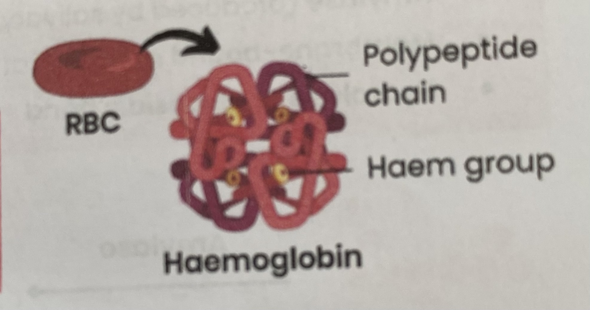
Describe the structure of haemoglobin
Protein with a quaternary structure
Made of 4 polypeptide chains
Each chain contains a Haem group containing an iron ion (Fe2+)
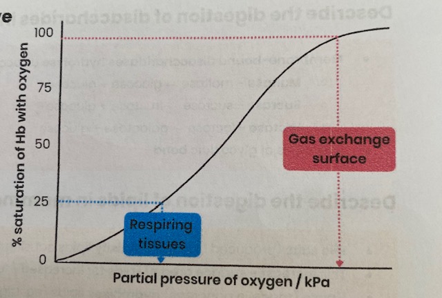
Describe the loading, transport, and unloading of oxygen in relation to the oxyhaemoglobin dissociation curve (area with low pO2= respiring tissue)
Hb has a low affinity for O2
so O2 readily unloads/ dissociates with Hb
so % saturation is low

Describe the loading, transport, and unloading of oxygen in relation to the oxyhaemoglobin dissociation curve (area with high pO2= gas exchange surfaces)
Hb has a high affinity for O2
so O2 readily loads/ associates with Hb
so % saturation is high
Explain how the cooperative nature of oxygen binding results in an S-shaped (sigmoid) oxyhaemoglobin dissociation curve
Binding of first oxygen changes tertiary/ quaternary structure of haemoglobin
This uncovers Haem group binding sites, making further binding of oxygens easier
Describe evidence for the cooperative nature of oxygen binding
At low pO2, as oxygen increases there is little/ slow increase in % saturation of Hb with oxygen
when first oxygen is binding
At higher pO2, as oxygen increases there is a big/ rapid increase in % saturation of Hb with oxygen
showing it has got easier for oxygens to bind
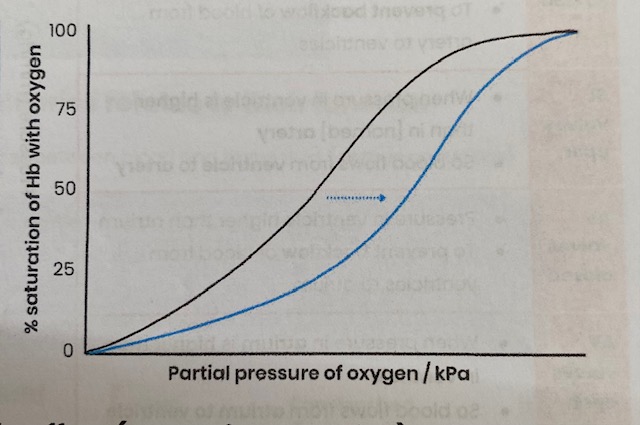
What is the Bohr effect?
Effect of CO2 conc on dissociation of oxyhaemoglobin= curve shifts to right
Explain effect of CO2 conc on the dissociation of oxyhaemoglobin
Increasing blood CO2 e.g. due to increased rate of respiration
Lowers blood pH (more acidic)
Reducing Hb’s affinity for oxygen as shape/ tertiary/ quaternary structure changes slightly
So more/ faster unloading of oxygen to respiring cells at a given pO2
Explain the advantage of the Bohr effect (e.g. during exercise)
more dissociation of oxygen= faster aerobic respiration/ less anaerobic respiration= more ATP produced
Explain why different types of haemoglobin can have different oxygen transport properties
Different types of Hb are made of polypeptide chains with slightly different amino acid sequences
Resulting in different tertiary/ quaternary structures/ shape= different affinities for oxygen
Explain how organisms can be adapted to their environment by having different types of haemoglobin with different oxygen transport properties (curve shift LEFT)
LEFT= Hb has higher affinity for O2
More O2 associates with Hb more readily
At gas exchange surfaces where pO2 is lower
e.g. organisms in low O2 environments- high altitudes, underground, or foetuses
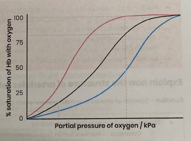
Explain how organisms can be adapted to their environment by having different types of haemoglobin with different oxygen transport properties (curve shift RIGHT)
RIGHT= Hb has lower affinity for O2
More O2 dissociates from Hb more readily
At respiring tissues where more O2 is needed
e.g. organisms with high rates of respiration/ metabolic rate (may be small or active)

Describe the general pattern of blood circulation in a mammal
Closed double circulatory system= blood passes through heart twice for every circuit around body:
Deoxygenated blood in right side of heart pumped to lungs; oxygenated returns to left side
Oygenated blood in left side of heart pumped to rest of body; deoxygenated returns to right
Suggest the importance of a double circulatory system
Prevents mixing of oxygenated/ deoxygenated blood
so blood pumped to body is fully saturated with oxygen for aerobic respiration
Blood can be pumped to body at a higher pressure (after being lower from lungs)
substances taken to/ removed from body cells quicker/ more efficiently
Draw a diagram to show the general pattern of blood circulation in a mammal, including the names of key blood vessels
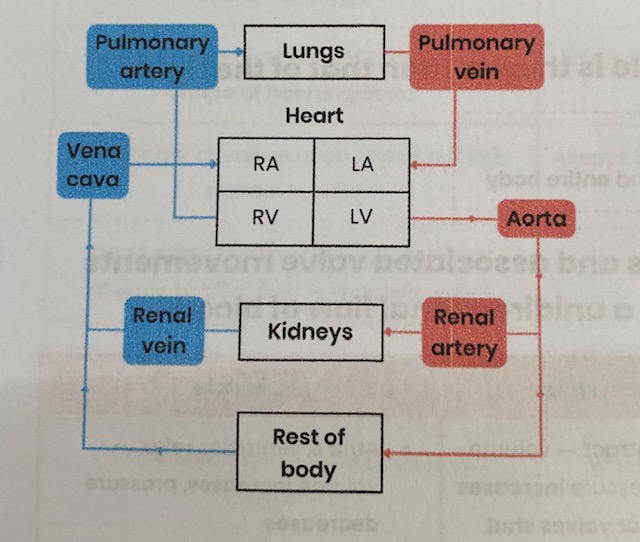
Name the blood vessels entering and leaving the heart and lungs
Vena cava= transports deoxygenated blood from respiring body tissues to heart
Pulmonary artery= transports deoxygenated blood from heart to lungs
Pulmonary vein= transports oxygenated blood from lungs to heart
Aorta= transports oxygenated blood from heart to respiring body tissues
Name the blood vessels entering and leaving the kidneys
Renal arteries= oxygenated blood to kidneys
Renal veins= deoxygenated blood to vena cava from kidneys
Name the blood vessels that carry oxygenated blood to the heart muscle
coronary arteries= located on surface of the heart, branching from aorta
Label a diagram to show the gross structure of the human heart (inside)
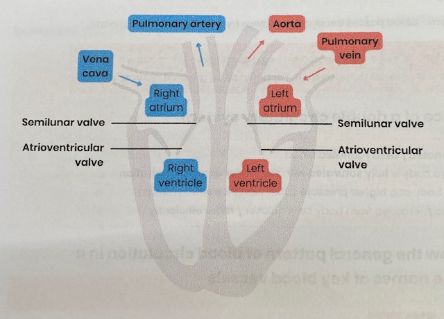
Suggest why the wall of the left ventricle is thicker than that of the right
Thicker muscle to contract with greater force
to generate higher pressure to pump blood around entire body
Explain the pressure & volume changes and associated valve movements during the cardiac cycle that maintain an unidirectional flow of blood (Atrial systole)
Atria contract= volume decreases, pressure increases
Atrioventricular valves open when pressure in atria exceeds pressure in ventricles
Semilunar valves remain shut as pressure in arteries exceeds pressure in ventricles
so blood pushed into ventricles
Explain the pressure & volume changes and associated valve movements during the cardiac cycle that maintain an unidirectional flow of blood (Ventricular systole)
Ventricles contract= volume decreases, pressure increases
Atrioventricular valves shut when pressure in ventricles exceeds pressure in atria
Semilunar valves open when pressure in ventricles exceeds pressure in arteries
so blood pushed out of heart through arteries
Explain the pressure & volume changes and associated valve movements during the cardiac cycle that maintain an unidirectional flow of blood (Diastole)
Atria & ventricles relax= volume increases, pressure decreases
Semilunar valves shut when pressure in arteries exceeds pressure in ventricles
Atrioventricular valves open when pressure in atria exceeds pressure in ventricles
so blood fills atria via veins & flows passively to ventricles
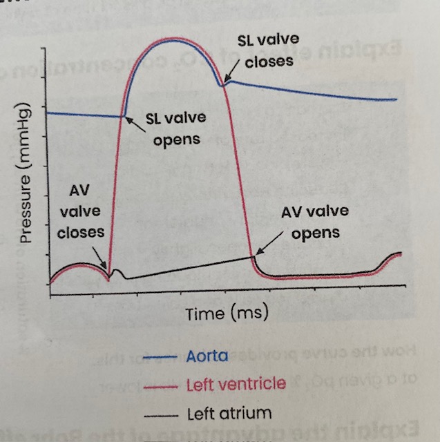
Explain how graphs showing pressure or volume changes during the cardiac cycle can be interpreted, e.g. to identify when valves are open/ closed
SL valves closed:
Pressure in … artery higher than in ventricle
to prevent backflow of blood from artery to ventricles
SL valves open:
When pressure in ventricle is higher than in … artery
so blood flows from ventricle to artery
AV valves closed:
Pressure in ventricle higher than atrium
to prevent backflow of blood from ventricles to atrium
AV valves open:
When pressure in atrium is higher than in ventricle
so blood flows from atrium to ventricle
Describe the equation for cardiac output
Cardiac output (volume of blood pumped out of heart per min)= stroke volume (volume of blood pumped in each heart beat) x heart rate (number of beats per min)
How can heart rate be calculated from cardiac cycle data?
Heart rate (beats/ min)= 60 (seconds) / length of 1 cardiac cycle (seconds)
Explain how the structure of arteries relates to their function
Function= carry blood away from heart at high pressure
Thick smooth muscle tissue= can contract and control/ maintain blood flow/ pressure
Thick elastic tissue= can stretch as ventricles contract and recoil as ventricles relax, to reduce pressure surges/ even out blood pressure/ maintain high pressure
Thick wall= withstand high pressure/ stop bursting
Smooth/ folded endothelium= reduces friction/ can stretch
Narrow lumen= increases/ maintains high pressure
Explain how the structure of arterioles relates to their function
Function= (division of arteries to smaller vessels which can) direct blood to different capillaries/ tissues
Thicker smooth muscle layer than arteries
contracts= narrows lumen (vasoconstriction)= reduces blood flow to capillaries
relaxes= widens lumen (vasodilation)= increases blood flow to capillaries
Thinner elastic layer= pressure surges are lower (as further from heart/ ventricles)
Explain how the structure of veins relates to their function
Function= carry blood back to heart at lower pressure
Wider lumen than arteries= less resistance to blood flow
Very little elastic and muscle tissue= blood pressure lower
Valves= prevent backflow of blood
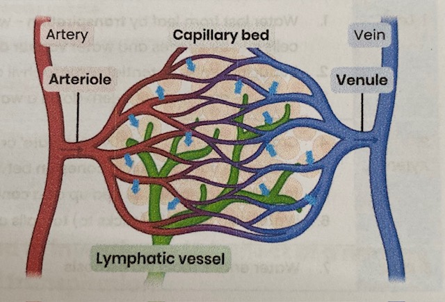
Explain how the structure of capillaries relates to their function
Function= allow efficient exchange of substances between blood and tissue fluid (exchange surface)
Wall is a thin (one cell) layer of endothelial cells= reduces diffusion distance
Capillary bed is a large network of branched capillaries= increases surface area for diffusion
Small diameter/ narrow lumen= reduces blood flow rate so more time for diffusion
Pores in walls between cells= allow larger substances through
Explain the formation of tissue fluid
At the arteriole end of capillaries:
Higher blood/ hydrostatic pressure inside capillaries (due to contraction of ventricles) than tissue fluid (so net outward force)
Forcing water (and dissolved substances) out of capillaries
Large plasma proteins remain in capillary

Explain the return of tissue fluid to the circulatory system
At the venule end of capillaries:
Hydrostatic pressure reduces as fluid leaves capillary (also due to friction)
(Due to water loss) an increasing concentration of plasma proteins lowers water potential in capillary below that of tissue fluid
Water enters capillaries from tissue fluid by osmosis down a water potential gradient
Excess water taken up by lymph capillaries and returned to circulatory system through veins
Suggest and explain causes of excess tissue fluid accumulation
Low conc of protein in blood plasma
water potential in capillary not as low= water potential gradient is reduced
so more tissue fluid formed at arteriole end/ less water absorbed at venule end by osmosis
High blood pressure (e.g. caused by high salt conc)= high hydrostatic pressure
increases outward pressure from arterial end AND reduces inward pressure at venule end
so more tissue fluid formed at arteriole end/ less water absorbed at venule end by osmosis
lymph system may not be able to drain excess fast enough
What is a risk factor? Give examples for cardiovascular disease
An aspect of a person’s lifestyle or substances in a person’s body/ environment
That have been shown to be linked to an increased rate of disease
Examples- age, diet high in salt or saturated fat, smoking, lack of exercise, genes
RP5- What is RP5?
Dissection of animal or plant gas exchange system or mass transport system or of organ within such a system
RP5- Describe precautions that should be followed when performing a dissection
Cover any cuts with a waterproof dressing
When using a scalpel, cut away body onto a hard surface
When using a scalpel, use a sharp blade
When using a scalpel, carry with blade protected/ pointing down
Wear disposable gloves and disinfect hands/ wash with soap
Disinfect surfaces/ equipment
Safe disposal- put gloves/ paper towels/ organ in a separate bag/ bin to dispose
If poisonous chemicals/ toxins involved, work in a well ventilated environment
RP5- Suggest an ethical consideration when dissecting animals
Morally wrong to kill animals just for dissection
so use animals for dissection that have already been killed (humanely) for meat
RP5- Describe how you could prepare a temporary mount of a piece of plant tissue for observation with an optical microscope
Add a drop of water to glass slide
Obtain a thin section of specimen and place on a slide
Stain (e.g. with iodine/ potassium iodide to view starch)
Lower coverslip at angle using mounted needle without trapping air bubbles
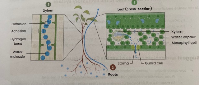
Describe the function of xylem tissue
transports water (and mineral ions) through the stem, up the plant to leaves of plants
Suggest how xylem tissue is adapted for its function
Cells joined with no end walls forming a long continuous tube= water flows as a continuous column
Cells contain no cytoplasm/ nucleus= easier water flow/ no obstructions
Thick cell walls with lignin= provides support/ withstand tension/ prevents water lots
Pits in side walls= allow lateral water movements
Explain the cohesion- tension theory of water transport in the xylem
1- LEAF:
Water lost from leaf by transpiration- water evaporates from mesophyll cells into air spaces and water vapour diffuses through (open) stomata
Reducing water potential of mesophyll cells
So water drawn out of xylem down a water potential gradient
2- XYLEM:
Creating tension (negative pressure/ pull) in xylem
Hydrogen bonds result in cohesion between water molecules (stick together) so water is pulled up as a continuous column
Water also adheres (sticks to) walls of xylem
3- ROOT:
Water enters roots via osmosis
Describe how to set up a potometer
Cut a shoot underwater at a slant= prevent air entering xylem
Assemble potometer with capillary tube end submerged in a beaker of water
Insert shoot underwater
Ensure apparatus is watertight/ airtight
Dry leaves and allow time for shoot to acclimatise
Shut tap to reservoir
Form an air bubble- quickly remove end of capillary tube from water
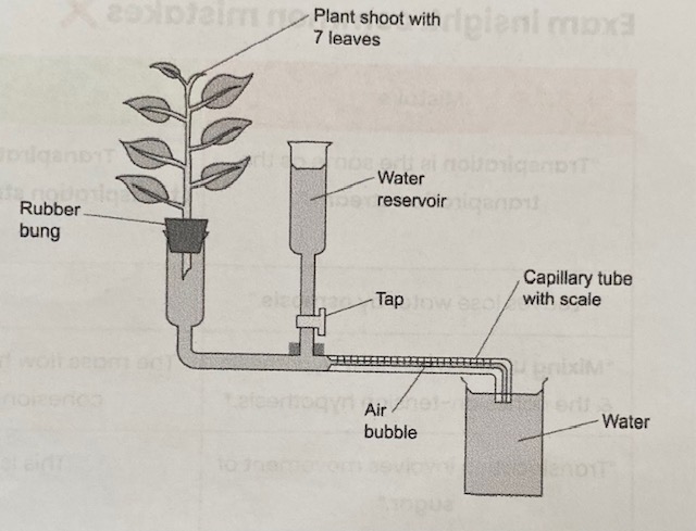
Describe how a potometer can be used to measure the rate of transpiration
Potometer estimates transpiration rate by measuring water uptake:
Record position of air bubble
Record distance moved in a certain amount of time (e.g. 1 min)
Calculate volume of water uptake in a given time:
Use radius of capillary tube to calculate cross-sectional area of water (pi r²)
Multiply this by distance moved by bubble
Calculate rate of water uptake- divide volume by time taken
Describe how a potometer can be used to investigate the effect of a named environmental variable on the rate of transpiration
Carry out the above, change one variable at a time (wind, humidity, light, temp)
e.g. set up a fan OR spray water in a plastic bag and wrap around the plant OR change distance of a light source OR change temperature of room
Keep all other variables constant
Suggest limitations in using a potometer to measure rate of transpiration
Rate of water uptake might not be same as rate of transpiration
water used for support/ turgidity
water used in photosynthesis and produced during respiration
Rate of movement through shoot in potometer may not be same as rate of movement through shoot of whole plant
shoot in potometer has no roots whereas a plant does
xylem cells very narrow
Suggest how different environmental variables affect transpiration rate (light intensity)
Increases rate of transpiration:
Stomata open in light to let in CO2 for photosynthesis
allowing more water to evaporate faster
Stomata close when it’s dark so there is a low transpiration rate
Suggest how different environmental variables affect transpiration rate (temperature)
Increases rate of transpiration:
Water molecules gain kinetic energy as temperature increases
so water evaporates faster
Suggest how different environmental variables affect transpiration rate (wind intensity)
Increases rate of transpiration:
Wind blows away water molecules from around stomata
decreasing water potential of air around stomata
increasing water potential gradient so water evaporates faster
Suggest how different environmental variables affect transpiration rate (humidity)
Decreases rate of transpiration:
More water in air so it has a higher water potential
decreasing water potential gradient from leaf to air
water evaporates slower
Describe the function of phloem tissue
transports organic substances e.g. sucrose in plants
Suggest how phloem tissue is adapted for its function
Sieve tube elements
no nucleus/ few organelles= maximise space for/ easier flow of organic substances
end walls between cells perforated (sieve plate)
Companion cells
many mitochondria= high rate of respiration to make ATP for active transport of solutes
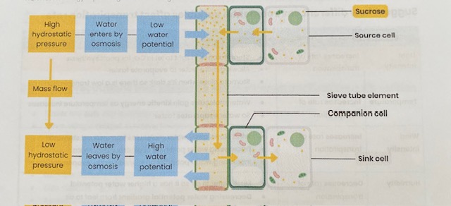
What is translocation?
Movement of assimilates/ solutes such as sucrose
from source cells (where made e.g. leaves) to sink cells (where used/ stored e.g. roots) by mass flow
Explain the mass flow hypothesis for translocation in plants
At source, sucrose is actively transported into phloem sieve tubes/ cells
By companion cells
This lowers water potential in sieve tubes so water enters (from xylem) by osmosis
This increases hydrostatic pressure in sieve tubes (at source)/ creates a hydrostatic pressure gradient
So mass flow occurs- movement from source to sink
At sink, sucrose is removed by active transport to be used by respiring cells or stored in storage organs
Describe the use of tracer experiments to investigate transport in plants
Leaf supplied with a radioactive tracer e.g. CO2 containing radioactive isotope 14C
Radioactive carbon incorporated into organic substances during photosynthesis
These move around plant by translocation
Movement tracked using autoradiography or a Geiger counter
Describe the use of ringing experiments to investigate transport in plants
Remove/ kill phloem e.g. remove a ring of bark
Bulge forms on source side of ring
Fluid from bulge has a higher conc of sugars than below- shows sugar is transported in phloem
Tissues below ring die as cannot get organic substances
Suggest some points to consider when interpreting evidence from tracer & ringing experiments and evaluating evidence for/ against the mass flow hypothesis
is there evidence to suggest the phloem (as opposed to xylem) is involved?
is there evidence to suggest respiration/ active transport is involved?
is there evidence to show movement if from source to sink? what are these in the experiment?
is there evidence to suggest movement is from high to low hydrostatic pressure?
could movement be due to another factor e.g. gravity?