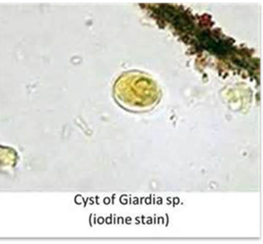
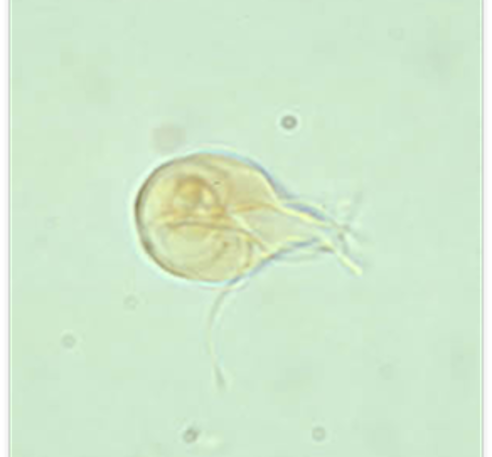
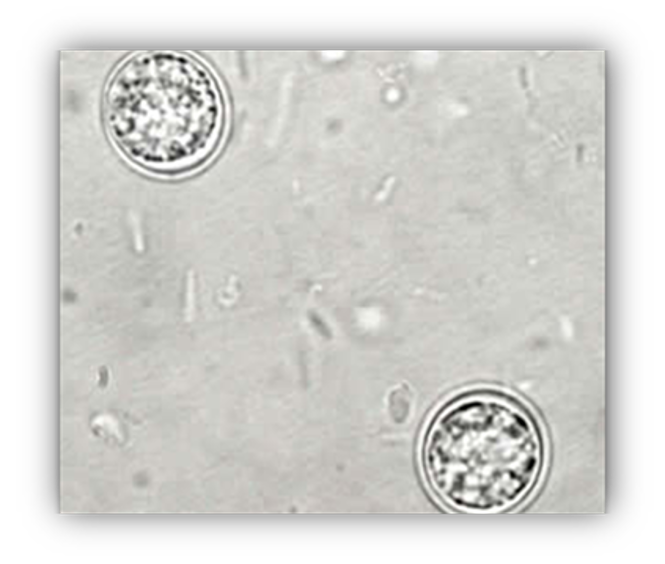
Giardia species
Host: dogs, cats, ruminants Location: intestinal mucosa Transmission: ingestion of oocysts Diagnosis: standard fecal float Treatment: metronidazole, fenbendazole Common name: giardia zoonotic? yes key fact: diarrhea will occur in 5 days if testing fecal float, results are more prominent in one week shether's solution and zinc sulfate are both good flotation solutions
Entamoeba histolytica
Host: canine, feline, primates Location: large intestine Transmission: ingestion of amoeba Diagnosis: direct smear Clinical signs: diarrhea and sporadic infections Common name: Entamoeba Zoonotic? yes
Balantidium coli
Host: canines and swine Location: cecum and colon in canine and large intestines in swine Transmission: ingestion of cysts Diagnosis: direct smear Clinical signs: diarrhea and sporadic infections Common name: Balantidium Zoonotic? yes
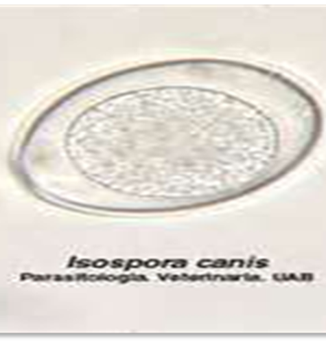
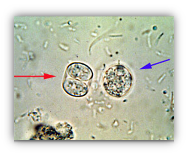
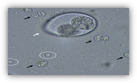
Cystoisospora species
Host: canines, felines, and swine Location: small intestines Transmission: ingestion of oocysts Diagnosis: fecal float of fresh feces and direct smear Clinical signs: loose stool to watery diarrhea, dehydration, even death Treatment: Sulfadimethoxine (albon) Common name: Coccidia Zoonotic? no
Toxoplasma gondii
Host: feline (def host) Location: intestines Transmission: ingestion of sporulated oocyst Diagnosis: standard fecal float Zoonotic? YES Key fact: it is very zoonotic to pregnant women and their fetus it is so important to tell mother that it can be infective to the mother and cross the placenta to cause brain damage, mental disorder, and blindness they should avoid changing the litter box or use gloves when they need too also to avoid garden work and put gloves on when washing veggies and fruit
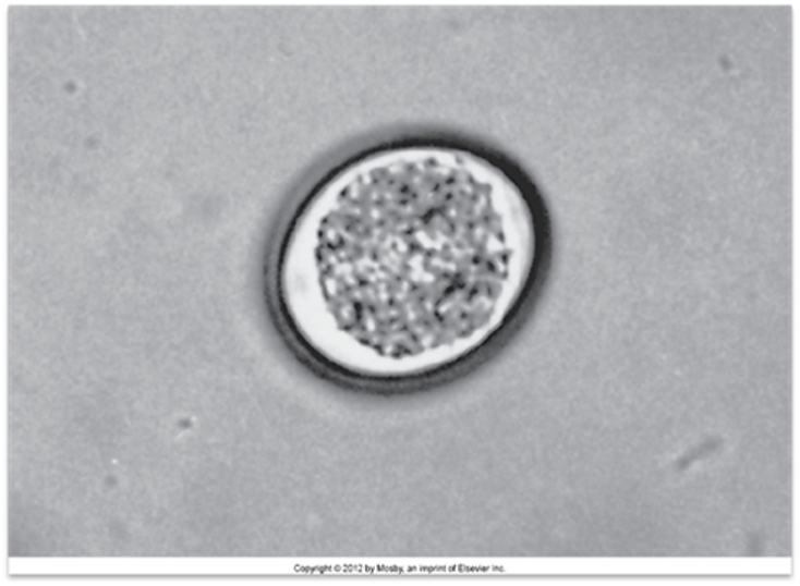
Cryptosporidium species
Host: canines, felines, guinea pigs, snakes Location: small intestine Transmission: ingestion of oocysts Diagnosis: standard fecal float Treatment: supportive care Zoonotic? yes Key fact: very small and may not be observed in same plane of focus as other eggs. acid fast stain or lugol's iodine helps most common zoonotic species
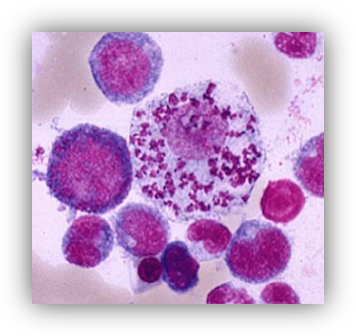
Leishmania species
Host: canines Location: phlebotomine sand flies Transmission: bite by infected intermediate host Location: reticuloendothelial cells of capillaries and spleen as well as other internal organs and WBC's Treatment: allopurinol Common name: Leishmania Zoonotic? zoonotic potential Key fact: can be found histopathologically
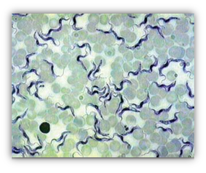
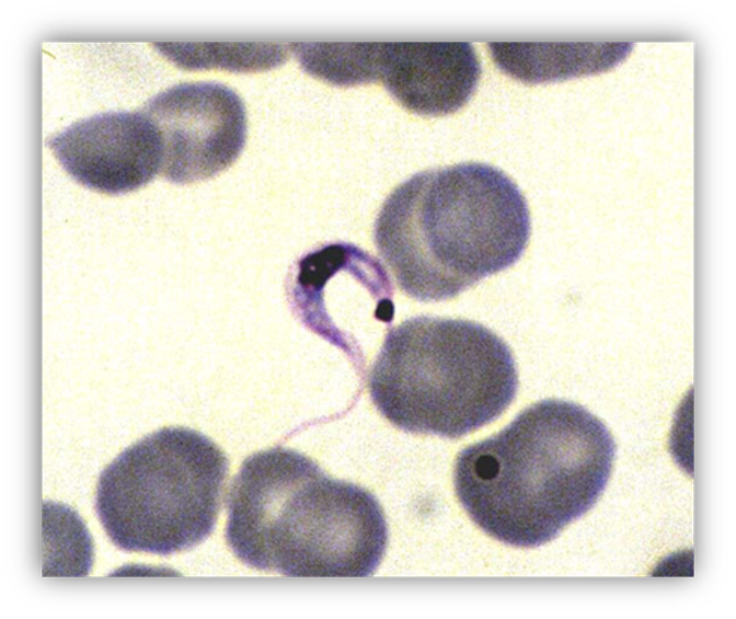
Trypanosoma cruzi
Host: humans and occasionally canines Location: peripheral blood Transmission: ingestion of intermediate host, reduviid bug or feces of reduviid bug left on mucus membranes of final host Diagnosis: direct blood smear Key fact: this is a hemoprotozoan it is extracellular and swims within the blood. the resting cyst stage may be found in cardiac muscle and other tissues like the esophagus
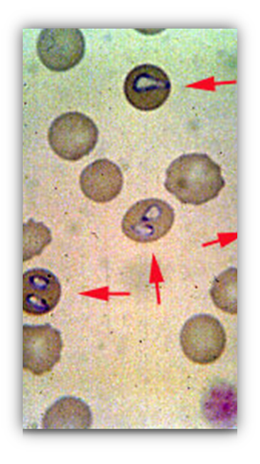
Babesia canis
Host: canine Intermediate host: ticks Location: within the RBC's Transmission: bite from an infected intermediate host Diagnosis: stained blood smears Clinical signs: pale mucus membranes, icterus (jaundice) anorexia Common name: Babesia Zoonotic? no Key fact: death can occur rapidly
Cytauxzoon felis
Host: feline Intermediate host: tick Location: within the RBC's Transmission: bite from the infective intermediate host Clinical signs: fever, icetrus, anemia, dehydration, death, prognosis is poor Diagnosis: stained blood smears Treatment: blood transfusions Common name: Cytauxzoon
Hepatozoon canis
Host: canines Intermediate host: Rhipicephalus sanguineus (brown dog tick) Locations: Gamonts are within the WBC's Transmission route: ingestion of infective intermediate host Diagnosis: H. canis is commonly seen in peripheral blood smears Treatment: TCP + Decoquinate (Trimethoprim/sulfa (antibiotic) Pyrimethamine (antiparasitic) and clindamycin (antibiotic) 2 weeks then Decoquinate Common name: Hepatozoon
Hepatozoon americanum
Host: canines Intermediate host: Amblyomma maculatum (gulf coast tick) Locations: gamonts are within the WBCs Transmission: ingestion of infective intermediate host Clinical signs: produces a violent and frequently fatal course of disease Diagnosis: rare to see. forms onion cysts (multiple layers) in the skeletal muscle of dogs Treatment: TCP+ Decoquinate (Trimethoprim/sulfa (antibiotic) Pyrimethamine (antiparasitic) and clindamycin (antibiotic) 2 weeks then Decoquinate Common name: Hepatozoon
Eimeria species ( Eimeria bovis and Eimeria zuernii)
Host: ruminants Locations: cecum and colon Transmission: ingestion of oocysts Clinical signs: diarrhea, neurological signs, death Diagnosis: fecal float Treatment: decoquinate (anticoccidial) lasalocid (anticoccidial) monesin (antibitotic) Common name: Coccidia
Cryptosporidium species
Host: canines, felines, bovines, swine, avians, guinea pigs, snakes, mice Locations: small intestine Transmission: ingestion of oocysts Diagnosis: fecal float and stained fecal smears Sheather's sugar solution Treatment: supportive care Common name: Crypto Key fact: zoonotic
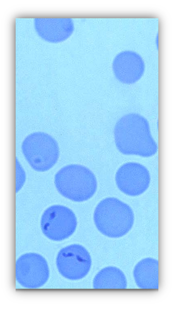
Babesia bigemina
Host: bovines Intermediate host: Boophilus annulatus (blue cattle tick) Locations: within the RBC's Transmission: bite of an infective intermediate host Clinical signs: fever, depression, icterus, anorexia, hemoglobinuria, pale mucus membranes, weakness, splenomegaly Diagnosis: stained blood smear Treatment: dip for ticks, Dimiazene aceturate (Berenil), Phenamidine Common name: Babesia
Tritrichomonas foetus
Host: bovine Locations: prepuce of bulls and vagina, cervix, and uterus of cows Transmission: sexual transmission Clinical signs: abortion Diagnosis: finding the protozoan in fluid from stomach of aborted fetus, from uterine discharges, or washings of vagina and prepuce Treatment: Metronidazole Common name: Trichomonas
Sarcocystis neurona
Host: equine Locations: asexual stage found in the nervous system Transmission: ingestion of oocyst from opposum feces Clinical signs: neurological signs Diagnosis: Histopathologic examination Treatment: trimethoprim-sulfadiazine and pyrimethamine Common name: Sarcocystis Key fact: can invade the CNS causing a condition called equine protozoal myeloencephalitis
Cystoisospora suis
Host: swine Locations: small intestine Transmission: ingestion of oocysts Clinical signs: diarrhea, dehydration, and weight loss Diagnosis: fecal float Treatment: decoquinate to sows before and after farrowing Common name: coccidia
Giardia psittaci
Host: avians Locations: intestinal mucosa Transmission: ingestion of oocysts Diagnosis: fresh saline mounts can use Lugol's gram's iodine, and wright's stain Key fact: the most common protozoan seen in pet birds allergic to skin conditions can be associated with giardiasis
Histomonas meleagridis
Host: turkeys, peafowl, chickens, and pheasants Intermediate host: Heterakis gallinarum Locations: liver Transmission: ingestion of intermediate host Diagnosis: necropsy and histopath of the liver Common name: blackhead
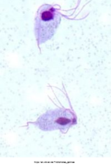
Trichomonas gallinae
Host: pigeons, doves, poultry, and raptors that feed on pigeons Location: crop Transmission: direct contact with contaminated water or infected birdDiagnosis: crop washes and swabs Treatment: dimetridazole, ipronidazole, metronidazole Common name: Trichomonas
Haemoproteus species
Host: cockatoos, green winged macaws, and some conures Locations: within the blood cells Transmission: bite by infeced Culicoides species or Chrysops species of fly Key fact: rarely clinical but can infect wild waterfowl and may cause death in these birds
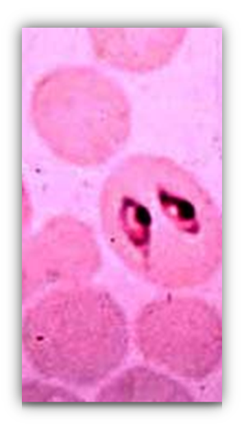
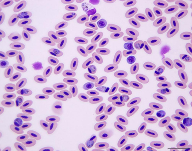
Plasmodium species
Host: avians Intermediate host: mosquito Locations: within the RBC's Transmission: bite of an infected mosquito Common name: Avian malaria Key fact: this is the etiologic agent of avian malaria (mosquito born disease of birds)
Leucocytozoon species
Host: raptors Locations: within the WBCs Transmission: bite of the infected black flies (Simulium species) Key fact: may be associated with leukocytosis very large and greatly distort the appearance of the WBC
Eimeria species
Host: lagomorph (rabbits) Locations: small intestine in most E. media also affects the large intestine E. stiedai affects the bile ducts Transmission: ingestion of oocysts Diagnosis: recognition of oocysts along with clinical signs Clinical signs: severe diarrhea, excessive thirst, dehydration Key fact: magna and irresidua are highly pathogenic
Ichthyophithirius multifiliis
Host: freshwater and ornamental fish Locations: skin, gills, fins, and eyes Transmission: contact with the infective stage Diagnosis: observation of typical lesions, is also possible to preform skin scrapings Key fact: causes a condition called ichthyophthiriosis or ICH characterized by tiny white spots over the exposed surfaces of fish