Microanatomy - Brain
1/84
Name | Mastery | Learn | Test | Matching | Spaced |
|---|
No study sessions yet.
85 Terms
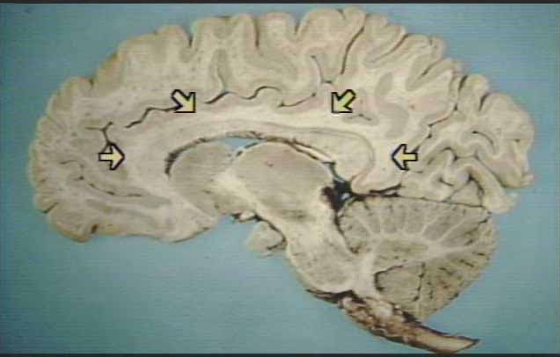
What are the arrows pointing at?
Limbic Region in the cingulate gyrus
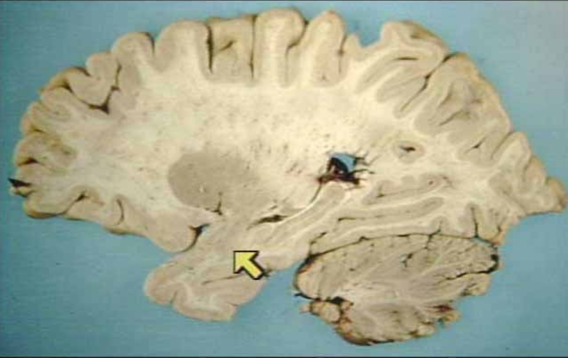
What is the arrow pointing at?
Hippocampus
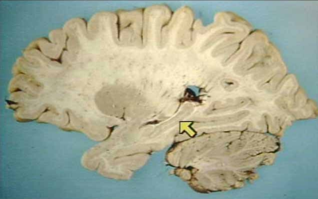
What is the arrow pointing at?
Amygdala
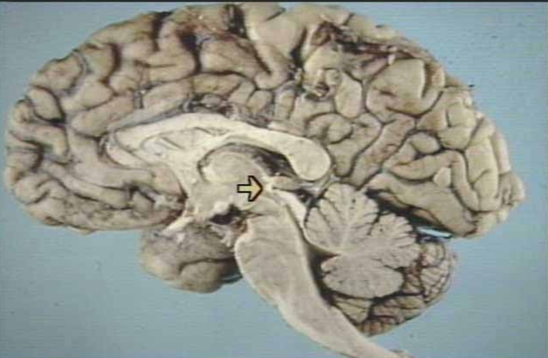
What is the arrow pointing at?
Posterior Commissure (arrow)
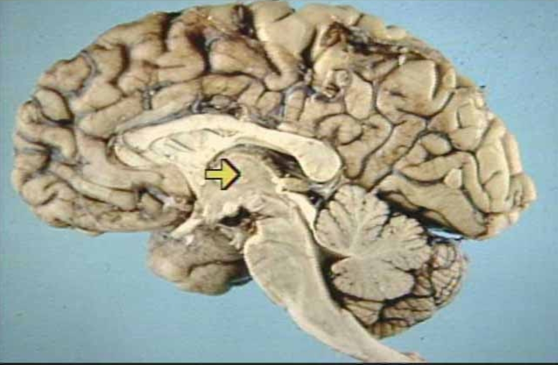
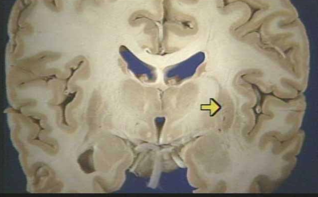
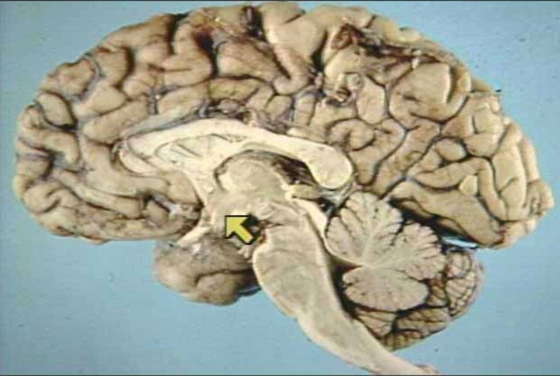
What is the arrow pointing at?
Thalamus (Arrow)

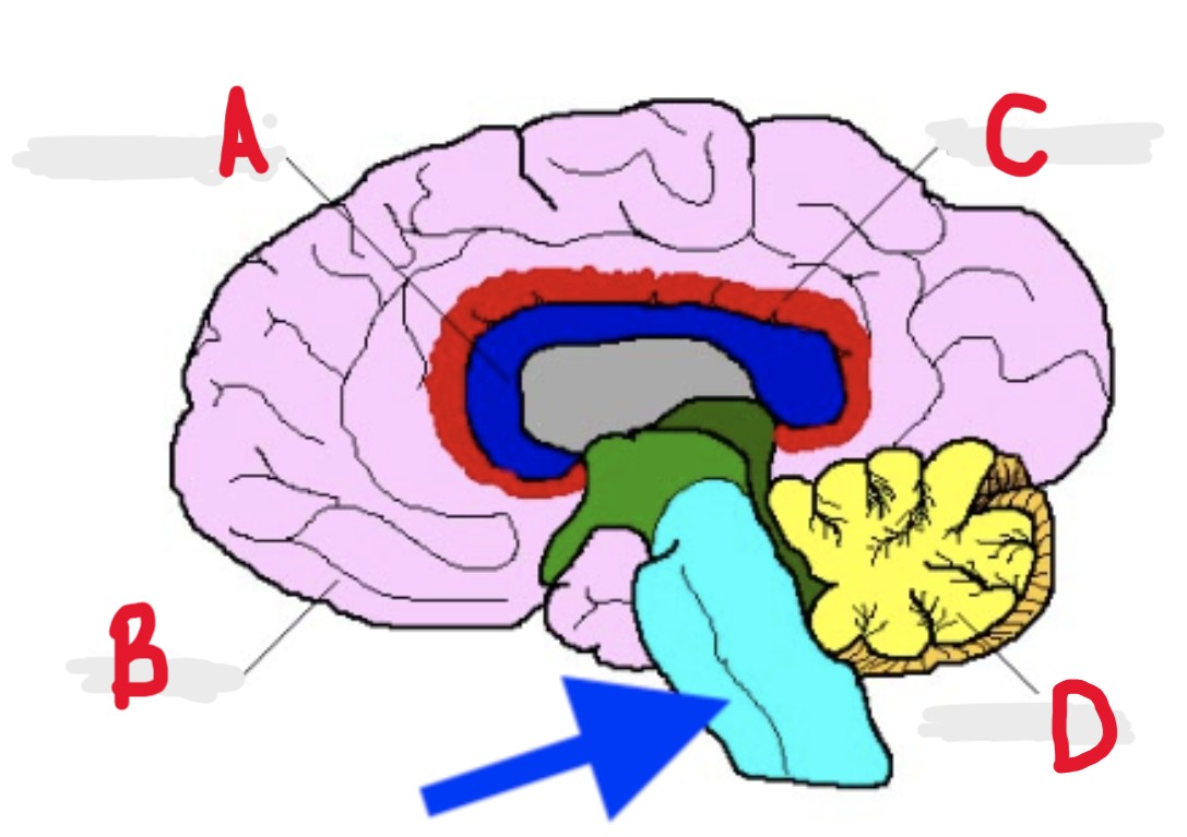
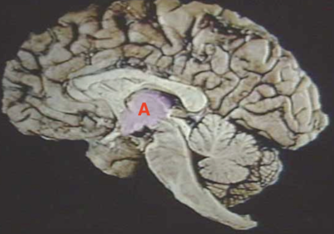
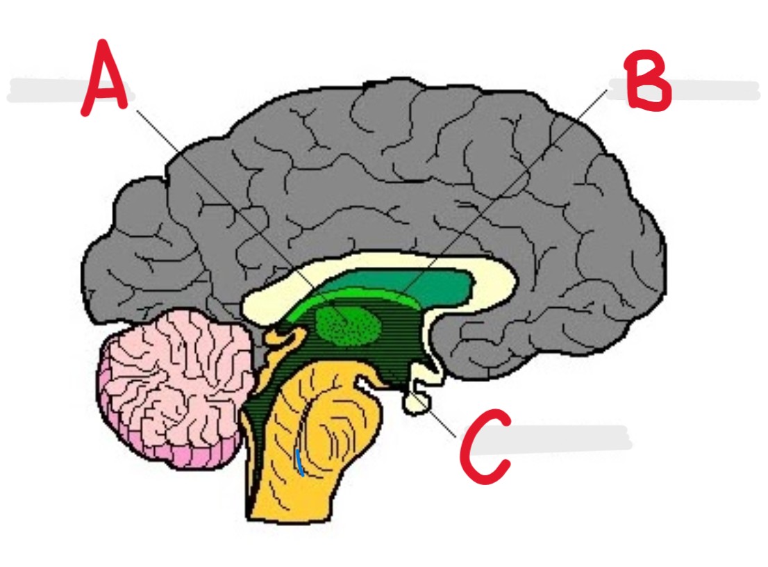
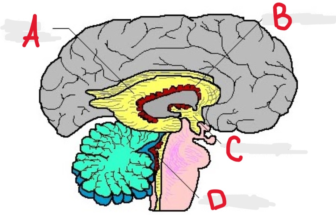
A
Hypothalamus + Thalamus

B
Midbrain

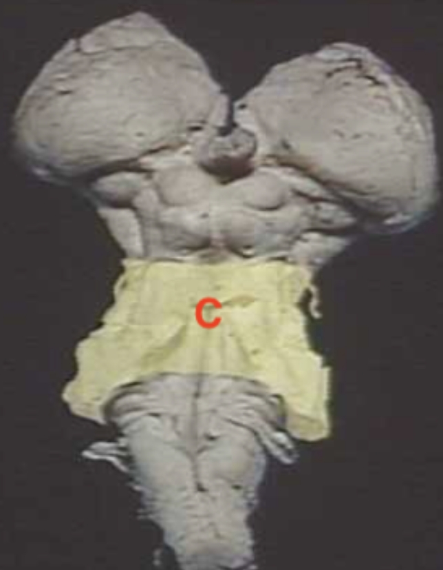
C
Pons
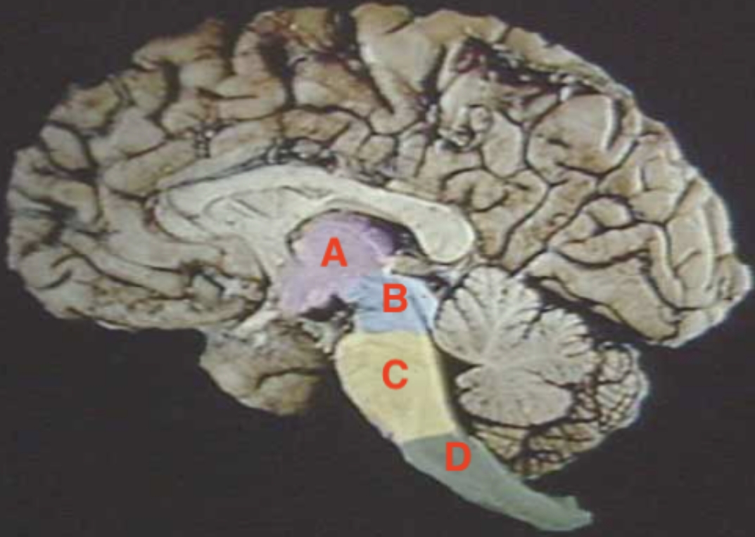
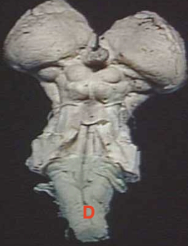
D
Medulla oblongata
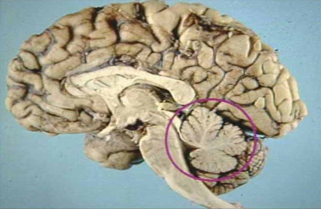
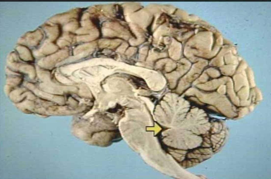
What is in the circle?
Cerebellum
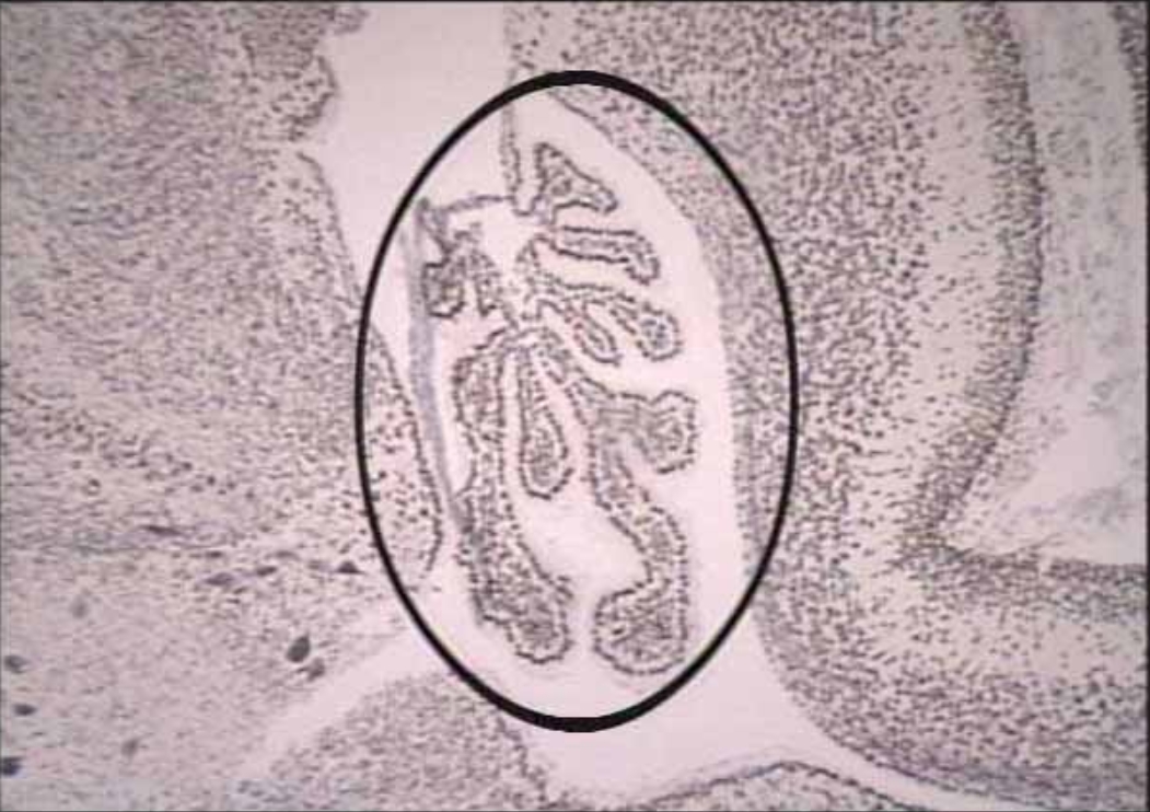
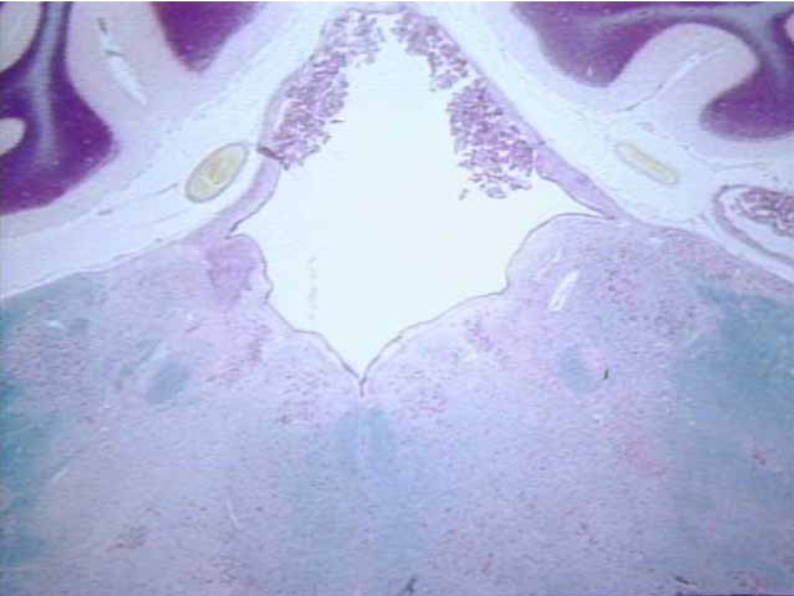
What is circled?
Choroid Plexus of a newborn
What is the arrow pointing at?
Fourth Ventricle (arrow)
What is the arrow pointing at?
Pia Matter (Inner Arrow)
What is the arrow pointing at?
Arachnoid Membrane (Arrow)
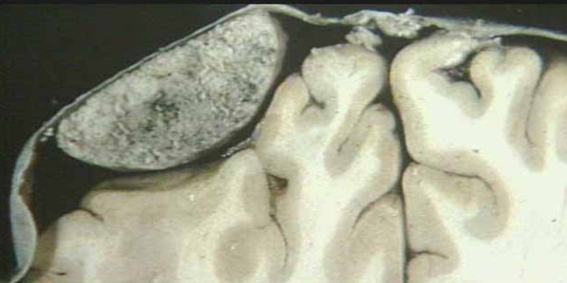
What is this?
Cerebral Meningioma

A
Premotor Cortex

B
Precentral Gyrus
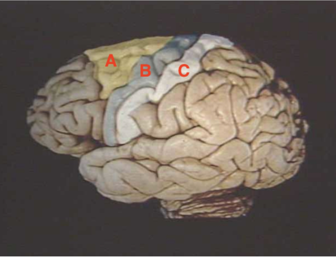
C
Postcentral Cyrus
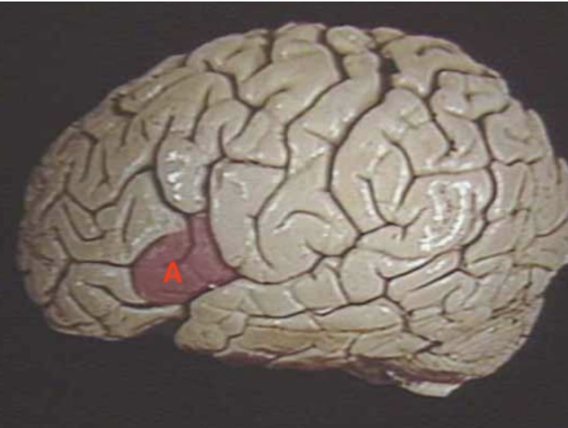
A
Broca’s Area
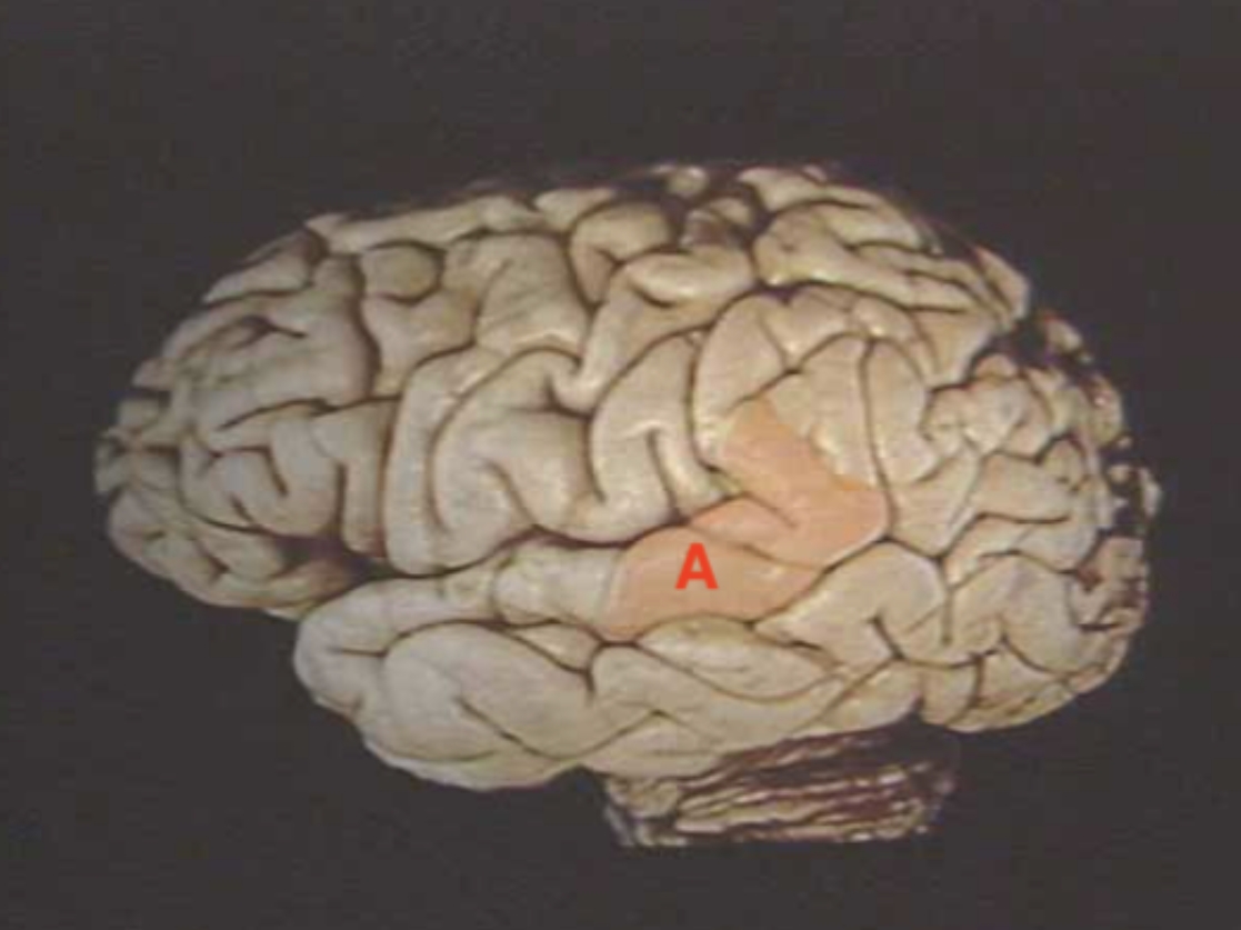
A
Wernicke’s Area
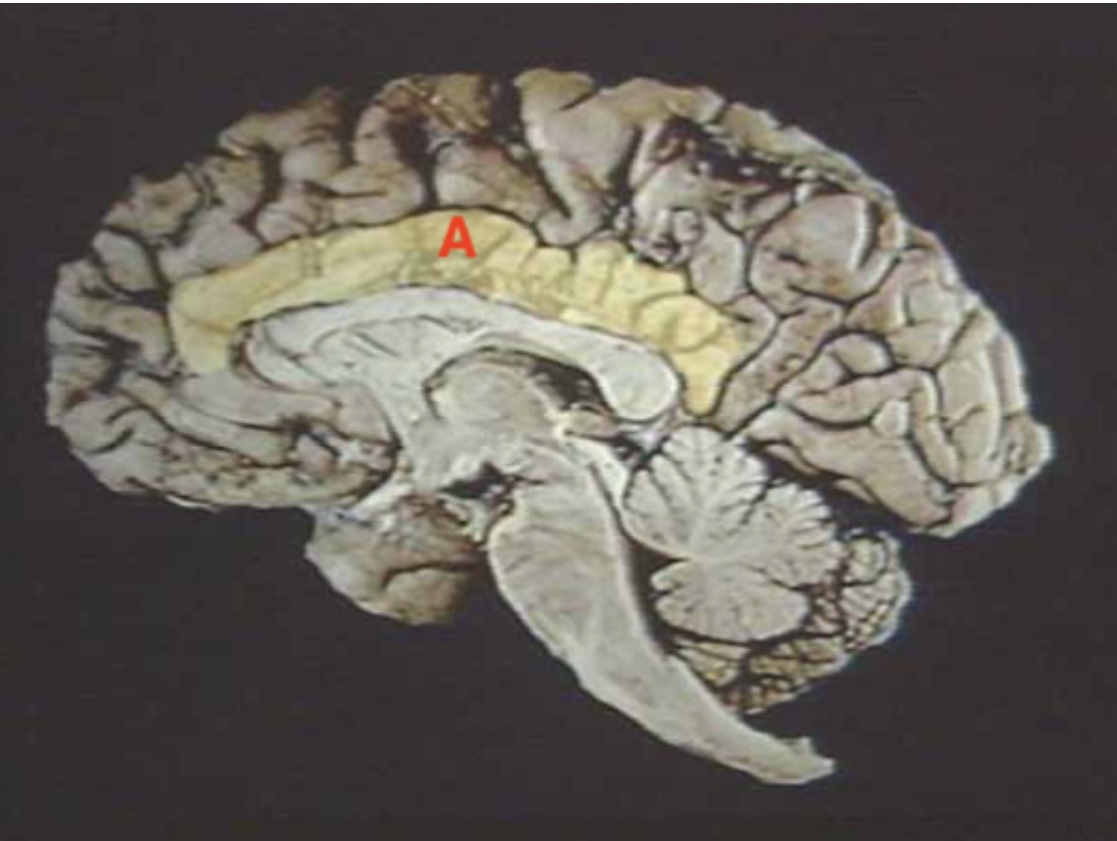
A
Corpus Callosum
A
Corpus Callosum (colorful illustration)

B
Cerebrum (colorful illustration)

C
Limbic Cortex (Colorful illustration)

D
Cerebellum (Colorful Illustration)
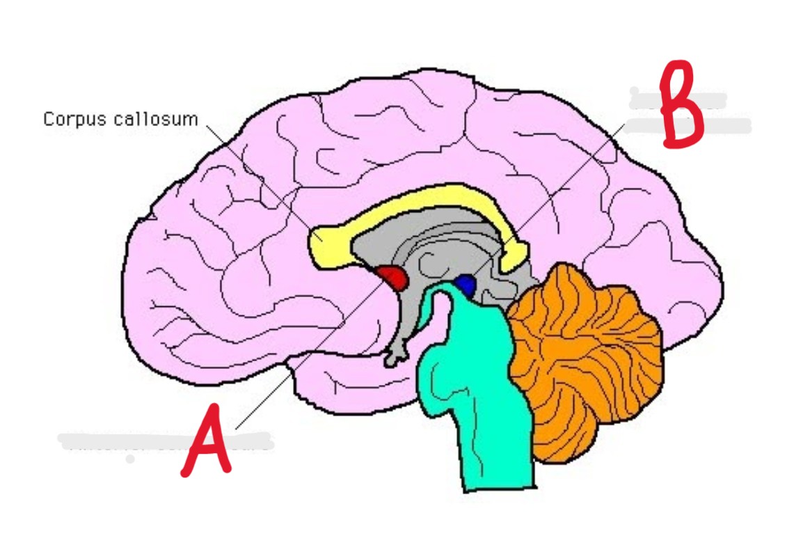
A
Anterior Commissure (colorful medial illustration)

B
Posterior Commissure (Colorful medial Illustration)
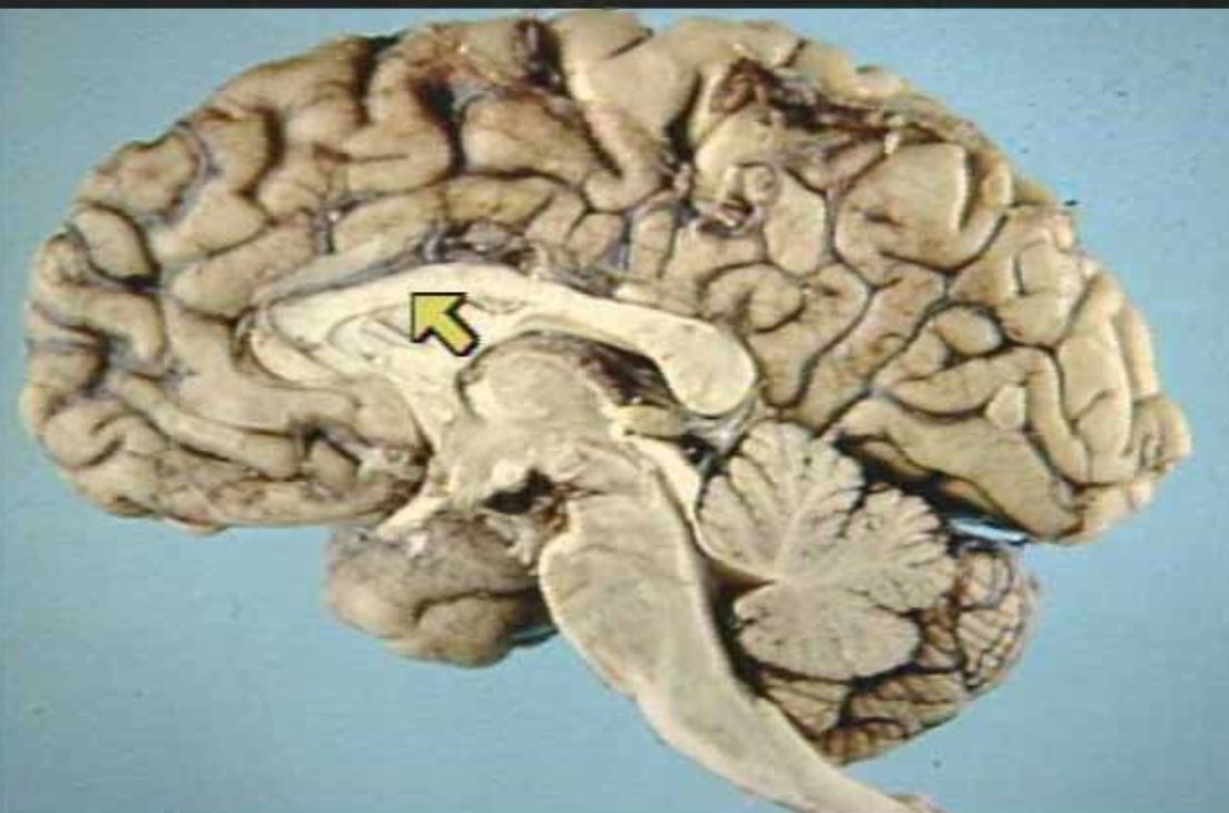
What is the arrow pointing at?
Corpus Callosum (Real Medial View of Brain)

What is the arrow pointing at?
Anterior Commissure (Real Medial View of Brain)
What is the arrow pointing at?
Basal Ganglia (Real Inferior View of Brain)
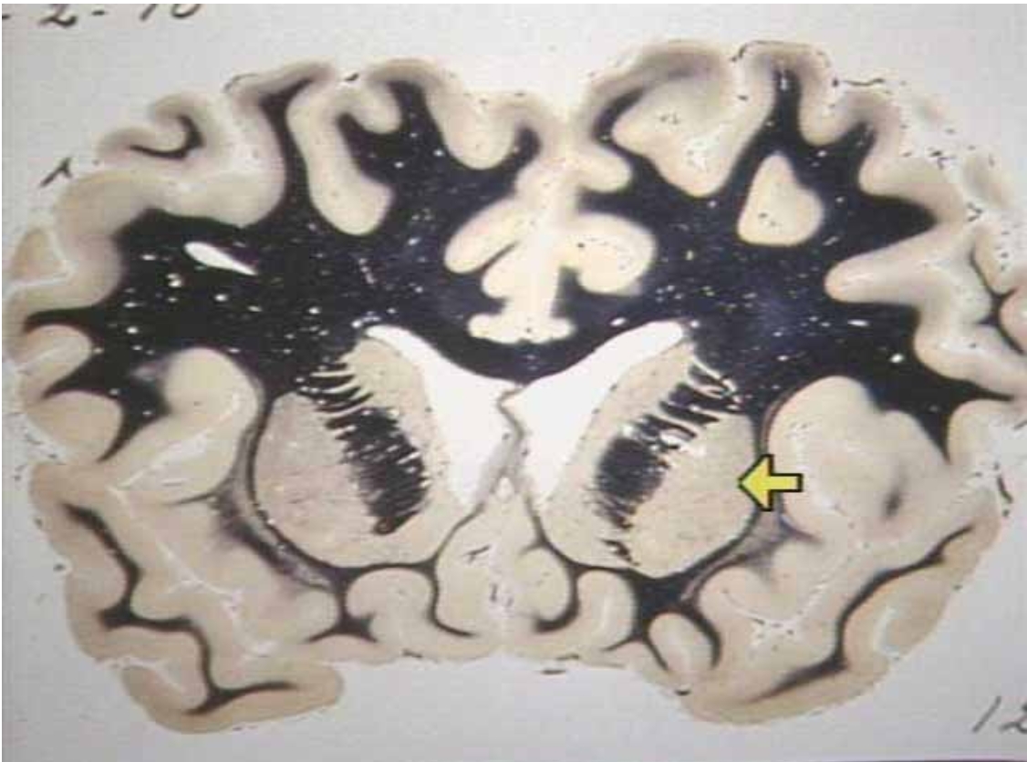
What is the arrow pointing at?
Lentiform Nucleus (Real view of brain)
A
diencephalon (Real medial view of brain)

A
Thalamus (Medial colorful illustration)

B
Epithalamus (Medial Colorful Illustration)
C
Hypothalamus (Medial Colorful Illustration)
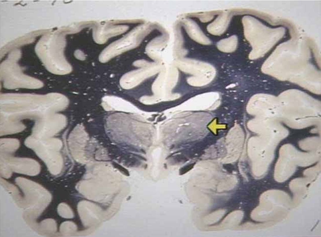
What is the arrow pointing at?
Thalamus (Frontal section view)
What is the arrow pointing at?
Hypothalamus (Real medial view of brain)
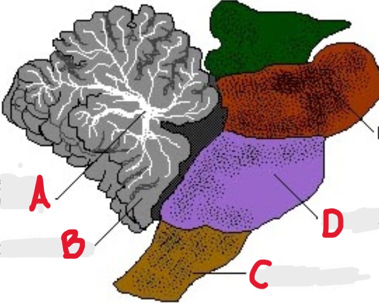
A
Pineal Gland (Real view)
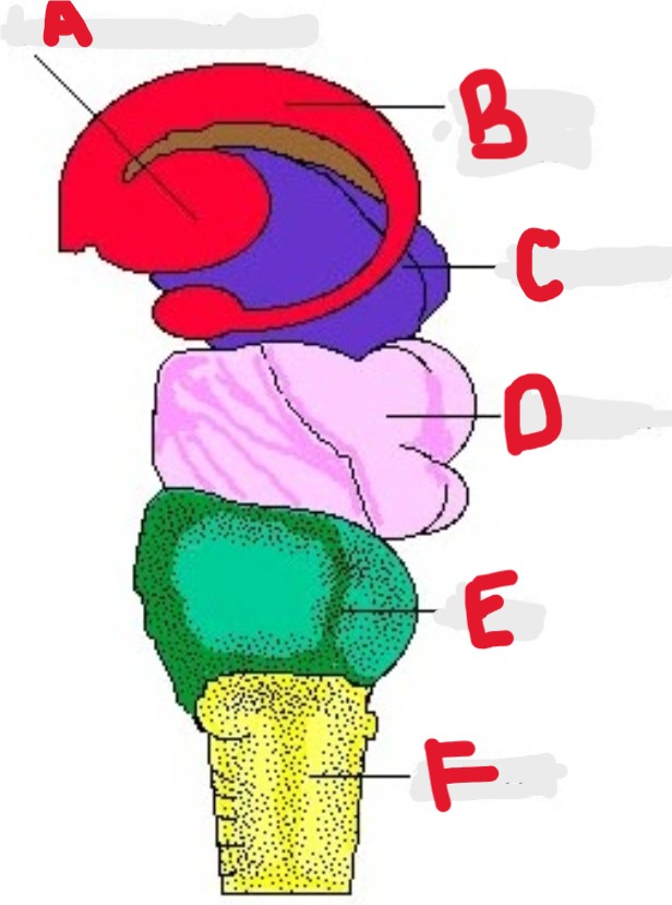
A
Lentiform Nucleus (Colorful side illustration)

B
Caudate Nucleus (Colorful side illustration)

C
Thalamus (Colorful side illustration)

D
Midbrain (Colorful side illustration)

E
Pons (colorful side illustration)

F
Medulla (Colorful side view)
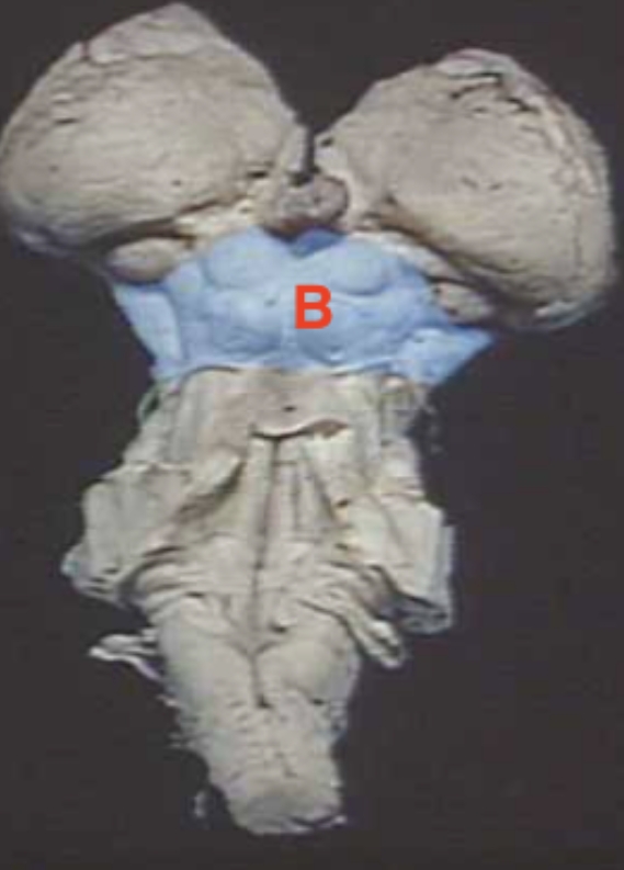
B
Midbrain (Real view)
C
Pons (Real view)
D
Medulla Oblongata (Real View)
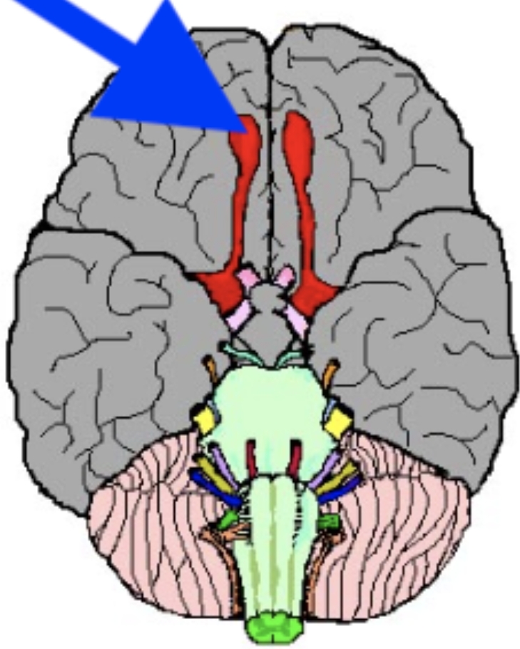
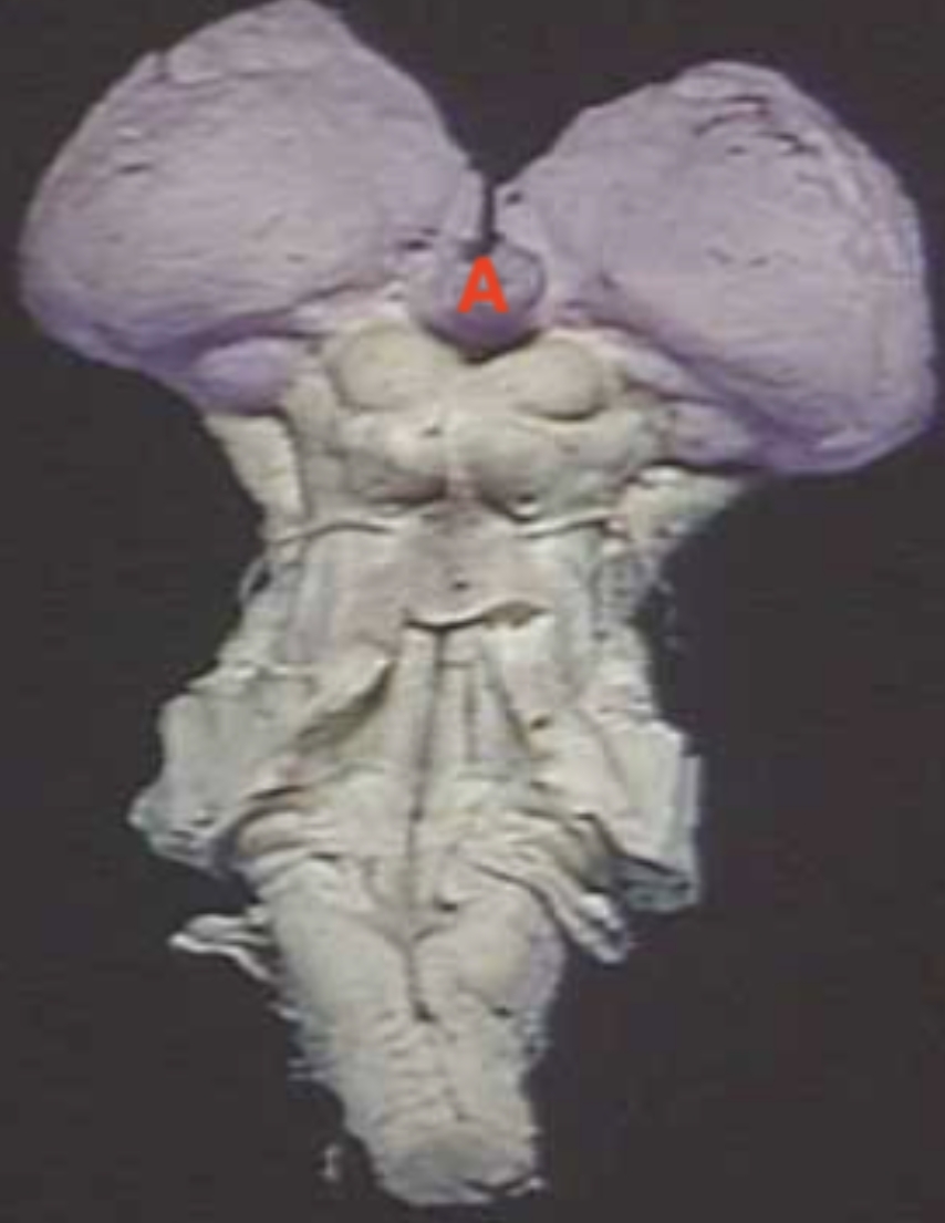
What is the arrow pointing at?
Olfactory Nerve (Inferior View Colorful Illustration)
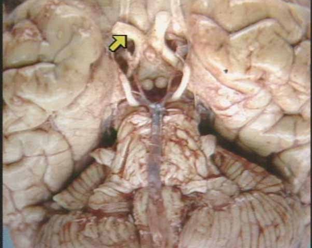
What is the arrow pointing at?
Optic Nerve (real inferior brain view)

A
Arbor Vitae (Colorful side illustration

B
cerebellum (colorful side illustration)

C
Spinal cord (Colorful Side illustration view)
D
medulla (Colorful Side illustration view)
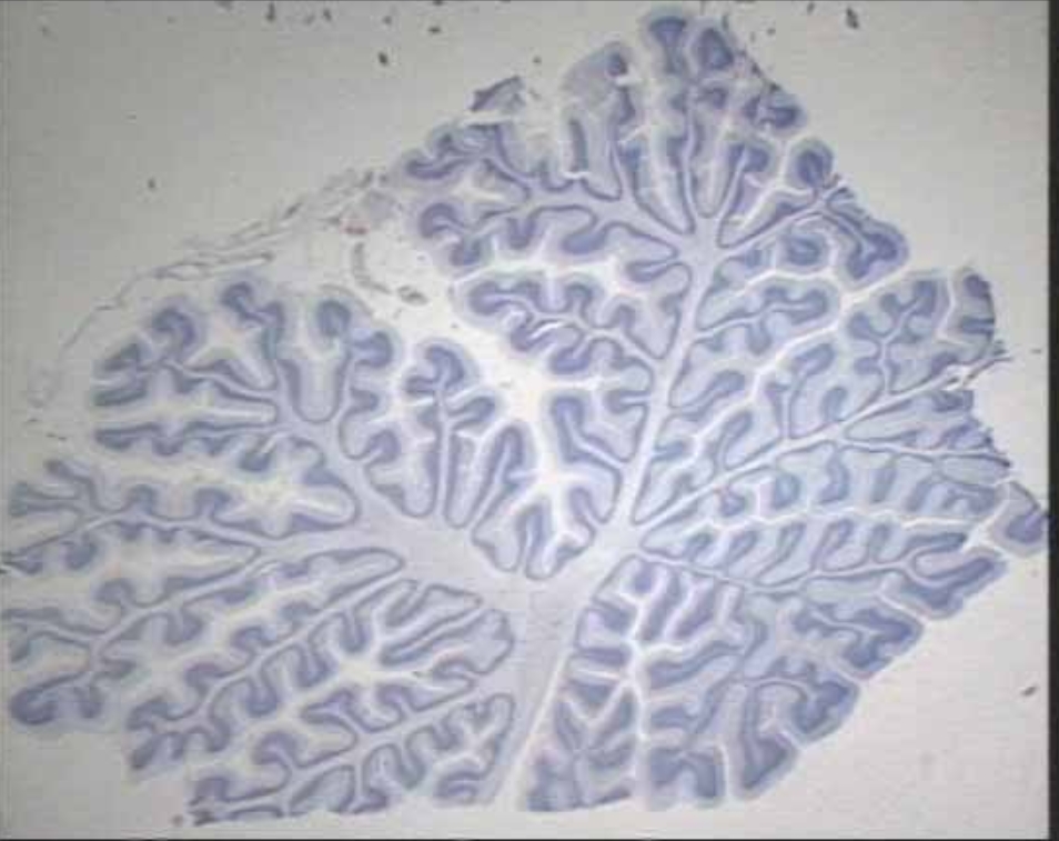
What is this
Midsagittal section of cerebellum (Microscopic real view)

A
Lateral Ventricle (Medial colorful illustration)

B
Choroid Plexus (Medial colorful illustration)

C
3rd Ventricle (Medial colorful illustration)
D
4th Ventricle (Medial colorful illustration)
What is this ?
Choroid Plexus (Microscopic real view)
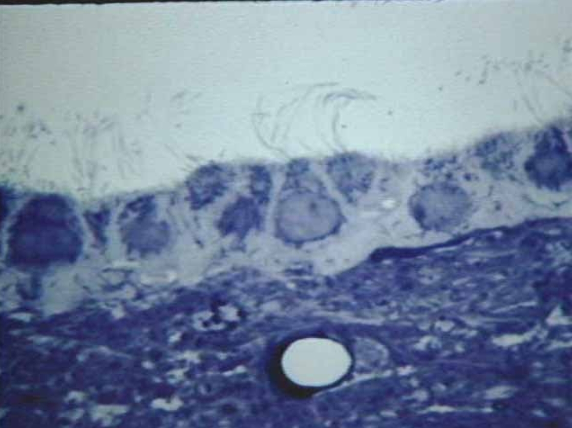
What is this?
Ependymal Cells (Microscopic real view)
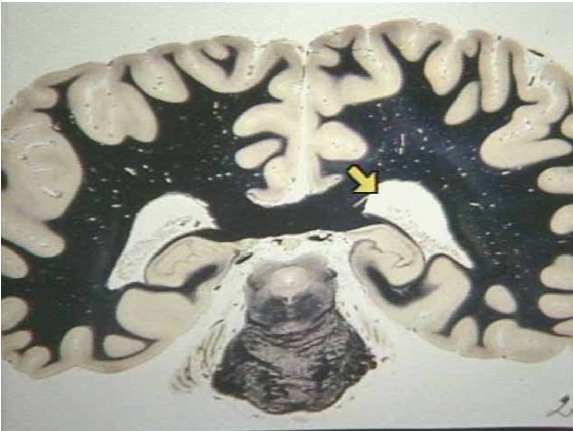
What is the arrow pointing at?
Occipital Horns of the left lateral ventricle (Real brain view)
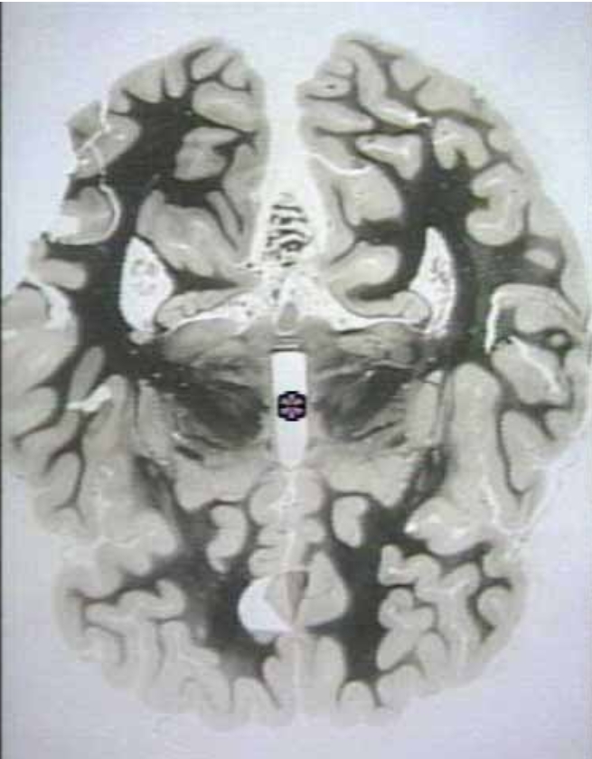
What area is the star in?
Third Ventricle (Real brain view)

A
Dura Mater (meninges illustration)

B
Arachnoid Mater (meninges illustration)

C
Pia Mater (meninges illustration)
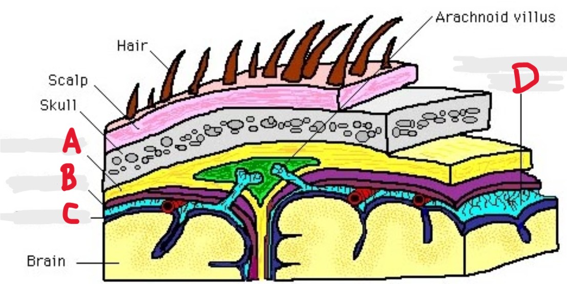
D
Subarachnoid space (meninges illustration)

A
Cervical Enlargement

B
Lumbar Enlargement

C
Conus Medullaris

D
Cauda Equina

E
Filum Terminale

F
Coccygeal
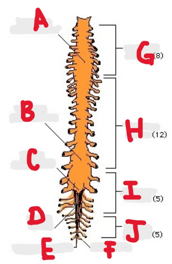
G
Cervical Nerves

H
Thoracic Nerves

I
Lumbar Nerves

J
Sacral Nerves

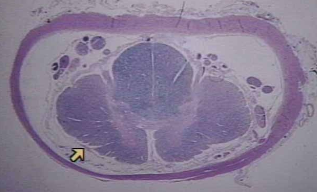
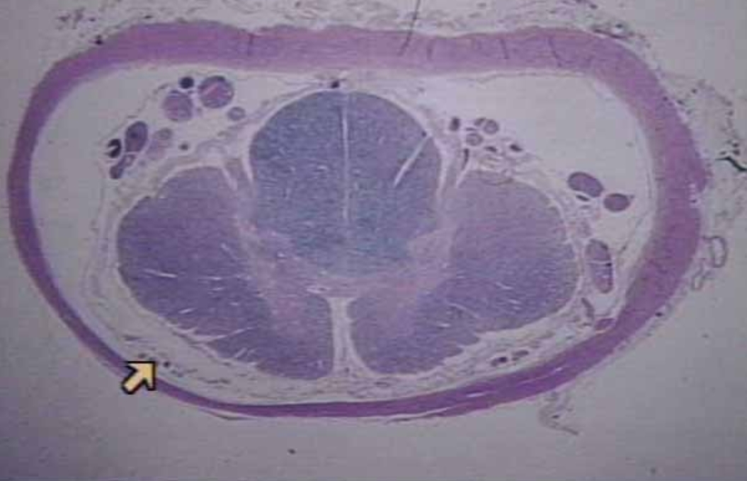
A
Dura Mater (Microscopic Real View)

B
Gray Mater (Microscopic Real View)
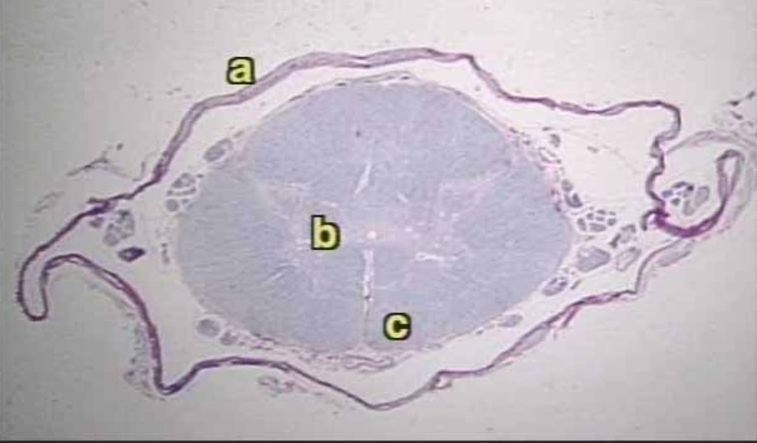
C
White Mater (Microscopic Real View)
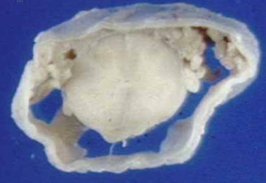
What is this?
Spinal Cord with Dura Mater (real view)
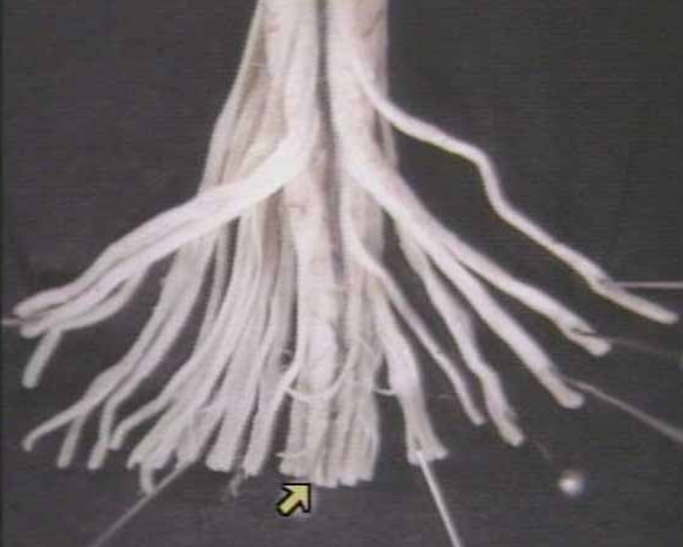
What is this?
Cauda Equina and conus Medullaris

What is the arrow pointing at?
Peripheral Nerve (Microscopic real view)
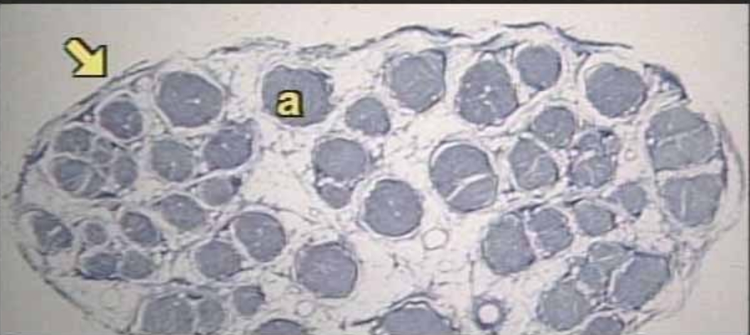
A
Fascicle inside peripheral nerve?
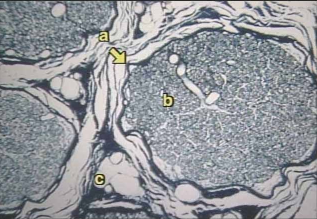
A
Perineurium of Fascicle (Microscopic live View)
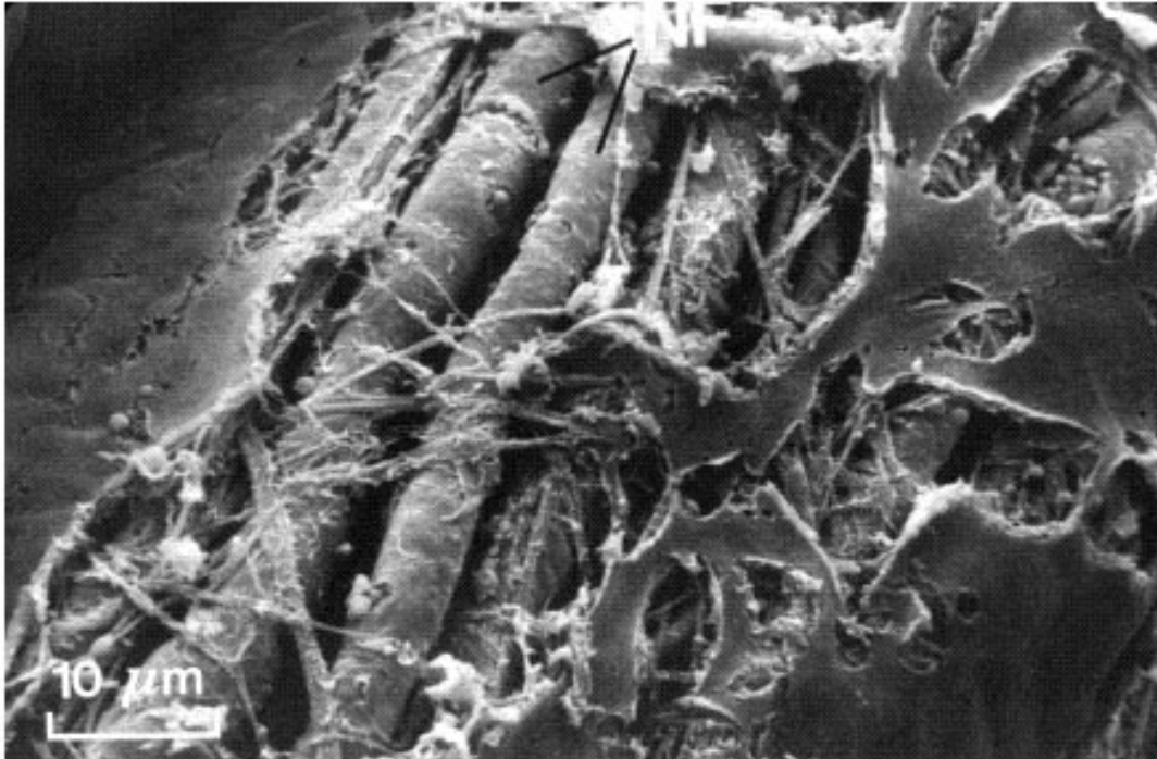
What is this?
myelinated nerve fibers (Microscopic real view)