Lecture 2 - Cell Types and Protein Components
1/34
There's no tags or description
Looks like no tags are added yet.
Name | Mastery | Learn | Test | Matching | Spaced | Call with Kai |
|---|
No analytics yet
Send a link to your students to track their progress
35 Terms
What are the 5 myeloid cells (innate)?
Macrophage, dendritic cell, neutrophil, basophil, eosinophil
What are the 3 phagocytes?
Macrophage, dendritic cell, neutrophil
Innate cells: phagocytes
Function to surround, engulf, and break down microbes, small particles, and apoptotic host cells
Leukocytes =
Myeloid cells + Lymphoid cells
What are the 4 granulocytes?
Neutrophil, basophil, eosinophil, mast cell
Cytoplasmic granules
Pre-filled with inflammatory and mediators
Degranulation
Kills microbes in the area
Amplifies immune responses by recruiting more immune cells
What are the first cells on the scene of infection?
Neutrophils (most abundant)
Neutrophils (phagocyte and granulocyte)
Rapidly migrate to the site of infection and inflammation
Release their DNA as they die to trap bacteria to limit dissemination
Netosis
DNA forms sticky “nets”
Eosinophils (granulocyte)
Express cytoplasmic granules containing enzymes that are harmful to the cell walls of parasites
High in parasitic worm infections and allergy
Basophils (granulocyte)
Involved in allergic responses and help fight parasitic worms
Bind IgE, when IgE sees it’s antigen it causes histamine release
Mast cells (granulocyte)
Rapidly secrete pro inflammatory factors (histamine)
Activated when IgE binds to antigen
Antigen
A substance (from a microbe or self) that stimulates an immune response
Antigen presenting cell (APC)
Capture antigens and process them for presentation to initiate the adaptive T cell responses
Antigen presentation
Phagocytosis of bacteria by APC (antigen presenting cell)
Digestion of microbial antigens
Presentation of antigen on MHC to T cell
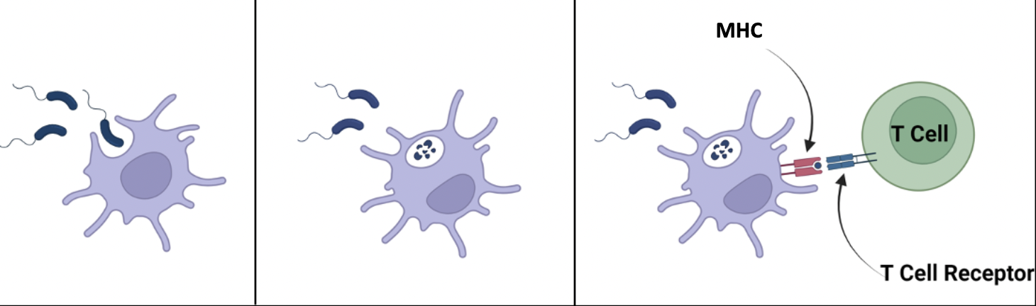
Dendritic cells (phagocytes)
Most expert at antigen presentation
Macrophages (phagocytes)
Pretty good at antigen presentation
What are the 3 lymphoid cells?
T cells, B cells, NK cells
Each B and T cell cell has a unique surface receptor that responds to a:
Specific antigen
Natural Killer cells (lymphoid)
Lack a variable antigen receptor and function like innate cells
Antimicrobial peptides (AMPs)
Small proteins secreted by epithelial cells, phagocytes, and keratinocytes that are toxic to m icrboes
Antibodies
Secreted version of the B cell antigen receptor
Do not directly kill, instead tag foreign antigens for removal by other immune cells or protein factors
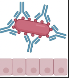
Neutralization
Process where antibodies block pathogens
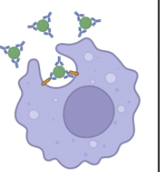
Opsononization
Process where antibodies (mainly IgG) coat the surface of pathogens, acting as "tags" (opsonins) that enhance recognition and destruction by phagocytes, such as macrophages and neutrophils
Complement activation
Process where C1q binds to antibody-antigen complexes (mainly IgM or IgG subclasses) on pathogens, initiating a protease cascade
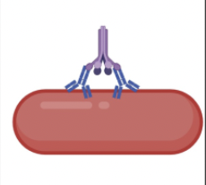
Classical pathway
Antigen-antibody complexes
Starts when IgG or IgM binds to pathogen
These antibodies trigger an enzymatic cascade that cleaves C3
C3
Tags (opsonizes) pathogens to be eaten
Activates C5, causing a membrane attack complex to form on the pathogen
C3b
Opsonizes pathogens
C3A (inflammation) and C5a
Recruit more immune cells
C5b
Initiates membrane attack complex that makes holes and kills microbes
Alternative (Spontaneous) Pathway
Antibody independent
Spontaneous hydrolysis of C3
What are the 3 ways the complement system controls infection?
Amplifies inflammation
Increases phagocytosis
Directly kills pathogens
Cytokines
Responsible for cell to cell communication during an immune response
Chemokines
Direct traffic - draw immune cells towards sites of infection or inflammation