Lecture 5 DDX of Neoplasms
1/45
There's no tags or description
Looks like no tags are added yet.
Name | Mastery | Learn | Test | Matching | Spaced |
|---|
No study sessions yet.
46 Terms
Ivory vertebra
-Idiopathic
-Hodgkin’s Lymphoma
-Osteoblastic METS
-Paget’s
FEGNOMASHIC
-Fibrous Dysplasia
-Enchondroma
-Giant Cell tumor
-Non-Ossifying fibroma/Fibrous cortical defect
-Osteoblastoma
-Metastasis/Myeloma (blowout mets)
-Aneurysmal bone cyst (ABC)
-Simple bone cyst
-Intraosseous Lipoma
-Chondroblastoma
FEGNOMASHIC Under 25 Lesions
-Enchondroma
-NOF/FCD
-Osteoblastoma
-ABC
-Simple Bone Cyst
-Chondroblastoma
-Lipoma
FEGNOMASHIC 20 to 40 years old lesions
-Enchondroma
-Giant cell tumor
-Fibroxanthoma
-Lipoma
FEGNOMASHIC Over 40 Lesions
-Enchondroma
-Mets (blowout)
-Myeloma
-Lipoma
FEGNOMASHIC w/ Pain
-Giant cell tumor
-Osteoblastoma
-Mets
-Myeloma
-ABC
-Chondroblastoma
-Lipoma (dull)
FEGNOMASHIC w/ no pain
-Enchondroma
-NOF/FCD
-Mets
-Myeloma
-Simple Bone cyst
-Lipoma
Enchondroma (Summary)
-Geo lytic appearance is more common in hands/ feet than long bones
-10 to 30 years old but have it forever
-No pain
-MC tumor of hands/feet
-Benign cartilaginous tumor
-50% show stippled calcification
Giant Cell Tumor (Summary)
-20 to 40 years old
-Painful
-no sclerotic margin
-Subarticular extension (up to the joint)
NOF/FCD (Summary)
-4 to 20 years old (FCD 4-8; NOF 8-20)
-Painless
-Eccentric
-Hazy, ground glass matrix
-Most common in proximal/distal tibia and distal femur
NOF/FCD on X-ray

Osteoblastoma (Summary)
-10 to 25 years old
-Painful
-Like a giant osteoid osteoma without as much pain or sclerosis
-Most common in the spine and long bones
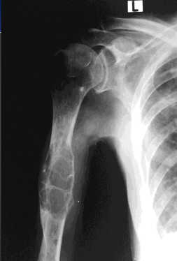
Metastasis (Blowout)
-Over 50 years old
-May or may not have pain
-From renal, adrenal, thyroid, skins (RATS)
-In axial skeleton and extremities proximal to elbow/knees
Blowout Mets on X-ray
Myeloma (Summary)
-Over 40 years old
-May or may not have pain
-Solitary version of multiple myeloma
-Most common in axial skeleton and extremities proximal to elbows/knees
-Much less common than mets
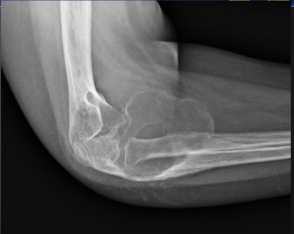
ABC (Summary)
-Less than 20 years old
-Painful
-Rapidly expansile
-Most common in long bones, spine, and pelvis
-Often seen in other lesions (GCT most common)
-Indicated by fluid fluid levels on advanced Imaging
ABC on X-ray
Simple Bone Cyst (summary)
-3 to 14 years old
-Painless until fracture (2/3 present with fracture)
-Most common in proximal humerus and femur as well as calcaneus
-Central within the long bone
-Often fracture (will have pain in this case) with possible fallen fragment
Intraosseous Lipoma (Summary)
-5 to 70 years old
-Dull pain or painless
-Most common in calcaneus and proximal long bones of lower extremity
-Possible central calcification (cockade sign)
Chondroblastoma (Summary)
-10 to 25 years old
-Painful
-In apophyses/epiphyses
-50% show punctate calcifications
Benign Lesions in posterior elements
-ABC
-GCT
-Osteoblastoma
-Osteochondroma
-Osteoid Osteoma
ABC age?
-Less than 20 years old
GCT age?
-20 to 40
Osteoblastoma age?
-10 to 25
Osteochondroma age?
-Less than 20
Osteoid Osteoma age?
-10 to 25
Most common primary malignancies to show geographic lytic pattern (Blowout mets)
-Renal
-Adrenal (Pheochromocytoma)
-Thyroid
-Skin (melanoma)
Most Common malignancies to metastasize to bone?
-Prostate
-Breast
-Kidney
-Thyroid
-Lung
(PB KTL)
Most Common Primary Osseous Malignancies in order
-Multiple Myeloma
-Osteosarcoma
-Chondrosarcoma
-Ewing Sarcoma
Multiple Myeloma Buzzwords
-Raindrop skull
-Bence Jones Proteinuria
-M spike on electrophoresis
-Vertebra plana
Osteosarcoma (Summary)
-Most common primary malignancy in kids
-Primary 10 to 25
-Secondary is over 50
-Blastic, mixed, or lytic lesion in around the knee
-Large osseous soft tissue mass
-Codman’s Triangle
-Sunburst periostitis
Chondrosarcoma (Summary)
-30 to 60 years old
-ICE lesion if intramedullary
-Most common in proximal long bones and pelvis
-Large soft tissue mass with arcs and rings calcification
-Most common malignancy to form from enchondroma and osteochondroma
Ewing Sarcoma
-10 to 25
-Diaphyseal
-Clinically and radiographically mimic infection
-Laminated periostitis
-Large soft tissue mass
ICE Lesion
-Punctate/Stippled/Flocculant calcification in the medullary canal of a Long bone
If Ice lesions is present lead with chondrosarcoma if:
-Over 40 years old
-over 5 cm in length
-Endosteal scalloping >2/3 width of cortex
-Pain not due to Fx
-Overt signs of aggression
Osteolytic (Geographic)
-Narrow zone of transition
-Possible sclerotic border
-Sharp margin/well-circumscribed
-Indicates slower growth (Likely benign)
Osteolytic (Motheaten)
-Wide zone of transition (Poorly defined margins)
-Multiple osteolytic punched out lesions
-Indicates more rapid growth/spread (Likely aggressive)
Osteolytic (Permeative)
-Wide zone of transition (poorly defined margins)
-Indicates more rapid growth/spread (likely aggressive)
Narrow Zone of Transiiton
-Well-defined border
-Seen with geographic lysis. Also common with blastic lesions
-Typically non-aggressive
Wide Zone of transition
-Indistinct margins
-Seen with moth eaten and permeative lysis
-Typically aggressive (Malignant or infection)
Periosteal Reaction
-Stimuli cause periosteum to react in various ways depending on degree of aggression
-10 to 21 day latency
Solid Periosteal Reaction
-Least aggressive pattern
-Caused by Osteoid osteoma, stress fracture, venous stasis, HOA, Intracortical abscess
Laminated Periosteal Reaction
-AKA lamellated, onion skin, layered
-Caused by cyclic exposure to stimuli
-may become solid
-Classically associated with Ewing sarcoma and infection
Sunburst Periosteal Reaction
-AKA hair on end, perpendicular brushed whiskers
-Seen in more aggressive process (Osteosarcoma)
Codman’s Triangle
-Most aggressive periosteal reaction
-Triangle of new bone with subperiosteal extension of aggressive process
-Seen in Osteosarcoma
Most common osseous site of infection and tumor?
-Metaphysis