muscles PART 1 [types of muscles and structure of skeletal muscle]
1/14
There's no tags or description
Looks like no tags are added yet.
Name | Mastery | Learn | Test | Matching | Spaced | Call with Kai |
|---|
No analytics yet
Send a link to your students to track their progress
15 Terms
key terms for muscle structure (2)
-multinucleate or uninucleate
-striated
types of muscle
→found where?
→function
→structure
→arrangement
→contraction speed and contraction duration
→control
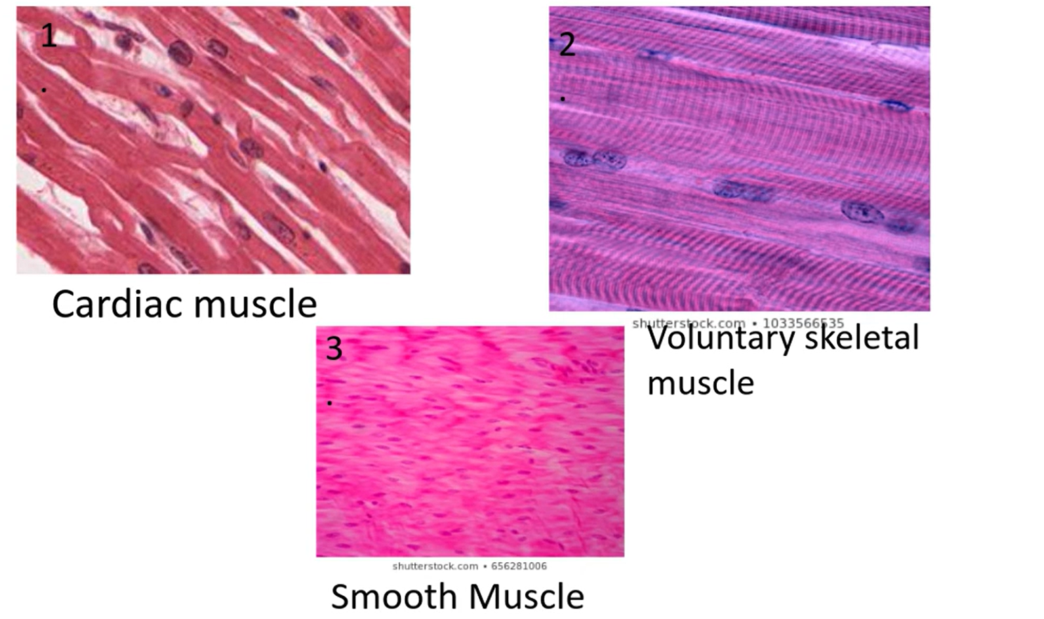
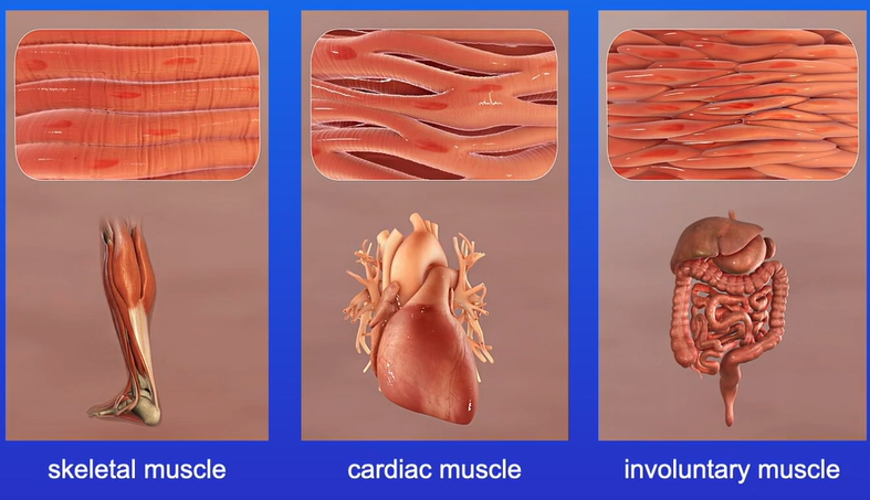
CARDIAC MUSCLE
found where?
-found only in the heart
function
-contracts to pump blood around the body
structure
-striated (fainter than skeletal)
-contain branched cells
-fibres are uninucleate
-fibres are connected by intercalated discs
arrangement
-arranged in a branched network
contraction speed and contraction length
-intermediate contraction speed
-intermediate contraction length
control
-involuntary
SKELETAL MUSCLE (voluntary muscle)
found where?
-attached to bone e.g. found intercoastal muscle (between the ribs)
function
-moves bones to allow movement
structure
-striated due to bands of actin & myosin
-contain cylindrical cells
-fibres are multinucleate
arrangement
-regular and parallel arrangement of fibres so muscle contraction occurs in one direction
contraction speed and contraction length
-fast contraction speed
-short contraction length
control
-voluntary
INVOLUNTARY MUSCLE (smooth muscle)
found where?
-bronchioles, arteries etc [airways, blood vessels]
function
-control diameter of bronchioles
-controls pupil size
-involved in peristalsis
structure
-unstriated [tip: smooth so no stripes]
-contain long spindle-shaped cells
-fibres are uninucleate
arrangement
-no regular arrangement of fibres so muscle contraction occurs in different directions
contraction speed and contraction length
-slow contraction speed
-long contraction length
control
-involuntary
why are cardiac muscle fibres branched?
to allow nerve impulses to spread quickly through the muscle
![<p>name muscles [microscope images]</p>](https://assets.knowt.com/user-attachments/974e8254-ef10-4382-b14c-1873163ede8c.png)
name muscles [microscope images]
![<p>labelled cardiac muscle</p><p>[intercalated discs are the faint lines that connect the fibres]</p>](https://assets.knowt.com/user-attachments/867ff0ed-2881-43e1-89c5-73d27cf3787b.png)
labelled cardiac muscle
[intercalated discs are the faint lines that connect the fibres]
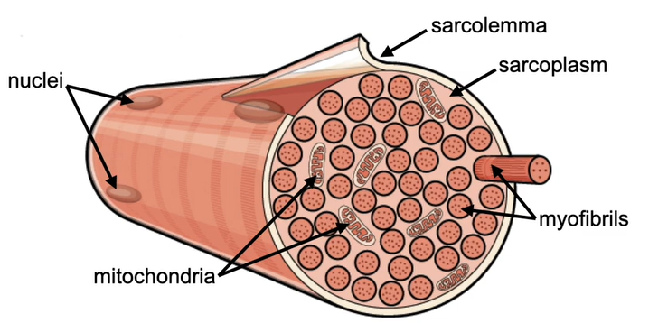
skeletal muscle consists of what
bundles of long, cylindrical muscle fibres
how are muscle fibres formed and why?
cells fuse together to form long muscle fibres, ensuring there's no points of weakness between cells and allows contraction over a greater distance
what do muscle fibres contain? (5)
label diagram
myofibrils
sarcolemma- plasma membrane of muscle fibre
sarcoplasm- cytoplasm of muscle fibre
mitochondria
sarcoplasmic reticulum
what are myofibrils made of
made up of repeating units called sarcomeres
what do sarcomeres contain? [describe structures]
contains protein filaments:
myosin
→thick myofilament [tip: has more letters= thick]
→rod shaped with bulbous heads
actin
→thin myofilament
→two strands twisted around each other
![<p>contains protein filaments:</p><p><strong>myosin</strong></p><p>→thick myofilament [tip: has more letters= thick]</p><p>→rod shaped with bulbous heads</p><p><strong>actin</strong></p><p>→thin myofilament</p><p>→<span>two strands twisted around each other</span></p>](https://assets.knowt.com/user-attachments/b4cbea43-460e-4beb-bb4e-6757ceed701a.png)
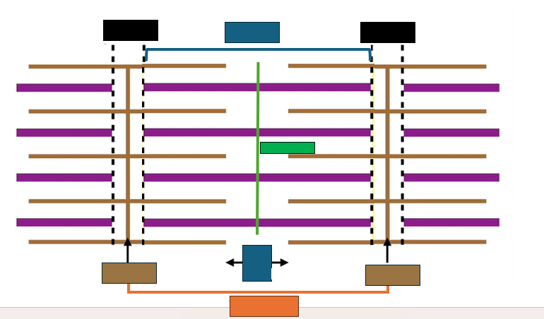
A band (dark band)
-contains myosin and overlapping actin filaments [tip: dark= A band]
I band (light band)
-contains only actin filaments [tip: light= I band]
Z line
-marks the ends of each sarcomere (central line within the I band)
M line
-central line of sarcomere
H zone
-contains only myosin filaments
![<p><strong>A band (dark band)</strong></p><p>-contains <span>myosin and overlapping actin filaments [tip: d<strong><u>a</u></strong>rk= <strong>A </strong>band]</span></p><p><span><strong>I band (light band)</strong></span></p><p><span>-contains only actin filaments [tip: l<strong><u>i</u></strong>ght= <strong>I</strong> band]</span></p><p><span><strong>Z line</strong></span></p><p><span>-marks the ends of each sarcomere (central line within the I band)</span></p><p><span><strong>M line</strong></span></p><p><span>-central line of sarcomere</span></p><p><span><strong>H zone</strong></span></p><p><span>-contains only myosin filaments</span></p>](https://assets.knowt.com/user-attachments/edee97c0-b750-4875-a64d-5eb60ef0545c.png)
what happens to patterns when skeletal muscle contracts?
-Z line moves closer
-sarcomere becomes shorter
-A band remains the same size [myosin filaments do not shorten]
-I band and H zone become smaller
![<p>-Z line moves closer</p><p>-sarcomere becomes shorter</p><p>-A band remains the same size [myosin filaments do not shorten] </p><p>-I band and H zone become smaller </p>](https://assets.knowt.com/user-attachments/c6e13388-a749-474c-9de9-41fe2cce39a5.png)
why does H zone become smaller?
-contains only myosin
-during contraction, actin filaments are pulled closer by myosin, increasing the overlap between actin and myosin
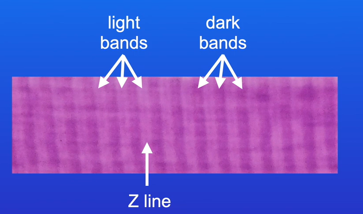
Why A and I bands appear different under a microscope
A band contains myosin filaments
I band contains only actin filaments
